

“What is an MRI Triple Whole Abdomen With Contrast? Purpose, Procedure & Benefits”
MRI Triple Whole Abdomen With Contrast: Complete Guide for Patients
An MRI triple whole abdomen with contrast is a detailed imaging scan that captures three separate phases of your abdominal organs using a special dye to enhance visibility. This comprehensive test helps doctors diagnose liver disease, kidney problems, pancreatic disorders, and other abdominal conditions that standard imaging might miss.
This guide is for patients scheduled for a triple phase MRI abdomen scan, family members supporting someone through the process, and anyone wanting to understand what this abdominal MRI procedure involves.
We’ll walk you through the medical conditions this whole abdomen MRI procedure can detect, from liver tumors to kidney dysfunction. You’ll also learn about the essential MRI contrast preparation steps you need to take before your appointment, including dietary restrictions and medication adjustments. Finally, we’ll break down what happens during the actual abdominal MRI scan and when you can expect your abdomen MRI results.
Understanding MRI Triple Whole Abdomen Scanning Technology
Advanced imaging capabilities for comprehensive organ visualization
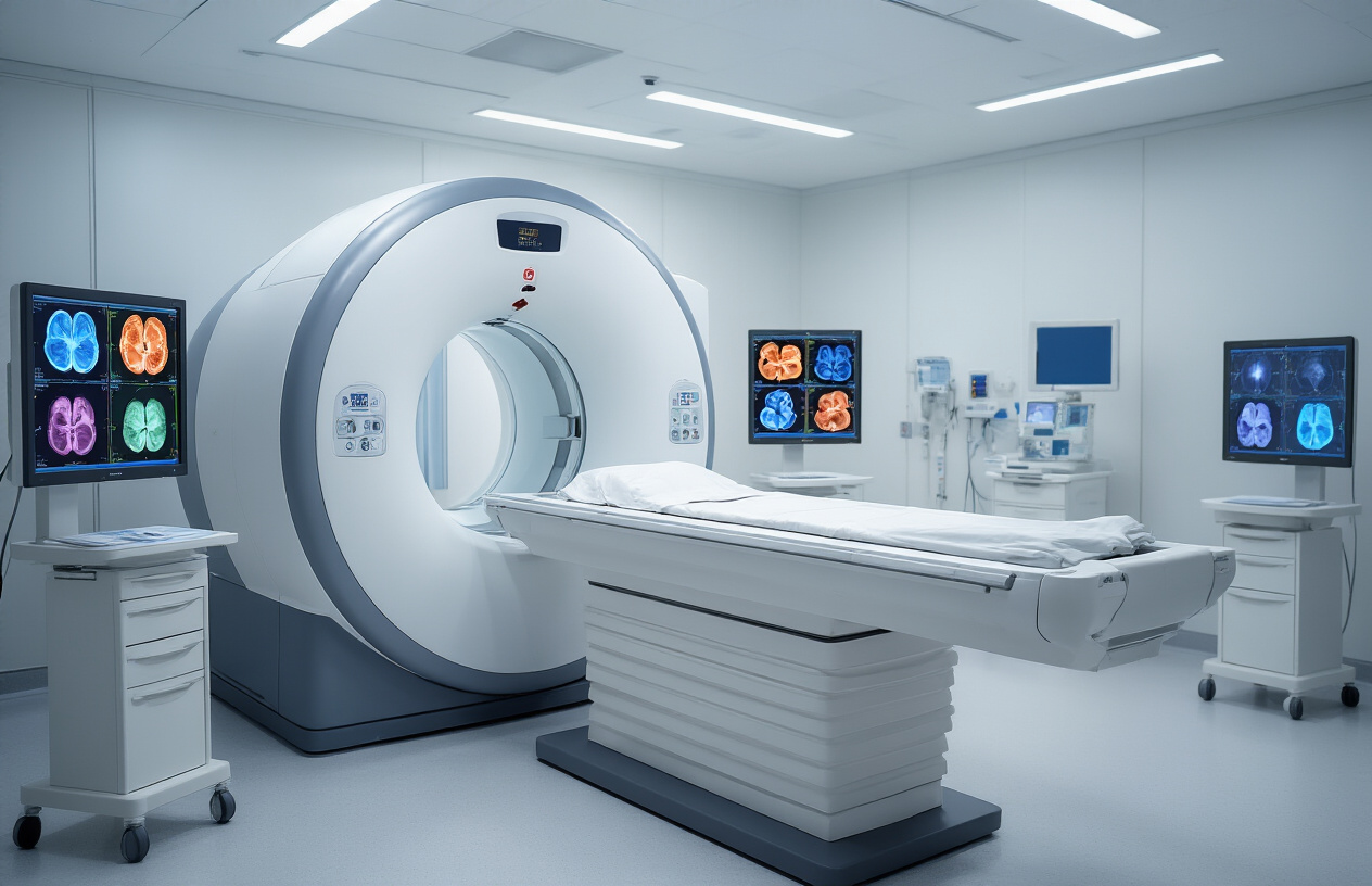
MRI triple whole abdomen scanning represents a breakthrough in medical imaging technology that captures detailed pictures of all abdominal organs simultaneously. This sophisticated approach examines the liver, kidneys, pancreas, spleen, gallbladder, and surrounding tissues in a single comprehensive session. The technology uses powerful magnetic fields and radio waves to create cross-sectional images with exceptional clarity, allowing doctors to spot abnormalities that might be missed with traditional imaging methods.
The scanning process generates multiple image sequences from different angles and planes, providing radiologists with a three-dimensional understanding of organ structure and function. This comprehensive visualization helps identify conditions ranging from tumors and cysts to inflammation and structural abnormalities. The high-resolution images reveal even subtle changes in tissue composition, making early detection of diseases possible.
Modern MRI equipment can differentiate between various tissue types based on their water content and molecular structure. This capability proves invaluable when examining complex abdominal anatomy where organs sit close together and share similar densities on conventional imaging studies.
Contrast enhancement benefits for detailed tissue differentiation
Contrast agents transform the diagnostic power of abdominal MRI scans by highlighting specific tissues and blood vessels. These specialized dyes, typically containing gadolinium, circulate through the bloodstream and accumulate in different organs at varying rates. This selective enhancement creates distinct contrast patterns that help radiologists distinguish between healthy and abnormal tissues.
The contrast material reveals blood flow patterns within organs, highlighting areas with increased or decreased circulation. Tumors often display unique enhancement patterns that help doctors determine their nature and aggressiveness. Inflammatory conditions also show characteristic enhancement patterns that aid in accurate diagnosis.
Contrast-enhanced imaging proves especially valuable for liver evaluation, where the enhancement pattern can differentiate between various types of lesions. The kidneys also benefit significantly from contrast enhancement, as it allows assessment of kidney function and detection of structural abnormalities. Blood vessels become clearly visible with contrast, enabling doctors to identify blockages, aneurysms, or other vascular problems.
Triple sequence methodology for maximum diagnostic accuracy
The triple sequence approach captures images at three distinct time points after contrast injection, providing comprehensive information about organ function and pathology. The pre-contrast phase establishes baseline tissue characteristics without any enhancement. The arterial phase, captured 15-25 seconds after injection, shows early arterial enhancement and reveals highly vascular structures and lesions.
The portal venous phase, occurring 60-80 seconds post-injection, demonstrates peak organ enhancement and optimal visualization of most abdominal structures. Some protocols include a delayed phase at 3-5 minutes, which helps characterize certain lesions and assess kidney function.
This methodical timing allows radiologists to observe how different tissues respond to contrast over time. Malignant tumors often show rapid early enhancement followed by quick washout, while benign lesions typically demonstrate more gradual and sustained enhancement patterns. The triple phase methodology significantly improves diagnostic confidence and reduces the need for additional imaging studies.
Each phase provides unique diagnostic information that contributes to the overall assessment. The arterial phase excels at detecting small liver lesions and assessing pancreatic pathology, while the portal venous phase offers optimal visualization for most routine abdominal evaluations.
Medical Conditions Diagnosed Through Triple Abdominal MRI

Liver diseases and hepatic lesion detection
The MRI triple whole abdomen with contrast excels at detecting liver abnormalities that might be missed by other imaging techniques. This advanced scanning method reveals hepatocellular carcinoma in its earliest stages, often before patients experience symptoms. The triple-phase approach captures arterial, portal venous, and delayed phases, creating a comprehensive picture of liver vascularity and function.
Doctors rely on abdominal MRI scan technology to identify liver metastases from other primary cancers, especially colorectal and breast cancers. The contrast enhancement patterns help distinguish between benign liver cysts and malignant lesions. Cirrhosis patients benefit greatly from regular triple phase MRI abdomen monitoring, as this method tracks disease progression and screens for developing tumors.
Fatty liver disease, hepatitis complications, and bile duct obstructions show up clearly on these detailed images. The scan reveals liver texture changes, helping doctors assess fibrosis levels without requiring invasive biopsies.
Pancreatic disorders and tumor identification
Pancreatic cancer diagnosis presents unique challenges due to the organ’s deep abdominal location, but MRI abdomen with contrast provides exceptional clarity for detecting these serious conditions. The imaging captures subtle changes in pancreatic tissue density and vascular patterns that indicate malignancy.
Chronic pancreatitis, pancreatic cysts, and neuroendocrine tumors appear distinctly different on contrast-enhanced images. The scan reveals pancreatic duct abnormalities and identifies blockages that might cause recurring abdominal pain. Abdominal imaging MRI helps surgeons plan complex pancreatic procedures by showing exact tumor locations and their relationship to surrounding blood vessels.
Inflammatory conditions like acute pancreatitis show characteristic enhancement patterns that help doctors determine severity and guide treatment decisions.
Kidney abnormalities and renal function assessment
Kidney function evaluation reaches new heights with whole abdomen MRI procedure technology. The scan detects kidney stones, tumors, and structural abnormalities with remarkable precision. Renal cell carcinoma appears as distinct masses with specific enhancement characteristics that help doctors plan appropriate treatment approaches.
The imaging reveals polycystic kidney disease progression and monitors transplanted kidney function. Vascular abnormalities affecting kidney blood supply become visible, helping doctors diagnose conditions like renal artery stenosis. Triple contrast MRI shows how kidneys filter and process contrast material, providing valuable insights into overall renal function.
Congenital kidney anomalies, scarring from previous infections, and drug-related kidney damage all show distinct patterns on these comprehensive scans.
Gastrointestinal tract complications and inflammatory conditions
Abdominal MRI diagnosis extends beyond solid organs to evaluate the entire digestive system. Inflammatory bowel diseases like Crohn’s disease and ulcerative colitis show characteristic wall thickening and enhancement patterns that help doctors monitor disease activity and treatment response.
Bowel obstructions, whether mechanical or functional, appear clearly on these detailed images. The scan identifies strictures, adhesions, and other structural problems that might require surgical intervention. Gastrointestinal bleeding sources become visible through careful contrast timing and imaging protocols.
Appendicitis complications, diverticulitis severity, and abdominal abscess locations are precisely mapped using this technology. The images help surgeons plan minimally invasive procedures and guide interventional radiologists during drainage procedures.
Pre-Examination Preparation Requirements

Dietary restrictions and fasting guidelines
Your MRI triple whole abdomen with contrast requires specific dietary preparation to get the clearest possible images. You’ll need to stop eating solid foods 4-6 hours before your scan. This fasting period helps reduce bowel movement and gas, which can interfere with image quality during the triple phase MRI abdomen procedure.
Clear liquids like water, apple juice, or black coffee are typically allowed up to 2 hours before your appointment. Avoid dairy products, carbonated drinks, and anything that might cause gas or bloating for at least 12 hours beforehand. Some facilities provide oral contrast agents that you’ll need to drink at specific intervals before your scan – usually starting 1-2 hours prior to the procedure.
If you’re diabetic, discuss your medication timing with your doctor since fasting can affect blood sugar levels. Your healthcare team will provide modified instructions to keep you safe while still achieving optimal imaging results for your abdominal MRI scan.
Medication adjustments and contrast allergy screening
Before your whole abdomen MRI procedure, your medical team will review all medications and supplements you’re taking. Certain drugs can interfere with contrast agents or affect image quality. Blood thinners, diabetes medications, and some heart medications may need timing adjustments.
The contrast preparation phase is critical for patient safety. You’ll complete a detailed questionnaire about previous allergic reactions to contrast dyes, iodine, or gadolinium. If you have kidney problems, blood tests may be required to check your kidney function before receiving contrast material. Patients with severe kidney disease might need alternative imaging approaches.
Tell your doctor about any history of asthma, multiple allergies, or previous reactions to MRI contrast. Some facilities may prescribe antihistamines or steroids before your scan if you’re at higher risk for contrast reactions. Don’t skip any prescribed pre-medications – they’re essential for your safety during the abdominal imaging MRI.
Clothing recommendations and metal object removal
Comfortable, loose-fitting clothing without metal components works best for your MRI abdomen with contrast. Leave jewelry, watches, and metal accessories at home. Even small metal items can create artifacts that compromise image quality or pose safety risks in the powerful magnetic field.
Remove all metal objects including coins, keys, credit cards, hearing aids, and removable dental work. Underwire bras, belt buckles, and clothing with metallic threads or snaps need to come off too. Most facilities provide hospital gowns, but you can wear your own metal-free athletic wear if preferred.
Special attention goes to medical implants and devices. Inform your technologist about pacemakers, artificial joints, surgical clips, or any metal implants. While many modern implants are MRI-safe, some older devices require special precautions or may prevent the scan altogether. Body piercings, even small ones, must be removed to prevent burns or image distortion during your triple contrast MRI procedure.
Step-by-Step Procedure Process
Initial positioning and IV contrast administration
When you arrive for your MRI triple whole abdomen scan, the technologist will first help you get positioned correctly on the scanning table. You’ll lie flat on your back with your arms typically placed above your head or at your sides, depending on your comfort and the specific imaging requirements. The technologist will place a receiver coil over your abdomen area – this looks like a lightweight cage or frame that helps capture the MRI signals.
Before the scanning begins, a qualified healthcare professional will establish IV access, usually in your arm or hand. The MRI abdomen with contrast requires gadolinium-based contrast material to be injected during specific phases of the scan. This contrast agent helps highlight blood vessels, organs, and any abnormal tissue, making the images much clearer and more diagnostic. The IV insertion feels similar to a routine blood draw, with just a brief pinch as the needle goes in.
Multiple scanning sequences and breath-holding instructions
The triple phase MRI abdomen gets its name from the three different timing sequences used to capture images after contrast injection. Each phase shows your abdominal organs at different stages of contrast enhancement, providing comprehensive information about blood flow and tissue characteristics.
During the whole abdomen MRI procedure, you’ll hear various knocking, buzzing, and tapping sounds as the machine captures different types of images. The technologist will communicate with you through an intercom system, giving you clear instructions about when to hold your breath. These breath-holding periods typically last 15-25 seconds and are crucial for getting sharp, motion-free images of your abdominal organs.
You’ll be asked to take a deep breath and hold it multiple times throughout the scan. The technologist will always warn you beforehand, saying something like “Take a deep breath and hold it” followed by “You can breathe normally now” when each sequence is complete. Don’t worry if you can’t hold your breath for the full duration – just do your best, and let the technologist know if you’re having trouble.
Real-time monitoring and safety protocols
Throughout your abdominal MRI scan, the medical team continuously monitors your safety and comfort. The technologist maintains visual contact with you through a window and audio contact through the intercom system. They can see and hear you at all times, and you’ll have a call button to press if you need assistance or feel uncomfortable.
Your vital signs and overall condition are monitored, especially during contrast injection phases. The medical team watches for any signs of allergic reactions to the contrast material, though serious reactions are quite rare with MRI contrast agents. If you experience any unusual sensations like nausea, dizziness, or tingling, you should immediately alert the technologist.
The MRI machine has built-in safety features that automatically stop the scan if any problems are detected. The magnetic field strength and radiofrequency energy are carefully controlled and monitored throughout the procedure to ensure they remain within safe limits.
Examination duration and patient comfort measures
A complete triple contrast MRI of the whole abdomen typically takes 45-60 minutes from start to finish. This includes the time needed for positioning, IV placement, and all three phases of imaging. The actual scanning time varies depending on your specific medical situation and how well you can follow the breathing instructions.
To make your experience more comfortable, you’ll be given earplugs or headphones to reduce the noise from the MRI machine. Some facilities offer music or even entertainment systems to help you relax during the longer scanning sequences. The room temperature is kept comfortable, and you’ll have blankets if needed.
The technologist will check on your comfort level regularly and can adjust your position slightly if you’re experiencing discomfort. While you need to remain still during active scanning, there are brief breaks between sequences when you can move slightly and get more comfortable. If you experience claustrophobia, the medical team can provide strategies to help you cope, and in some cases, mild sedation may be available.
Post-Scan Recovery and Results Timeline

Immediate aftercare instructions and hydration recommendations
After your MRI triple whole abdomen scan, you can resume normal activities right away. The procedure doesn’t require any recovery time, and you’re free to drive home, return to work, or continue with your daily routine. The magnetic fields and radio waves used during the scan don’t cause any lasting effects on your body.
Staying well-hydrated becomes crucial after your abdominal MRI with contrast. Drinking plenty of water – aim for at least 8-10 glasses throughout the day – helps your kidneys process and eliminate the contrast material more efficiently. This extra fluid intake supports your body’s natural filtration system and reduces the small risk of contrast-related complications.
Skip alcohol for the remainder of the day, as it can interfere with proper hydration and may stress your kidneys while they’re working to clear the contrast agent. Light meals are perfectly fine, but heavy or greasy foods might make you feel sluggish as your body processes the contrast material.
Some patients experience mild fatigue or a slight headache after the procedure. These symptoms typically resolve within a few hours and respond well to adequate rest and hydration. If you received sedation for anxiety or claustrophobia during the scan, arrange for someone else to drive you home and avoid making important decisions for several hours.
Contrast elimination process and potential side effects
The gadolinium-based contrast used in your whole abdomen MRI procedure gets filtered out through your kidneys and eliminated in your urine. Most healthy individuals clear approximately 95% of the contrast material within 24 hours, with complete elimination occurring within 2-3 days.
Your kidneys handle this process naturally, but drinking extra water speeds up the elimination timeline. The contrast agent doesn’t accumulate in healthy kidney tissue and passes through your system without causing permanent changes to your organs or metabolism.
Most people experience no side effects from the MRI contrast preparation, but some mild reactions can occur. A small percentage of patients report a metallic taste in their mouth during or immediately after contrast injection. This sensation typically fades within 30-60 minutes and poses no health concerns.
Nausea affects roughly 2-3% of patients, usually appearing within the first hour after the scan. This mild queasiness generally resolves on its own without treatment. Eating a light snack and staying hydrated often helps settle your stomach.
Serious allergic reactions to MRI contrast remain extremely rare, occurring in fewer than 0.1% of cases. Watch for signs like severe itching, difficulty breathing, or widespread rash. These symptoms require immediate medical attention, though they’re much less common than reactions to CT contrast materials.
Patients with kidney disease face different considerations, as their bodies may eliminate contrast more slowly. Your radiologist reviews your kidney function tests before the procedure to ensure safe contrast administration.
Report generation timeframe and follow-up scheduling
Your triple phase MRI abdomen results typically become available within 24-48 hours after the scan. The radiologist needs time to carefully analyze the multiple image sequences captured during different phases of contrast enhancement, comparing pre-contrast images with arterial, portal venous, and delayed phases.
Complex cases involving detailed abdominal imaging MRI may require additional review time, especially if the radiologist needs to consult with subspecialty colleagues or compare your current scan with previous imaging studies. Emergency situations receive priority processing, with critical findings communicated to your physician immediately.
Most healthcare facilities send preliminary findings to your referring doctor within 24 hours, followed by a comprehensive written report. Your physician receives both digital images and the detailed interpretation, allowing them to correlate the MRI findings with your symptoms and medical history.
Your doctor’s office typically contacts you within 2-5 business days to discuss the abdomen MRI results and plan next steps. Some practices offer patient portals where you can access results directly, though reviewing findings with your physician provides valuable context and explanation.
Follow-up scheduling depends entirely on what the scan reveals. Normal results might only require routine monitoring, while abnormal findings could prompt additional testing, specialist referrals, or treatment discussions. Your referring physician determines the appropriate timeline for any follow-up care based on the specific findings and your overall health status.
Urgent findings trigger immediate communication between the radiologist and your doctor, sometimes resulting in same-day phone calls or expedited appointment scheduling. This rapid response system ensures that time-sensitive conditions receive prompt attention and appropriate medical intervention.
Comparing MRI to Alternative Abdominal Imaging Options
CT Scan Limitations Versus MRI Advantages
CT scans rely on X-ray technology and ionizing radiation, which creates potential health concerns with repeated exposure. The MRI triple whole abdomen procedure eliminates this risk entirely by using magnetic fields and radio waves. CT imaging struggles with soft tissue contrast, making it challenging to distinguish between different types of abdominal organs and detect subtle abnormalities in liver, pancreas, and kidney tissues.
Abdominal MRI scan technology provides superior soft tissue resolution, allowing radiologists to identify early-stage tumors, inflammation, and organ dysfunction that might be missed on CT scans. The triple phase MRI abdomen captures images before, during, and after contrast injection, revealing vascular patterns and organ enhancement characteristics that CT cannot match. This detailed visualization proves especially valuable when evaluating liver lesions, pancreatic masses, and complex abdominal pathologies.
MRI’s ability to perform functional imaging sets it apart from CT technology. The MRI abdomen with contrast can assess organ perfusion, bile duct flow, and tissue metabolism – information that remains invisible on CT scans. Additionally, MRI doesn’t require iodinated contrast agents, reducing allergic reaction risks for patients with contrast sensitivities.
Ultrasound Capabilities and Diagnostic Scope Differences
Ultrasound imaging offers real-time visualization and remains the most accessible abdominal imaging option. However, its diagnostic scope pales compared to comprehensive abdominal imaging MRI capabilities. Ultrasound waves cannot penetrate gas-filled bowel loops or dense bone structures, creating significant blind spots throughout the abdomen.
The whole abdomen MRI procedure overcomes these limitations by providing complete visualization of all abdominal organs, retroperitoneal structures, and vascular systems. While ultrasound excels at detecting gallstones, fluid collections, and basic organ assessment, it cannot evaluate tissue characteristics, detect early malignancies, or assess complex anatomical relationships with the precision of triple contrast MRI.
Ultrasound remains operator-dependent, with image quality varying significantly based on technician skill and patient body habitus. MRI produces standardized, reproducible images regardless of operator experience. The detailed cross-sectional anatomy provided by MRI enables accurate staging of abdominal cancers, precise measurement of organ dimensions, and comprehensive evaluation of treatment response.
Cost-Benefit Analysis for Comprehensive Abdominal Evaluation
Abdominal MRI diagnosis typically costs 2-3 times more than CT scans and significantly exceeds ultrasound expenses. However, the comprehensive nature of MRI often eliminates the need for multiple follow-up imaging studies, potentially reducing overall healthcare costs. A single MRI triple whole abdomen examination can replace several targeted imaging procedures.
The diagnostic accuracy of MRI reduces false-positive and false-negative results, preventing unnecessary surgical interventions and repeat biopsies. This precision translates to cost savings through reduced complications, shorter hospital stays, and more targeted treatment approaches. The abdomen MRI results provide definitive diagnosis in many cases where other imaging modalities yield inconclusive findings.
Insurance coverage varies, but many providers recognize MRI’s value for complex abdominal conditions. The initial investment in comprehensive MRI evaluation often proves cost-effective when considering the complete clinical picture, reduced need for repeat imaging, and improved treatment planning capabilities that lead to better patient outcomes and reduced long-term healthcare expenditures.
MRI triple whole abdomen scans with contrast offer doctors a comprehensive view of your internal organs, helping them spot everything from liver disease to kidney problems with remarkable clarity. The preparation might seem like a hassle with the fasting and drinking contrast solution, but it’s worth the temporary inconvenience for the detailed images this technology provides. Unlike CT scans that use radiation, MRI gives you detailed pictures without the health risks, making it the safer choice for repeated examinations.
Getting your results typically takes a few days, but remember that waiting doesn’t mean bad news – radiologists need time to carefully review every detail. If your doctor recommends this scan, trust that they’re choosing the best tool to get answers about your health. Don’t let anxiety about the procedure hold you back from getting the care you need – most patients find the experience much easier than they expected.