
“DEXA Dual Femur Bone Densitometry: A Complete Guide to Bone Health Assessment”
A DEXA scan that focuses specifically on both hip bones offers crucial insights into your bone health and fracture risk. This dual femur bone density test is designed for adults over 50, postmenopausal women, people with family histories of osteoporosis, and anyone concerned about bone strength after medical treatments or lifestyle factors that affect bone density.
The femur DEXA scan provides precise measurements of your hip bone density, which doctors consider one of the most important indicators of overall bone health. Unlike basic bone density screening, this targeted approach examines both femur bones to give you a complete picture of your fracture risk.
We’ll walk you through what makes dual femur bone densitometry different from standard osteoporosis testing, what happens during the quick and painless DEXA bone density procedure, and how to understand your results. You’ll also learn when doctors recommend this specific type of femur bone scan and what steps you can take based on your DEXA dual femur results to protect your bone health going forward.
Understanding DEXA Dual Femur Bone Densitometry
What DEXA dual femur scanning measures in your hip bones
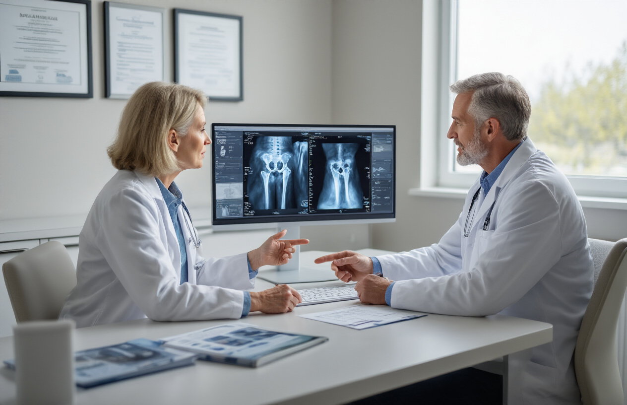
A DEXA dual femur scan specifically measures bone mineral density (BMD) in both your left and right hip bones, focusing on the femoral neck and total hip regions. This bone densitometry test analyzes how much calcium and other minerals are packed into your bone tissue, giving doctors a precise measurement of your bone strength and density.
The dual femur DEXA scan uses low-dose X-ray beams at two different energy levels to differentiate between bone, soft tissue, and fat. When these X-rays pass through your hip bones, the scanner measures how much radiation gets absorbed. Denser bones with more minerals absorb more radiation, while bones with lower density allow more radiation to pass through.
Your femur bone scan results are typically expressed as T-scores and Z-scores. The T-score compares your bone density to that of a healthy 30-year-old adult of the same gender – the age when bone density peaks. A T-score of -1.0 or above is considered normal, while scores between -1.0 and -2.5 indicate osteopenia (low bone density), and scores below -2.5 suggest osteoporosis.
The hip bone density test is particularly important because the femur experiences significant stress from weight-bearing activities. Hip fractures can be devastating, especially for older adults, making accurate measurement of femoral bone density crucial for preventing serious injuries.
How this advanced technology detects osteoporosis early
DEXA technology excels at catching bone loss before it becomes severe enough to cause fractures. The dual femur bone density test can detect as little as 2-3% bone loss, making it incredibly sensitive for early osteoporosis detection. This level of precision allows doctors to identify problems years before you might experience symptoms or suffer a fracture.
The femur DEXA scan provides a baseline measurement that doctors can track over time. By comparing your current bone density screening results to previous scans, medical professionals can determine if you’re losing bone mass and how quickly. This tracking capability makes DEXA scanning invaluable for monitoring the progression of bone loss and evaluating how well treatments are working.
Early detection through DEXA dual femur results gives you and your healthcare provider time to take preventive action. When osteoporosis is caught in its early stages, lifestyle changes like increased calcium intake, vitamin D supplementation, weight-bearing exercises, and medications can significantly slow or even reverse bone loss. The sooner bone density issues are identified, the more effective these interventions tend to be.
The DEXA bone density procedure’s ability to measure both hips separately also helps identify asymmetrical bone loss, which might indicate localized problems or the need for targeted treatment approaches. This comprehensive assessment makes dual femur scanning one of the most reliable methods for osteoporosis screening available today.
Key Benefits of Dual Femur DEXA Testing
Accurate Assessment of Both Hip Joints Simultaneously
Getting a comprehensive view of your hip bone health has never been more efficient. A dual femur DEXA scan measures bone density in both hip joints during a single appointment, providing doctors with a complete picture of your skeletal health in the most fracture-prone areas. This bilateral assessment proves especially valuable because bone density can vary between your left and right hip, sometimes significantly.
The femur bone scan technology captures precise measurements of your hip’s cortical and trabecular bone structures, areas that bear the most weight and stress during daily activities. By examining both hips simultaneously, healthcare providers can identify asymmetrical bone loss patterns that might indicate specific underlying conditions or biomechanical issues affecting one side more than the other.
This comprehensive approach eliminates the need for multiple appointments while delivering consistent, reproducible results. The DEXA dual femur results offer standardized T-scores and Z-scores for both hips, allowing for accurate comparison against age-matched populations and tracking changes over time with remarkable precision.
Early Detection Prevents Costly Fractures and Complications
Hip fractures represent one of the most serious consequences of untreated bone loss, with medical costs often exceeding $40,000 per incident. A dual femur bone density test can detect bone deterioration years before fractures occur, when interventions remain most effective and least invasive.
Bone density screening through dual femur analysis identifies individuals at high fracture risk while bones still respond well to lifestyle modifications, medications, and targeted therapies. This proactive approach prevents the cascade of complications that follow hip fractures, including extended hospital stays, rehabilitation costs, lost independence, and reduced quality of life.
The DEXA bone density procedure catches osteoporosis and osteopenia in their earliest stages, when treatment success rates remain highest. Early intervention can reduce fracture risk by up to 70% in high-risk patients, representing thousands of dollars in avoided medical expenses and immeasurable improvements in long-term health outcomes.
Who Should Get Dual Femur Bone Densitometry

Postmenopausal women at increased fracture risk
Women who have gone through menopause face a dramatically increased risk of bone loss due to declining estrogen levels. This hormonal shift accelerates bone density loss by approximately 3-5% per year during the first five years after menopause. A dual femur bone density test becomes essential for these women, particularly those with additional risk factors.
You should consider getting a DEXA dual femur scan if you’re postmenopausal and have a family history of osteoporosis, hip fractures, or bone breaks. Women who are thin or small-framed, have a history of eating disorders, or take certain medications like corticosteroids face even higher risks. Smoking, excessive alcohol consumption, and sedentary lifestyles also increase your chances of developing osteoporosis.
The femur DEXA scan specifically targets the hip area, which is one of the most common fracture sites in postmenopausal women. Early detection through bone density screening allows for timely intervention with lifestyle changes, calcium and vitamin D supplementation, or prescription medications that can slow bone loss.
Medical professionals typically recommend starting osteoporosis testing within the first few years after menopause, especially for women over 50. If you’ve already experienced a fracture after age 50, or if your mother broke a hip, don’t wait – schedule your hip bone density test right away. The results will help your doctor create a personalized plan to protect your bones and prevent future fractures.
Adults over 65 requiring routine bone health screening
Once you hit 65, regular bone densitometry becomes a standard part of your healthcare routine, regardless of gender. Both men and women over 65 should get baseline DEXA bone density measurements, as age-related bone loss affects everyone, though women typically experience more severe changes.
Your doctor will likely recommend a DEXA scan every two years after age 65, or more frequently if initial results show low bone density. Men over 65 often overlook bone health, but they account for about 20% of osteoporosis cases and face higher mortality rates after hip fractures compared to women.
Certain health conditions common in older adults make dual femur bone density tests even more critical. If you have rheumatoid arthritis, diabetes, kidney disease, or gastrointestinal disorders that affect nutrient absorption, you’re at higher risk for bone loss. Long-term use of medications like proton pump inhibitors, anticonvulsants, or blood thinners can also weaken bones over time.
The femur bone scan provides valuable information about your fracture risk and helps guide treatment decisions. Results from DEXA dual femur results are combined with other risk factors like age, weight, smoking history, and previous fractures to calculate your 10-year fracture probability. This comprehensive assessment helps you and your doctor make informed decisions about preventive treatments, exercise programs, and lifestyle modifications to maintain your independence and quality of life as you age.
What to Expect During Your DEXA Dual Femur Scan
Simple preparation steps for accurate test results
Getting ready for your dual femur DEXA scan requires minimal effort on your part. The night before your test, avoid taking calcium supplements as they can interfere with the bone density readings. Skip the multivitamins too if they contain calcium – check the label to be sure.
On the day of your femur DEXA scan, wear comfortable, loose-fitting clothes without metal zippers, buttons, or underwire bras. Metal objects show up on the scan and can block the view of your hip bones, potentially affecting your results. Athletic wear or yoga pants work perfectly for this type of bone densitometry test.
Remove all jewelry, watches, and belts before the procedure begins. If you’ve had recent contrast studies like CT scans or barium X-rays within the past week, let your technologist know. These contrast materials can linger in your system and impact the accuracy of your DEXA bone density procedure.
You can eat normally before your appointment – there’s no fasting required for this bone density screening. However, arrive about 15 minutes early to complete any necessary paperwork and allow time for these simple prep steps. The preparation is straightforward because the dual femur bone density test focuses specifically on measuring bone mineral density in your hip area without requiring any special dietary restrictions or medications.
Quick 15-minute painless scanning procedure
Your dual femur DEXA scan happens faster than most medical appointments. The entire bone densitometry procedure typically takes just 10 to 15 minutes from start to finish, making it one of the most efficient ways to assess your hip bone health.
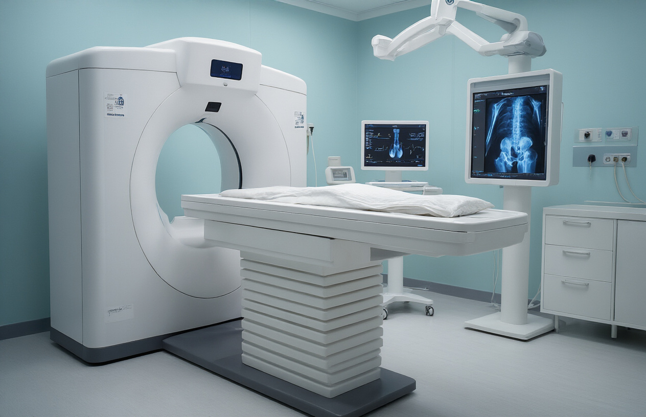
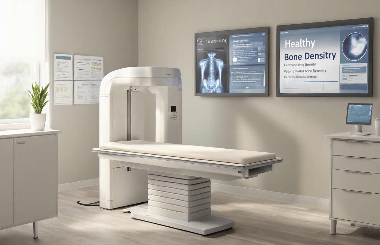
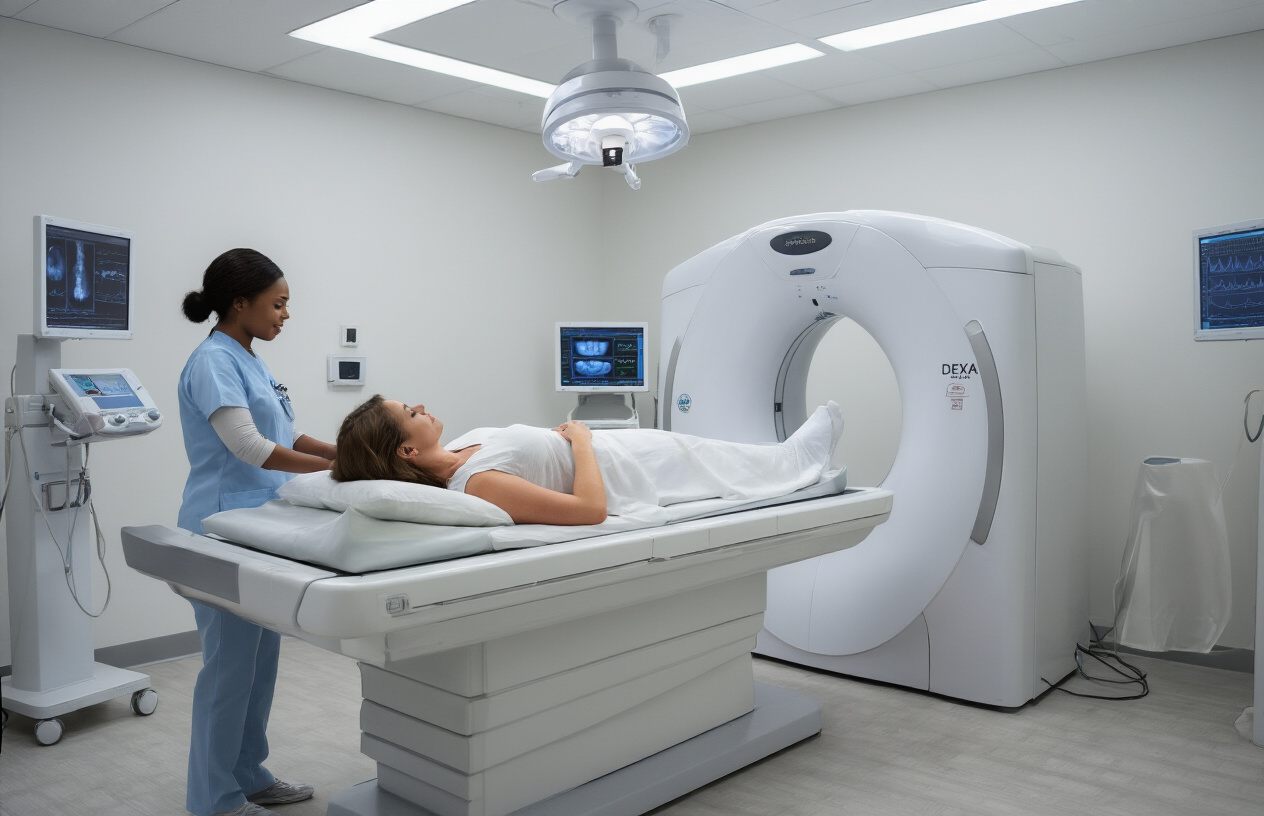
You’ll lie comfortably on a padded table while the DEXA scanner moves quietly overhead. The machine looks similar to an open MRI but much less intimidating – there’s no tunnel or enclosed space to worry about. During your femur bone scan, you simply need to stay still while the scanner takes precise measurements of both hip bones.
The scanning arm passes over your hip area twice, capturing detailed images of your bone density. You won’t feel anything during the actual scanning – no heat, cold, or vibrations. The low-dose radiation used in this osteoporosis testing is minimal, roughly equivalent to a cross-country airplane flight.
A trained technologist operates the equipment and stays nearby throughout your hip bone density test. They’ll position your legs slightly apart and may use a positioning device to ensure your femurs are properly aligned for accurate measurements. You can breathe normally during the scan, though they might ask you to hold your breath briefly for clearer images.
The painless nature of this DEXA dual femur procedure makes it suitable for people of all ages, including those with mobility concerns or chronic pain conditions.
Interpreting Your Dual Femur DEXA Results

Understanding T-scores and Z-scores for each hip
Your dual femur DEXA scan provides two distinct bone density measurements – one for your left hip and one for your right hip. Each measurement comes with both a T-score and Z-score that tell different parts of your bone health story.
T-scores compare your current bone density to that of a healthy 30-year-old of the same gender at peak bone mass. This comparison matters because bones naturally lose density with age, and the T-score shows how far you’ve deviated from optimal bone strength. A T-score of -1.0 or higher indicates normal bone density, while scores between -1.0 and -2.5 suggest osteopenia (low bone mass). Scores of -2.5 or lower indicate osteoporosis.
Z-scores, on the other hand, compare your bone density to other people your same age and gender. These scores help doctors determine if your bone loss is typical for your age group or if something else might be causing accelerated bone loss. A Z-score below -2.0 often prompts doctors to investigate underlying conditions that could be affecting your bones.
Don’t be surprised if your left and right hip show different results. Many people have slight variations between sides due to factors like dominant-side usage patterns, previous injuries, or natural anatomical differences. Your doctor will typically focus on the lower of the two scores when making treatment recommendations, as this represents your highest fracture risk area.
Recognizing normal versus concerning bone density levels
Reading your femur DEXA scan results becomes clearer when you understand the specific thresholds that separate normal bone health from conditions requiring attention or treatment.
Normal bone density falls within the T-score range of -1.0 and above. If your dual femur bone density test shows T-scores in this range for both hips, your bones maintain good strength and your fracture risk remains low. However, even with normal results, your doctor might recommend lifestyle changes to maintain bone health as you age.
Osteopenia represents the middle ground, with T-scores ranging from -1.1 to -2.4. This condition indicates your bones have become weaker than ideal but haven’t reached the osteoporosis threshold. Many people in their 50s and beyond fall into this category, especially postmenopausal women. While osteopenia doesn’t automatically require medication, it does signal the need for preventive measures and closer monitoring through regular bone density screening.
Osteoporosis diagnosis occurs when T-scores reach -2.5 or lower on your femur bone scan. This level indicates significantly weakened bones with substantially higher fracture risk. People with osteoporosis often need prescription medications along with lifestyle modifications to prevent fractures.
Your doctor will also consider additional factors beyond just the numbers. Your age, medical history, family history of fractures, current medications, and lifestyle factors all influence whether your bone density levels are concerning for your specific situation. Some people with borderline scores might need treatment if they have multiple risk factors, while others with similar scores might only need monitoring and lifestyle changes.
Taking Action Based on Your Test Results
Lifestyle modifications to strengthen bone density naturally
Your dual femur DEXA scan results don’t have to define your future bone health. Several natural approaches can help strengthen your bones and potentially improve your next bone density screening.
Weight-bearing exercises top the list of effective interventions. Activities like walking, jogging, dancing, and resistance training signal your bones to build new tissue. Aim for at least 30 minutes of weight-bearing activity most days of the week. Strength training with weights or resistance bands twice weekly targets the hip and femur areas specifically measured in your DEXA dual femur results.
Nutrition plays a crucial role in bone health. Calcium-rich foods like dairy products, leafy greens, and fortified alternatives provide the building blocks for strong bones. Vitamin D helps your body absorb calcium effectively – consider spending time outdoors or discussing supplements with your doctor. Protein supports bone structure, so include lean meats, fish, beans, and nuts in your daily meals.
Smoking cessation and limiting alcohol consumption can dramatically improve bone density over time. Both habits interfere with bone formation and accelerate bone loss. If you smoke, quitting now can help preserve your existing bone mass and support new bone growth.
Balance exercises reduce fall risk, protecting your bones from fractures. Simple activities like standing on one foot or practicing tai chi can improve stability and coordination.
Medical treatments available for low bone density
When lifestyle changes aren’t enough, medical interventions can effectively address low bone density revealed by your femur bone scan. Your healthcare provider will recommend treatments based on your specific DEXA bone density procedure results and individual risk factors.
Bisphosphonates represent the most commonly prescribed medications for osteoporosis. These drugs slow bone breakdown and allow bone-building cells to work more effectively. Options include weekly or monthly oral medications, or yearly intravenous infusions. Most patients tolerate these treatments well, though some experience mild side effects like stomach upset.
Hormone replacement therapy may benefit postmenopausal women with declining estrogen levels. Estrogen helps maintain bone density, and replacement can slow bone loss in the hip and femur areas measured during your DEXA scan. Your doctor will weigh the benefits against potential risks based on your medical history.
Newer medications like denosumab injections every six months offer alternatives for patients who can’t tolerate bisphosphonates. This treatment blocks cells that break down bone tissue.
Selective estrogen receptor modulators (SERMs) provide estrogen-like benefits for bones without affecting breast or uterine tissue. These medications can be particularly useful for women with a history of breast cancer.
Regular monitoring through repeat osteoporosis testing helps track treatment effectiveness. Most doctors recommend follow-up bone densitometry every two years to assess progress and adjust treatment plans accordingly.
Your healthcare team will create a personalized treatment strategy combining the most appropriate medical interventions with lifestyle modifications to optimize your bone health.
DEXA dual femur bone densitometry gives you a clear picture of your bone health, specifically focusing on your hip bones where fractures can be most devastating. This simple, painless test takes just minutes but provides valuable insights into your fracture risk and bone strength. Whether you’re over 65, have risk factors for osteoporosis, or are monitoring existing bone conditions, this scan helps catch problems early when treatment works best.
Your test results become your roadmap for protecting your bones moving forward. Work with your doctor to understand your T-scores and Z-scores, then take action based on what they reveal. This might mean starting calcium and vitamin D supplements, beginning weight-bearing exercises, or discussing medication options. Don’t wait for a fracture to happen – schedule your DEXA scan today and take control of your bone health for years to come.