
Deep vein thrombosis (DVT) affects millions of people each year, and catching it early can literally save your life. If you’re experiencing leg pain, swelling, or have risk factors for blood clots, understanding how Doppler ultrasound for DVT works is crucial for getting the correct diagnosis and treatment.
This guide is written for patients who suspect they might have DVT, healthcare professionals seeking practical insights, and anyone wanting to understand this common but serious condition. We’ll walk you through recognizing the warning signs that shouldn’t be ignored and explain how Doppler ultrasound has become the gold standard for detecting blood clots quickly and accurately.
You’ll also learn what actually happens during the examination process, so you know exactly what to expect when you walk into that ultrasound room.
Understanding Deep Vein Thrombosis and Its Hidden Dangers
What DVT Is and How Blood Clots Form in Deep Veins
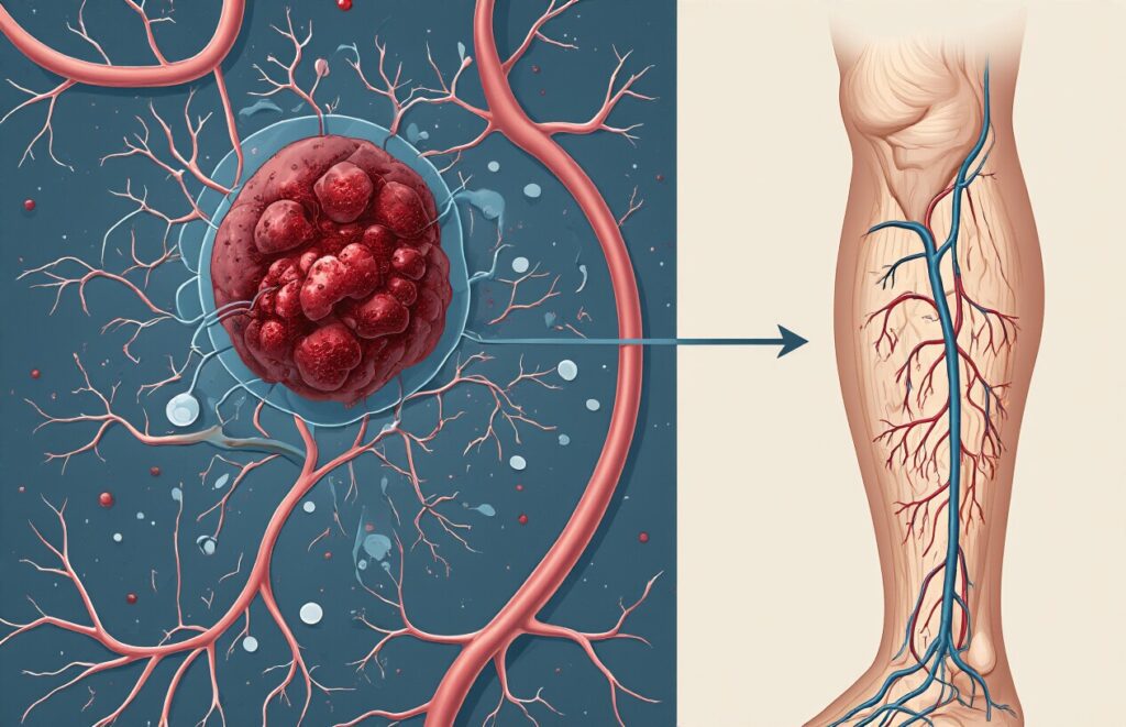
Deep vein thrombosis occurs when blood clots develop in the large veins deep within your legs, typically in the calf or thigh muscles. These clots form when blood flow slows, vessel walls are damaged, or your blood becomes thicker than usual. Unlike surface clots you can see, DVT occurs in veins deep in muscle tissue, making them invisible from the outside.
The clotting process starts when platelets stick together, and fibrin proteins create a mesh-like structure that traps blood cells. Usually, this mechanism helps stop bleeding from injuries, but in DVT, clots form unnecessarily inside healthy blood vessels, potentially blocking circulation completely.
Why DVT Often Goes Undetected Until Serious Complications Arise
DVT symptoms can be surprisingly subtle or completely absent in nearly half of all cases. When signs do appear, they often mimic common conditions like muscle strains, making diagnosis challenging. Mild leg swelling, cramping, or warmth might be dismissed as normal aches and pains, especially after prolonged sitting or physical activity.
The deep location of these clots means they don’t cause obvious visual changes like surface blood clots do. Many people continue their daily routines unaware that a potentially dangerous clot is growing larger and becoming more likely to break free and travel through their bloodstream.
The Life-Threatening Risk of Pulmonary Embolism
Pulmonary embolism represents the most feared complication of DVT, occurring when a blood clot breaks loose and travels to the lungs. This medical emergency can block blood flow to lung tissue, causing sudden shortness of breath, chest pain, rapid heart rate, and in severe cases, cardiac arrest. Statistics show that untreated DVT leads to pulmonary embolism in approximately 10% of cases.
The danger lies in how quickly this can happen – a clot can dislodge and reach the lungs within minutes. Even small clots can be life-threatening if they block critical blood vessels in the lungs, which is why early detection and treatment of DVT is absolutely crucial for preventing this devastating outcome.
Who Is Most at Risk for Developing DVT
Several factors dramatically increase DVT risk, with prolonged immobility being the most common trigger. Long flights, bed rest after surgery, or extended hospital stays create perfect conditions for blood clots to form. Age plays a significant role too – people over 60 face higher risks, though DVT can strike at any age.
Medical conditions like cancer, heart disease, and clotting disorders also elevate risk levels. Women taking birth control pills or hormone replacement therapy, pregnant women, and people with family histories of blood clots need extra vigilance: obesity, smoking, and recent major surgeries round out the primary risk factors that physicians carefully monitor.
Recognizing DVT Warning Signs Before It’s Too Late
Leg Pain and Swelling That Shouldn’t Be Ignored
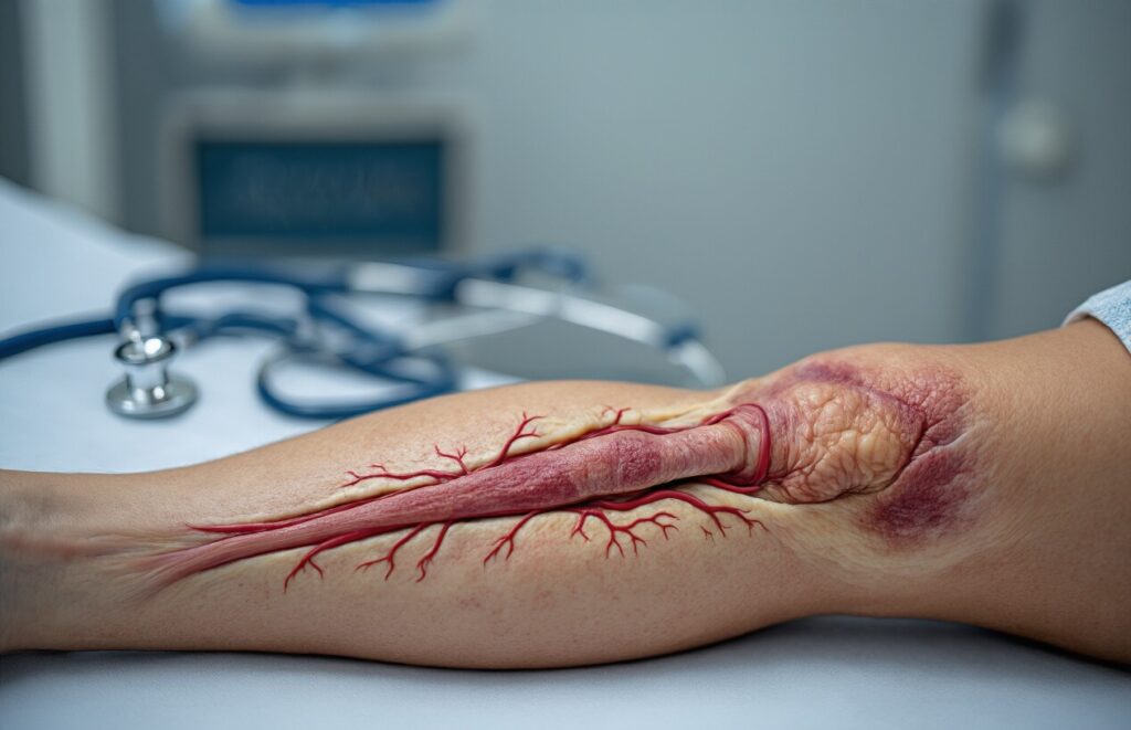
The most recognizable DVT symptom is sudden, unexplained leg pain that feels like a charley horse or muscle cramp that won’t go away. This pain typically worsens when you flex your foot upward or walk around. Swelling usually appears in one leg, making it noticeably larger than the other.
Skin Changes and Discoloration Around Affected Areas
Your skin might develop a bluish or reddish tint where the clot has formed, creating a stark contrast with your normal skin tone. The affected area often feels tight and stretched due to the underlying inflammation and reduced blood flow.
Warmth and Tenderness in the Affected Limb
The blocked vein causes localized inflammation, making the skin feel warm to the touch, almost as if you have a fever in that spot. Light pressure or even gentle touching can cause significant discomfort, making everyday activities like wearing tight clothing unbearable.
When Symptoms May Be Subtle or Completely Absent
Nearly half of all DVT cases produce no obvious symptoms, making them particularly dangerous because people don’t seek treatment. Some experience only mild achiness that they dismiss as workout soreness or aging. Silent DVT can progress undetected until a piece breaks off and travels to the lungs.
How Doppler Ultrasound Revolutionizes DVT Detection

The Science Behind Sound Waves Detecting Blood Flow Problems
Doppler ultrasound works by bouncing high-frequency sound waves off moving red blood cells in your veins. When blood flows normally, these waves return at predictable patterns. However, when a clot blocks or slows circulation, the reflected waves change dramatically. This shift in frequency, called the Doppler effect, allows technicians to map blood flow with incredible precision and identify blockages that traditional methods might miss.
Why Doppler Technology Outperforms Traditional Diagnostic Methods
Unlike invasive procedures that require contrast dyes or radiation, Doppler ultrasound provides immediate, accurate results through your skin. The technology detects even partial blockages that other diagnostic tools often overlook. Traditional methods like venography involve injecting dyes into veins and expose patients to radiation risks. Doppler ultrasound eliminates these concerns while delivering superior accuracy rates exceeding 95% for detecting DVT in major leg veins.
Real-Time Visualization of Blood Clots Without Radiation Exposure
The beauty of Doppler ultrasound lies in its ability to show blood flow happening right now, not just static images. Technicians can watch blood moving through your veins live on screen, immediately spotting areas where flow stops or slows unnaturally. This real-time capability means doctors can assess clot severity and location instantly. The complete absence of radiation makes it safe for pregnant women and allows for repeated monitoring without health risks.
What to Expect During Your Doppler Ultrasound Examination

Simple Preparation Steps That Maximize Test Accuracy
Doppler ultrasound requires minimal preparation, making it one of the most patient-friendly diagnostic tests available. You’ll wear comfortable, loose-fitting clothing that allows easy access to the area being examined. Most facilities recommend avoiding lotions or oils on your skin before the appointment, as these can interfere with the ultrasound gel’s effectiveness.
The Painless Procedure That Takes Minutes to Complete
The entire examination typically lasts 15-30 minutes and involves no discomfort whatsoever. You’ll lie on an examination table while the technician applies gentle pressure with the ultrasound probe. The process feels similar to a routine pregnancy ultrasound, with no needles, radiation, or invasive procedures required.
How Technicians Use Gel and Probes to Map Your Blood Flow
Cool, clear gel is applied to your skin to eliminate air pockets that could block sound waves. The technician moves a handheld transducer across the gel-covered area, sending high-frequency sound waves through your tissues. These waves bounce back from blood cells, creating detailed images of blood flow patterns and vessel structure on the monitor.
Understanding the Sounds and Images on the Monitor
The distinctive whooshing sounds you hear represent blood flowing through your veins and arteries. Different flow patterns create unique audio signatures that trained technicians can interpret instantly. The monitor displays colorful, real-time images showing blood movement, with red and blue colors typically representing flow direction toward or away from the probe.
Interpreting Your Doppler Results and Next Steps

What Normal Blood Flow Patterns Look Like
Normal blood flow appears as smooth, continuous waves on your Doppler ultrasound screen. Healthy veins show spontaneous flow that increases with breathing and responds to gentle compression. The blood moves freely without interruptions, creating distinct patterns that trained technologists recognize immediately.
How Doctors Identify Partial and Complete Blockages
Complete blockages eliminate all blood flow signals, creating silent areas on the ultrasound display. Partial clots appear as reduced flow patterns with altered wave shapes. Doctors also check for vein compressibility – healthy veins collapse easily under probe pressure, while clotted veins remain rigid and incompressible.
When Additional Testing May Be Necessary for Confirmation
Challenging cases may require CT venography or MRI imaging, especially when clots are suspected in deep pelvic veins or upper extremities. D-dimer blood tests help rule out DVT when ultrasound results are inconclusive. Repeat scanning after 48-72 hours can catch developing clots that weren’t initially visible.
Treatment Options Based on Your Specific Diagnosis
| Clot Location | Treatment Approach | Duration |
| Lower leg | Anticoagulation therapy | 3-6 months |
| Thigh/pelvis | Blood thinners + monitoring | 6-12 months |
| Extensive clots | Hospital admission + IV therapy | Variable |
Anticoagulant medications like warfarin or newer oral drugs prevent clot growth and reduce recurrence risk. Severe cases might need thrombolytic therapy to dissolve existing clots. Compression stockings provide additional support during recovery, improving circulation and reducing post-thrombotic syndrome risk.

Blood clots don’t wait for convenient timing, and recognizing DVT symptoms early can literally save your life. Doppler ultrasound has become the gold standard for detecting these potentially dangerous clots because it’s non-invasive, accurate, and gives doctors the clear picture they need to make quick treatment decisions. The swelling, pain, and warmth you might dismiss as a pulled muscle could actually be your body’s warning system telling you something more serious is happening.
Getting a Doppler ultrasound is straightforward and painless, but the information it provides is incredibly valuable. Your doctor can see precisely where clots are forming, how severe they are, and create a treatment plan that’s right for your specific situation. Don’t ignore leg pain or swelling that doesn’t seem normal for you – these symptoms deserve medical attention, and a simple ultrasound can give you the answers and peace of mind you need.