
MRI Breast With Contrast: What Makes It Different From Mammograms and Ultrasounds
If you’re facing breast health concerns or need advanced screening, understanding your imaging options can feel overwhelming. This guide is designed for women who’ve been recommended for breast MRI with contrast, those at high risk for breast cancer, and anyone wanting to understand how different breast imaging methods work.
MRI breast with contrast uses magnetic fields and a special dye to create detailed images of breast tissue. Unlike mammograms that use X-rays or ultrasounds that use sound waves, MRI can spot abnormalities that other methods might miss.
We’ll break down how MRI breast with contrast actually works and why doctors choose it for certain situations. You’ll also learn when each imaging method works best for different breast health scenarios, plus the real advantages and drawbacks of each technique so you can have informed conversations with your healthcare team.
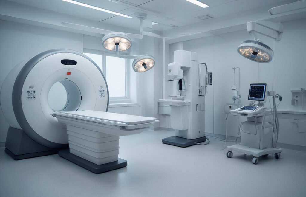
Understanding MRI Breast With Contrast Technology
How contrast agents enhance breast tissue visibility
Contrast agents work like a spotlight in breast MRI scans, making blood vessels and tissue differences pop out clearly. When injected into your bloodstream, these gadolinium-based compounds travel through your body and accumulate in areas with increased blood flow. Since cancerous tissues typically have more blood vessels than normal breast tissue, they light up brighter on the scan, creating a stark contrast that helps radiologists spot even tiny abnormalities.
Advanced imaging capabilities for detecting abnormalities
MRI breast scans capture incredibly detailed images from multiple angles, creating a 3D map of your breast tissue that’s far more comprehensive than traditional imaging methods. The technology can detect lesions as small as 2-3 millimeters and distinguish between different types of tissue based on how they respond to magnetic fields. This precision makes MRI particularly valuable for screening high-risk patients, evaluating the extent of known cancers, and monitoring treatment responses.
Safety protocols and patient preparation requirements
Before your MRI appointment, you’ll need to remove all metal objects including jewelry, bras with underwire, and any magnetic items that could interfere with the scan. The contrast injection requires a brief medical history review to check for kidney problems or previous allergic reactions. Most patients can eat normally before the procedure, though you should arrive wearing comfortable, metal-free clothing and expect to spend about 45 minutes in the scanning room lying face-down on a specialized table.
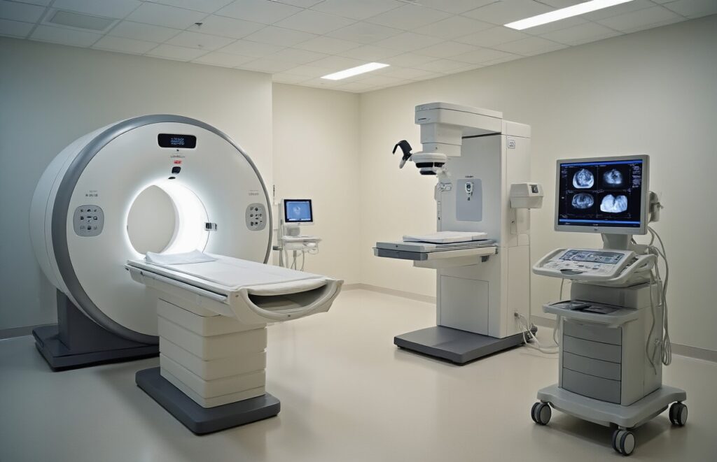
Comprehensive Comparison of Breast Imaging Methods
MRI breast scanning accuracy and detailed tissue analysis
MRI with contrast excels at detecting invasive cancers and provides the highest sensitivity among all breast imaging methods, reaching up to 95% accuracy for malignant tumors. The contrast agent highlights blood flow patterns, allowing radiologists to spot even small lesions that might escape detection through other methods.
Mammography effectiveness for routine breast cancer screening
Mammograms remain the gold standard for annual screening, particularly effective at identifying microcalcifications that may indicate early-stage ductal carcinoma in situ. Digital mammography works best for women over 40 with average risk factors, offering excellent cost-effectiveness for population-wide screening programs.
Ultrasound benefits for targeted breast examination
| Feature | MRI | Mammography | Ultrasound |
| Radiation | None | Low dose | None |
| Real-time imaging | No | No | Yes |
| Cyst differentiation | Excellent | Limited | Excellent |
| Dense tissue penetration | Superior | Challenging | Good |
Ultrasound shines when examining dense breast tissue and distinguishing between solid masses and fluid-filled cysts. The real-time imaging capability allows technicians to guide biopsies with precision while providing immediate results during the examination.
When Each Imaging Method Delivers Optimal Results
MRI Recommended Scenarios for High-Risk Patients
MRI breast imaging with contrast becomes the gold standard for women carrying BRCA gene mutations, those with strong family histories of breast or ovarian cancer, and patients who received chest radiation before age 30. High-risk patients benefit from MRI’s superior ability to detect small cancers that mammograms and ultrasounds might miss, particularly in dense breast tissue.
Mammogram Ideal Timing for Regular Preventive Care
Annual mammograms work best for routine screening in average-risk women aged 40-75, offering excellent detection of calcifications and architectural distortions that signal early cancer. This imaging method excels during regular preventive visits when no specific concerns exist, providing cost-effective population screening that catches most breast cancers before they become palpable lumps.
Ultrasound Perfect Situations for Follow-up Examinations
Ultrasound shines when investigating specific breast lumps found during physical exams or when mammogram results need clarification. This real-time imaging technique works perfectly for pregnant or breastfeeding women who cannot undergo radiation-based mammography, and for distinguishing between solid masses and fluid-filled cysts during diagnostic workups.
Key Advantages and Limitations of Each Technique
MRI Superior Sensitivity for Detecting Small Lesions
MRI breast imaging excels at finding tiny cancerous growths that other methods might miss, particularly in dense breast tissue. The contrast agent highlights blood vessel patterns around tumors, making even millimeter-sized lesions visible to radiologists.
However, this high sensitivity comes with a trade-off – MRI often flags benign tissue as suspicious, leading to unnecessary biopsies and patient anxiety. The procedure also requires a significant time commitment and costs substantially more than alternative imaging methods.
Mammography Proven Track Record for Early Cancer Detection
| Advantage | Limitation |
| Detects 85% of breast cancers | Less effective in dense breast tissue |
| Identifies calcifications early | Radiation exposure concerns |
| Widely available and affordable | Compression causes discomfort |
| Quick 15-minute procedure | Annual screening debates |
Ultrasound Radiation-Free Imaging for Pregnant Women
Ultrasound provides real-time imaging without any radiation risks, making it the go-to choice for pregnant and breastfeeding women. The technology works particularly well for distinguishing between solid masses and fluid-filled cysts, offering immediate results during the examination.
The main drawback involves operator dependency – results can vary significantly based on the technician’s skill level. Ultrasound also struggles to detect microcalcifications, which are early warning signs of certain cancer types that mammography catches easily.
Time Efficiency and Patient Comfort Factors
Procedure Duration:
- Mammography: 15-20 minutes
- Ultrasound: 20-30 minutes
- MRI: 45-90 minutes
Comfort Considerations:
- Mammography requires breast compression causing temporary discomfort
- Ultrasound involves gentle probe movement with warming gel
- MRI demands lying still in a confined space with loud noises
Making Informed Decisions About Your Breast Health Screening

Risk factors that influence imaging method selection
Your personal risk profile plays a huge role in determining which breast imaging method works best for you. Women with dense breast tissue often need an MRI or an ultrasound because mammograms can miss cancers hiding in dense areas. Family history of breast or ovarian cancer, genetic mutations like BRCA1/BRCA2, and previous chest radiation treatments all bump you into the high-risk category, where doctors typically recommend MRI screening.
The importance of doctor consultations for personalized recommendations
Combining multiple imaging techniques for comprehensive assessment
Your doctor might suggest using different imaging methods together to get the complete picture. Mammograms catch calcifications that MRI might miss, while MRI spots cancers that don’t show up on mammograms. Ultrasound helps determine if a suspicious area is solid or fluid-filled. This layered approach gives your medical team the best chance to detect problems early and avoid unnecessary biopsies.

MRI breast with contrast, mammograms, and ultrasounds each serve unique roles in breast health screening. MRI offers the most detailed images and can detect small abnormalities that other methods might miss, making it invaluable for high-risk patients and those with dense breast tissue. Mammograms remain the gold standard for routine screening and are excellent at spotting calcifications, while ultrasounds provide real-time imaging without radiation and work well for distinguishing between solid masses and fluid-filled cysts.
Your doctor will recommend the best imaging method based on your individual risk factors, age, breast density, and specific health concerns. Don’t hesitate to discuss which screening approach makes sense for your situation. Staying proactive about breast health means understanding your options and working with your physician to create a screening plan that gives you the best chance of early detection and peace of mind.