

“Volume rendering makes medical scans clearer, faster, and more accurate—here’s what it means for you.”
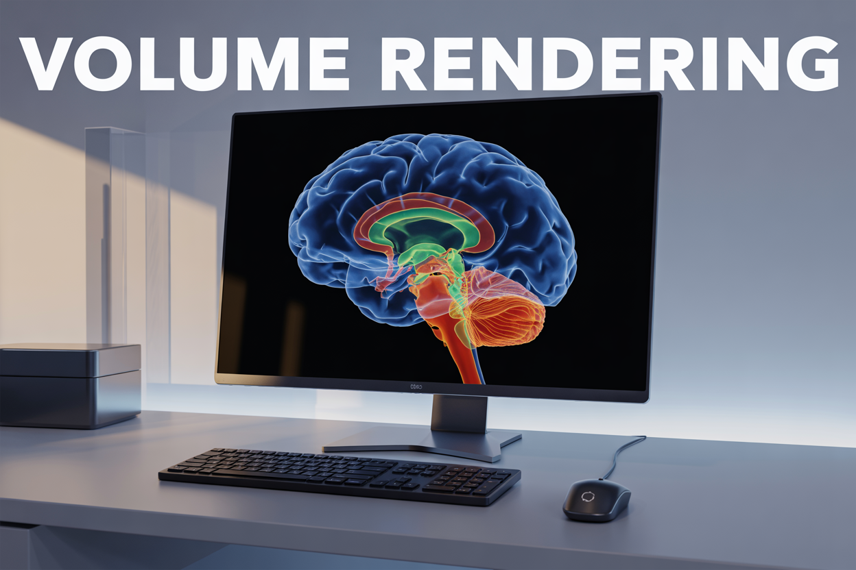
Volume rendering transforms 3D datasets into vivid visual representations by directly displaying volumetric data without converting it to surface models first. This powerful visualization technique lets you see inside complex structures like medical scans, scientific simulations, and geological formations.
This guide is perfect for developers, researchers, medical professionals, and visualization specialists who want to understand how volume rendering works and apply it to their projects. You’ll also benefit if you’re working with medical imaging, scientific visualization, or any field that deals with complex 3D data.
We’ll start by breaking down the core technical concepts that make volume rendering possible, including ray casting algorithms and direct volume rendering methods. Then we’ll explore real-world applications across industries like healthcare, scientific research, and entertainment. Finally, we’ll cover practical implementation approaches and performance optimization strategies to help you build efficient volume rendering systems.
Understanding Volume Rendering Fundamentals
Transform Complex 3D Data Into Visual Representations
Volume rendering takes massive amounts of three-dimensional data and transforms them into images you can actually see and understand. Think of it like having millions of invisible data points floating in space – volume rendering makes them visible by creating a comprehensive picture of what’s happening inside that 3D space.
This process works by sampling data throughout an entire volume rather than just looking at surfaces. Medical CT scans provide a perfect example: instead of just seeing the outline of organs, volume rendering reveals the internal structures, density variations, and relationships between different tissues. Each tiny cube of space (called a voxel) contains specific values that get translated into colors, opacity levels, and brightness.
3D volume rendering techniques handle this transformation through sophisticated algorithms that assign visual properties to data values. A dense bone might appear bright white and opaque, while soft tissue shows up as semi-transparent with different color mappings. The rendering engine processes millions of these data points simultaneously, creating smooth transitions and realistic depth perception.
The beauty of volume visualization methods lies in their ability to reveal hidden patterns and relationships within complex datasets. Scientists studying atmospheric conditions can visualize wind patterns, temperature gradients, and pressure systems all within a single rendered volume. Engineers examining stress distributions in materials can see exactly where potential failure points might occur, long before physical testing begins.
Differentiate Volume Rendering From Traditional Surface Rendering
Traditional surface rendering and volume rendering approach 3D visualization from completely different angles. Surface rendering focuses exclusively on the outer boundaries of objects – think of it like painting the shell of an egg while ignoring everything inside. This method works great for solid objects with well-defined surfaces, like architectural models or product designs.
Direct volume rendering, on the other hand, peers inside that egg and shows you every layer, gradient, and internal structure. Where surface rendering might show you a smooth sphere, volume rendering reveals the density variations, internal cavities, and material transitions that exist within that same sphere.
The data requirements differ dramatically between these approaches. Surface rendering needs geometric information – vertices, edges, and polygons that define object boundaries. Volume rendering consumes volumetric datasets where every point in 3D space has associated properties like density, temperature, or intensity values.
Processing methods also diverge significantly. Surface rendering relies on polygon rasterization and traditional graphics pipeline techniques that most modern GPUs handle efficiently. Volume rendering algorithms employ ray casting, ray marching, or texture-based approaches that sample data throughout the entire volume, making them more computationally intensive but far more informative.
Medical imaging volume rendering showcases this difference perfectly. A surface-rendered MRI might show you the outline of a tumor, but volume rendering reveals its internal composition, surrounding tissue interactions, and blood flow patterns. Surface rendering gives you the “what,” while volume rendering provides the “what, where, how much, and how dense.”
The visual results speak for themselves. Surface rendering produces clean, sharp edges and well-defined object boundaries. Volume rendering creates soft transitions, translucent effects, and the ability to see multiple layers of information simultaneously – like looking through colored glass with varying thickness and opacity.
Master Ray Casting Techniques for Data Sampling
Ray casting serves as the backbone of most volume rendering algorithms, working by shooting virtual rays from the camera through each pixel into the 3D dataset. Think of it like shining a flashlight through fog – the light beam samples the density of particles along its path to create the final image you see.
The process starts with determining ray direction and origin for each pixel on the screen. Each ray marches through the volume data using fixed or adaptive step sizes, sampling scalar values at regular intervals. These samples represent data points like tissue density in medical scans or temperature values in scientific simulations.
Smart sampling strategies make a huge difference in both quality and performance. Uniform sampling provides consistent results but wastes computation in empty regions. Adaptive sampling adjusts step sizes based on data variation – taking smaller steps in areas with rapid changes and larger steps in homogeneous regions. This approach dramatically reduces unnecessary calculations while preserving important details.
Empty space skipping represents another powerful optimization technique. By preprocessing the volume to identify empty regions, rays can jump over areas containing no meaningful data. This becomes especially valuable when working with sparse datasets like medical CT scans where much of the volume contains air or empty space.
Multi-resolution approaches allow rays to sample at different detail levels depending on distance from the camera or importance of the region. Close objects get high-resolution sampling while distant features use coarser sampling rates, creating an efficient balance between visual quality and computational load.
Implement Transfer Functions for Opacity and Color Mapping
Transfer functions act as the creative bridge between raw volume data and the final rendered image, determining how scalar values translate into visible colors and opacity levels. These functions essentially tell the rendering engine which parts of your data should appear solid, transparent, or completely invisible.
The most common approach uses 1D transfer functions that map scalar values directly to color and opacity. Imagine a simple medical scan where bone density values above a certain threshold appear white and opaque, while soft tissue remains semi-transparent with flesh tones. The transfer function creates these visual mappings through carefully designed curves or lookup tables.
Multi-dimensional transfer functions offer more sophisticated control by considering additional properties like gradient magnitude or local statistics. A 2D transfer function might map both density and gradient strength, allowing you to highlight tissue boundaries more effectively. High gradient areas could appear more opaque to emphasize edges, while low gradient regions stay transparent even at higher density values.
Interactive transfer function design has become essential for practical volume rendering applications. Users need real-time feedback when adjusting opacity curves or color gradients to achieve the desired visualization. Modern tools provide histogram-based interfaces where you can directly manipulate curves while seeing immediate results in the rendered volume.
Preprocessing transfer function data into optimized lookup tables significantly improves runtime performance. Rather than computing complex mathematical functions for every sample, the renderer can perform fast table lookups. Pre-integrated transfer functions take this concept further by precomputing the integral of the transfer function over small intervals, enabling more accurate rendering with fewer samples along each ray.
Essential Applications Across Industries
Medical imaging has transformed completely with volume rendering techniques. Radiologists now visualize complex anatomical structures in three dimensions, making diagnoses faster and more accurate than ever before. CT scans, MRI images, and ultrasound data come alive through 3D volume rendering techniques, allowing doctors to explore inside the human body like never before.
Medical imaging volume rendering shines brightest in surgical planning. Surgeons can navigate through a patient’s anatomy before making the first incision, identifying critical structures and planning optimal approaches. Brain tumor removal becomes safer when neurosurgeons can visualize blood vessels, neural pathways, and tumor boundaries in stunning detail. Cardiac surgeons rely on volumetric heart reconstructions to understand complex congenital defects and plan intricate repairs.
Cancer diagnosis and treatment planning benefit enormously from volume visualization methods. Oncologists track tumor growth, assess treatment response, and plan radiation therapy with precision that saves lives. The technology reveals subtle tissue changes that might escape detection in traditional 2D images.
Emergency medicine departments use volumetric rendering applications to quickly assess trauma patients. A motorcycle accident victim’s internal injuries become visible within minutes, guiding life-saving decisions about surgery priorities and treatment approaches. Blood vessel visualization helps identify internal bleeding sources rapidly.
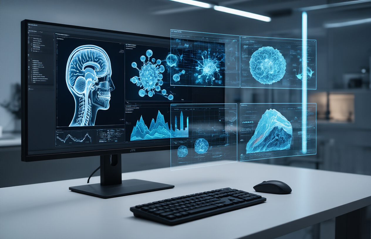
Enhance Scientific Research Through Volumetric Data Analysis
Research laboratories across the globe depend on volume rendering algorithms to unlock scientific mysteries. Biologists studying cell structures use confocal microscopy data rendered in three dimensions to understand protein interactions, cellular development, and disease mechanisms. What once required countless 2D slices now appears as complete, navigable 3D models.
Materials science researchers examine crystal structures, composite materials, and manufacturing defects through volumetric analysis. They can peer inside metal alloys to understand failure mechanisms or optimize new material designs. Aerospace engineers use these techniques to inspect critical components without destructive testing.
Climate scientists process massive atmospheric and oceanic datasets using direct volume rendering to visualize weather patterns, ocean currents, and climate change effects. Hurricane formation, temperature gradients, and pollution dispersion become visible phenomena that researchers can study interactively.
Archaeological teams reconstruct ancient artifacts and burial sites from CT scan data. They explore fragile mummies, sealed containers, and deteriorating manuscripts without physical damage. Volume rendering reveals hidden inscriptions, internal structures, and construction techniques from civilizations past.
Pharmaceutical companies accelerate drug discovery by visualizing molecular interactions and protein folding in three dimensions. Chemical engineers optimize reactor designs by studying fluid dynamics and heat transfer patterns through volumetric simulations.
Choose Between GPU and CPU-Based Rendering Solutions
The choice between GPU and CPU-based volume rendering significantly impacts your project’s performance and capabilities. Modern GPUs excel at volume rendering because they handle massive parallel computations that volumetric data demands. Graphics cards like NVIDIA’s RTX series or professional Quadro cards can process thousands of ray samples simultaneously, making them ideal for real-time volume visualization applications.
CPU-based solutions offer different advantages. They provide better precision for scientific computing and handle complex algorithms that don’t parallelize well. Multi-core processors work well for batch processing large volumetric datasets where rendering time isn’t critical. The CPU approach also gives you more control over memory management and algorithm customization.
GPU memory limitations often determine feasibility for large datasets. A graphics card with 8GB VRAM might struggle with high-resolution medical scans that require 16GB or more. In these cases, CPU implementations with system RAM access become necessary. Hybrid approaches combine both processors – CPUs handle preprocessing and data management while GPUs accelerate the actual rendering pipeline.
Consider your target platform carefully. Mobile devices rely heavily on integrated GPUs with limited capabilities, while workstations can leverage powerful dedicated cards. Cloud-based rendering services offer scalable GPU access without hardware investment, making them attractive for occasional high-performance needs.
Select Appropriate Software Frameworks and Libraries
VTK (Visualization Toolkit) stands as the most comprehensive open-source framework for volume rendering. It supports multiple rendering backends including OpenGL and provides extensive algorithms for medical imaging volume rendering applications. VTK handles complex data formats and offers both Python and C++ APIs, making it accessible for different developer preferences.
OpenGL and its newer counterpart Vulkan provide low-level graphics programming interfaces for custom volume rendering implementations. These APIs give maximum control over the rendering pipeline but require significant graphics programming expertise. DirectX serves similar purposes on Windows platforms and integrates well with Microsoft development tools.
Three.js brings volume rendering to web browsers through WebGL, enabling interactive volumetric visualization without plugin requirements. This JavaScript library democratizes access to 3D volume rendering techniques by running directly in modern browsers. WebGL 2.0 support has expanded capabilities for shader-based volume rendering approaches.
Commercial solutions like Amira, ParaView, and MeVisLab offer complete environments with built-in volume rendering algorithms. These platforms reduce development time but limit customization options. They excel for research environments where quick prototyping and standard visualization methods meet most requirements.
Specialized libraries target specific domains. ITK focuses on medical image processing with volume rendering capabilities, while OpenVDB handles sparse volumetric data efficiently for visual effects applications. CUDA and OpenCL frameworks enable custom GPU implementations when existing solutions don’t meet performance requirements.
Ray casting volume rendering implementations benefit from optimization libraries like Intel’s Embree for CPU ray tracing or OptiX for GPU acceleration. These libraries handle complex ray-surface intersection calculations that volumetric rendering depends on.
Performance Optimization Strategies
Reduce Memory Usage Through Data Compression Techniques
Memory management remains one of the biggest challenges in volume rendering performance optimization. Large volumetric datasets can easily consume gigabytes of RAM, causing system slowdowns and limiting the complexity of scenes you can render effectively.
Lossy compression techniques offer significant memory savings by reducing data precision in areas where visual quality won’t suffer noticeably. Block-based compression methods like Block Truncation Coding (BTC) divide volume data into small blocks and represent each block with fewer bits. This approach works particularly well for medical imaging volume rendering, where certain anatomical regions contain relatively uniform density values.
Lossless compression strategies preserve original data integrity while still achieving substantial memory reductions. Run-length encoding excels with datasets containing large homogeneous regions, while dictionary-based methods like LZ77 work better with more complex volume patterns. Progressive compression allows you to load base resolution data first, then stream higher detail levels as needed.
Adaptive compression schemes analyze local volume characteristics to choose optimal compression methods for different regions. Dense bone structures in CT scans might use different compression than soft tissue areas, maximizing both storage efficiency and rendering quality. Modern GPU-accelerated decompression enables real-time access to compressed volume data without significant performance penalties.
Texture compression formats like BC4 and BC5 provide hardware-accelerated decompression directly on graphics cards. These formats integrate seamlessly with volume rendering algorithms, allowing compressed data to remain in GPU memory throughout the entire rendering pipeline.
Accelerate Rendering Speed With Level-of-Detail Methods
Level-of-detail (LOD) techniques dramatically improve volume rendering performance by adapting data resolution based on viewing conditions and perceptual importance. Distance-based LOD reduces voxel resolution for volume regions far from the camera, where fine details become imperceptible anyway.
Octree-based hierarchical structures enable efficient multi-resolution volume representation. Each octree node stores volume data at different detail levels, allowing ray casting volume rendering algorithms to sample appropriate resolution levels during traversal. Empty space skipping becomes much more efficient when combined with octree structures, as entire subtrees can be bypassed when they contain no meaningful data.
Temporal coherence LOD exploits frame-to-frame similarities in dynamic volume visualization. Areas that showed little change between frames can use cached lower-resolution data, while regions with significant motion or lighting changes receive full-resolution updates. This selective updating approach maintains visual quality while reducing computational overhead.
Error-guided LOD systems measure visual importance using perceptual metrics and gradient information. Regions with high spatial frequency content or significant opacity changes maintain higher resolution, while smooth areas drop to lower detail levels. These adaptive systems often achieve better quality-to-performance ratios than fixed LOD schemes.
GPU-based LOD selection leverages parallel processing to evaluate detail requirements across the entire volume simultaneously. Modern direct volume rendering implementations can dynamically adjust resolution per-pixel, creating seamless transitions between detail levels without visible popping artifacts.
Volume rendering transforms complex 3D data into stunning visual representations that we can actually understand and work with. From medical imaging that helps doctors spot diseases early to scientific simulations that reveal hidden patterns in climate data, this technology bridges the gap between raw numbers and meaningful insights. The core methods like ray casting and volume ray tracing might sound technical, but they’re really just clever ways to make invisible data visible.
Getting started with volume rendering doesn’t have to be overwhelming. Whether you’re working with open-source tools like ParaView or commercial software like OsiriX, the key is matching your tool to your specific needs and data type. Remember that performance matters just as much as pretty pictures – smart optimization strategies can mean the difference between waiting hours for results and getting instant feedback. Start small, experiment with different techniques, and don’t be afraid to dive into the community forums where fellow developers share their best tricks and solutions.