
“USG Level II (5D Ultrasound) Explained: Detailed Fetal Anatomy & Growth Assessment”
USG LEVEL II SINGLE – 5D ULTRASOUND: Advanced Imaging for Modern Healthcare
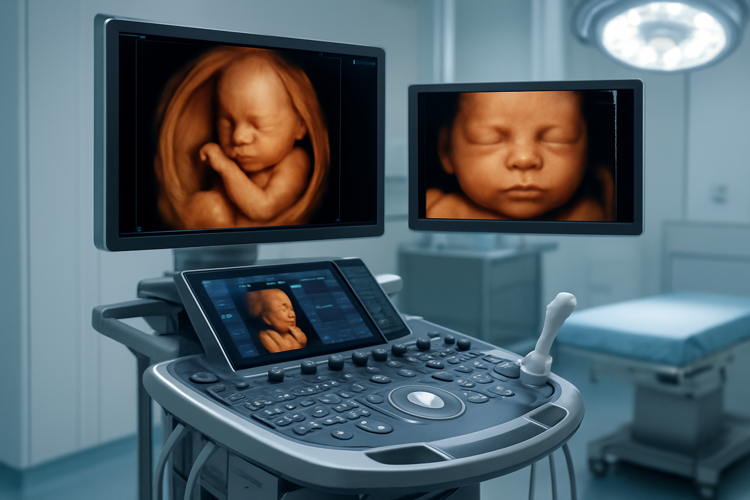
Expecting parents and healthcare providers looking for the most detailed prenatal imaging now have access to USG LEVEL II SINGLE – 5D ULTRASOUND technology. This advanced ultrasound imaging combines traditional diagnostic capabilities with cutting-edge 5D sonography benefits, delivering high-definition fetal imaging that goes beyond standard 2D scans.
Our CGHS empanelled facility in Sector 65, Gurugram, serves patients who want comprehensive prenatal diagnostic ultrasound services using modern ultrasound equipment that meets the highest clinical ultrasound applications standards. Digital reports are conveniently delivered via WhatsApp, while physical films can be collected from the centre at your preferred time.
We’ll explore the revolutionary features of 5D ultrasound technology and how it transforms diagnostic accuracy. You’ll also learn about the complete patient experience, including ultrasound safety guidelines and what makes this diagnostic imaging technology essential for modern prenatal care. Finally, we’ll break down the technical capabilities that make this advanced imaging possible and why it matters for your healthcare decisions.
Revolutionary 5D Ultrasound Features and Capabilities
Multi-dimensional imaging for comprehensive organ assessment
5D ultrasound technology transforms the way medical professionals visualize and assess internal structures, offering unprecedented depth in diagnostic imaging. Unlike traditional 2D ultrasounds that provide flat, single-plane images, advanced ultrasound imaging creates volumetric representations that capture organs and tissues from multiple angles simultaneously.
This multi-dimensional approach proves especially valuable during USG Level II single examinations, where detailed anatomical assessment is critical. The technology generates real-time, three-dimensional volumes while adding temporal dimension and enhanced tissue characterization capabilities. Medical practitioners can rotate, slice, and examine structures from virtually any angle, providing comprehensive organ assessment that was previously impossible with conventional methods.
The enhanced visualization particularly benefits complex anatomical regions where overlapping structures might obscure important details in traditional imaging. Cardiac assessments, for instance, benefit tremendously from this technology, as physicians can observe heart chambers, valves, and blood flow patterns with remarkable precision. Similarly, abdominal organ evaluation becomes more thorough, allowing for better detection of subtle abnormalities that might be missed with standard imaging techniques.
High-definition fetal imaging represents another breakthrough application, enabling detailed examination of developing structures and early detection of potential developmental concerns. The technology’s ability to capture fine anatomical details helps clinicians make more confident diagnoses and treatment decisions.
Superior image clarity and resolution quality
Modern ultrasound equipment incorporating 5D sonography benefits delivers exceptional image quality that significantly surpasses conventional ultrasound systems. The enhanced resolution stems from advanced beamforming algorithms, sophisticated signal processing, and improved transducer technology that work together to minimize artifacts and maximize diagnostic clarity.
The improved image quality becomes immediately apparent when comparing side-by-side examinations. Fine anatomical structures appear sharper and more defined, while tissue boundaries show enhanced contrast that makes differentiation between normal and abnormal tissues much clearer. This clarity proves especially valuable when examining small structures or detecting subtle pathological changes that might be overlooked with lower-resolution imaging.
| Image Quality Factor | Traditional 2D | 5D Technology |
|---|---|---|
| Resolution | Standard | Ultra-high definition |
| Depth Penetration | Limited | Enhanced |
| Artifact Reduction | Basic | Advanced algorithms |
| Contrast Resolution | Good | Exceptional |
| Real-time Processing | Standard | Multi-dimensional |
The superior resolution also reduces the need for repeat examinations, as initial scans capture sufficient detail for accurate diagnosis. This efficiency benefits both patients and healthcare facilities, reducing examination time while improving diagnostic confidence. The enhanced image quality particularly shines in challenging imaging scenarios, such as obese patients or areas with significant acoustic shadowing, where traditional methods often struggle to provide adequate visualization.
Diagnostic imaging technology continues advancing, and 5D ultrasound represents a significant leap forward in providing clinicians with the visual information needed for accurate, confident medical decisions.
Clinical Applications and Diagnostic Benefits
Obstetric and Gynecological Examination Advantages
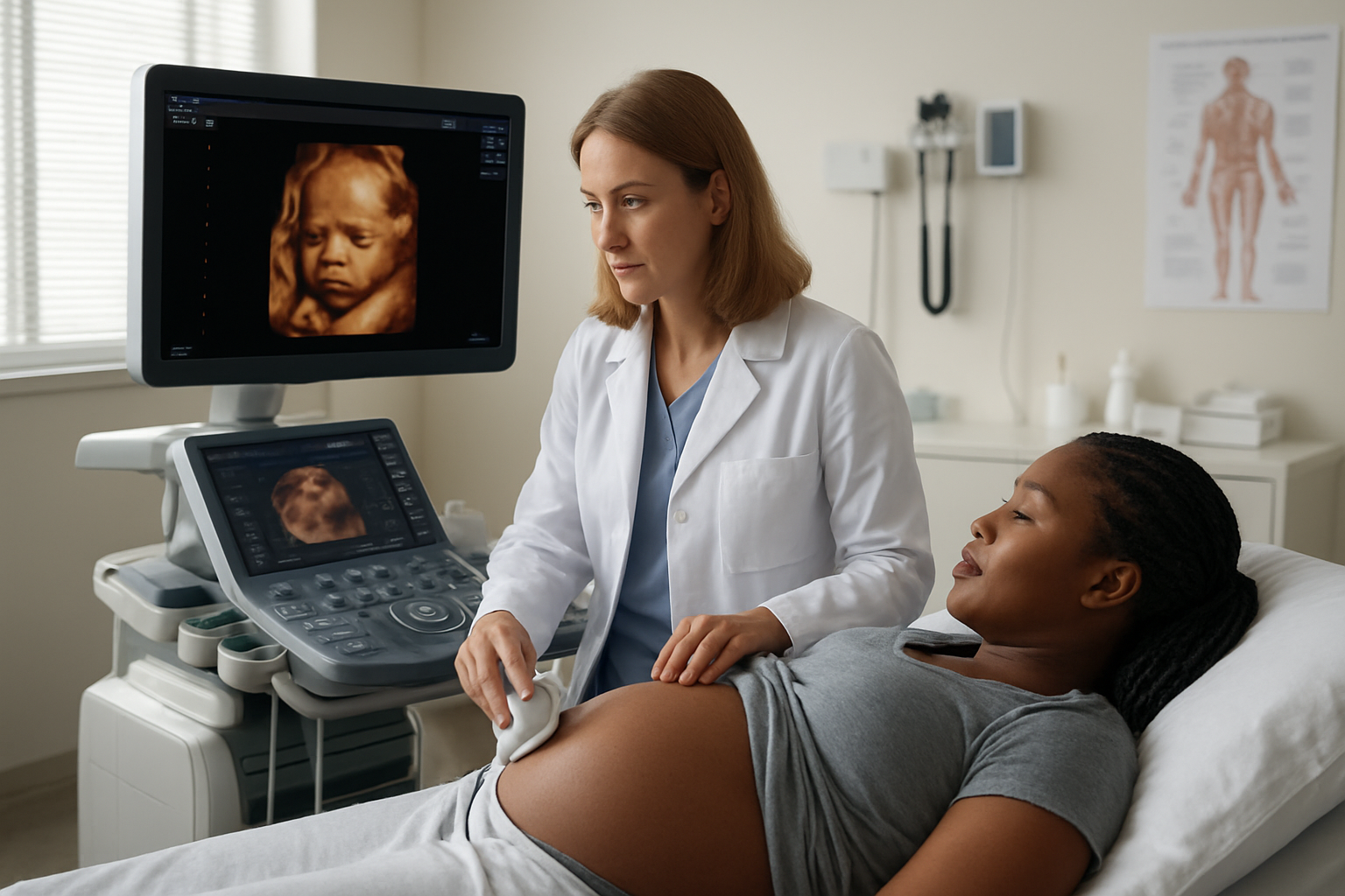
5D ultrasound technology transforms prenatal care by delivering incredibly detailed images that help doctors spot potential issues much earlier than traditional methods. The enhanced visualization capabilities allow medical professionals to examine fetal anatomy with remarkable precision, making USG Level II single examination an essential tool for comprehensive pregnancy monitoring.
During prenatal checkups, this advanced ultrasound imaging technology provides crystal-clear views of the developing baby’s organs, limbs, and facial features. Parents can see their baby’s expressions, movements, and even thumb-sucking behaviors in real-time. Beyond the emotional connection, these detailed images help detect structural abnormalities, heart defects, and neural tube problems that might require specialized care.
For gynecological assessments, 5D sonography benefits extend to evaluating ovarian cysts, uterine fibroids, and endometrial thickness with exceptional accuracy. The technology’s ability to capture multiple imaging planes simultaneously reduces examination time while improving diagnostic confidence. This proves particularly valuable for monitoring complex gynecological conditions that require regular follow-ups.
Our CGHS empanelled facility ensures that government employees can access these cutting-edge services without financial barriers. The high-definition fetal imaging capabilities help create detailed reports that specialists can review digitally through WhatsApp, though patients still need to collect physical films from our centre at their convenience.
Abdominal Organ Assessment and Pathology Detection
The clinical ultrasound applications of 5D technology extend far beyond obstetrics, revolutionizing how doctors examine internal organs and detect various pathological conditions. This diagnostic imaging technology provides unprecedented clarity when evaluating liver, gallbladder, kidneys, pancreas, and spleen structures.
Liver assessments benefit enormously from the enhanced resolution and multi-dimensional imaging capabilities. Doctors can detect fatty infiltration, cirrhosis, and space-occupying lesions with greater confidence. The technology’s ability to differentiate between solid and cystic masses helps determine appropriate treatment pathways more quickly.
Gallbladder examinations become more precise when checking for stones, polyps, or wall thickening. The modern ultrasound equipment captures subtle changes in organ texture and vascularity that traditional 2D imaging might miss. This improved detection capability means patients receive more accurate diagnoses and appropriate treatments sooner.
Kidney evaluations through 5D imaging excel at identifying hydronephrosis, renal stones, and structural abnormalities. The technology’s enhanced penetration and resolution help visualize deeper structures more clearly, even in patients with challenging body habitus.
Our centre in Sector 65, Gurugram, handles emergency cases requiring urgent abdominal imaging while maintaining the highest ultrasound safety guidelines. The non-invasive nature of prenatal diagnostic ultrasound and abdominal imaging makes it the preferred first-line investigation for many conditions, reducing patient anxiety and providing immediate diagnostic information that guides clinical decision-making.
Patient Experience and Safety Considerations
Non-invasive procedure with zero radiation exposure
5D ultrasound technology represents a breakthrough in patient safety, completely eliminating radiation exposure during prenatal examinations. Unlike X-rays or CT scans that use ionizing radiation, advanced ultrasound imaging relies entirely on sound waves to create detailed images of your developing baby. This makes the USG Level II single examination completely safe for both mother and child, with no cumulative radiation effects to worry about throughout pregnancy.
The sound waves used in 5D sonography benefits are the same frequency range as those used in traditional ultrasounds, but with enhanced processing capabilities that create remarkably clear images. Medical professionals can perform multiple scans without any safety concerns, allowing for comprehensive monitoring of fetal development. This technology has been extensively studied and approved by medical authorities worldwide, giving expecting parents complete peace of mind during their diagnostic journey.
For patients visiting our CGHS empanelled facility in Sector 65, Gurugram, this safety profile means you can focus entirely on the excitement of seeing your baby rather than worrying about potential health risks. The centre follows strict ultrasound safety guidelines established by international medical organizations, ensuring every procedure meets the highest standards of care.
Comfortable positioning and reduced examination time
Modern 5D ultrasound technology dramatically improves patient comfort through ergonomic examination tables and streamlined procedures. The advanced imaging capabilities mean technicians can capture the necessary diagnostic information much faster than traditional methods, typically reducing examination time by 30-40% compared to conventional ultrasound systems.
Patients experience minimal discomfort during positioning, as the high-definition fetal imaging system requires fewer probe adjustments and repositioning. The enhanced sensitivity of modern ultrasound equipment means clearer images can be obtained even when the baby is in challenging positions, reducing the need for extended examination sessions.
Our centre provides a relaxed environment where you can comfortably view real-time images of your baby on high-resolution monitors. The digital nature of our diagnostic imaging technology allows immediate image processing and quality assessment, eliminating the need for repeat scans in most cases.
After your examination, you’ll receive digital reports directly on WhatsApp for immediate access and sharing with your healthcare provider. While digital delivery ensures instant availability, you can collect your physical scan films from our facility at your convenience. This flexible approach accommodates busy schedules while maintaining the professional documentation needed for your medical records.
The combination of reduced examination time and comfortable positioning makes the entire prenatal diagnostic ultrasound experience more pleasant for expecting mothers, particularly during later stages of pregnancy when comfort becomes increasingly important.
Technical Specifications and Equipment Requirements
Advanced Transducer Technology and Probe Configurations
Modern 5D ultrasound technology relies on sophisticated transducer systems that deliver exceptional image quality through advanced crystal arrangements and multi-frequency capabilities. These cutting-edge transducers incorporate wide-band technology, allowing frequency ranges from 2-8 MHz for optimal penetration and resolution balance during USG Level II single examinations.
The probe configurations feature curved array designs with enhanced ergonomics for extended scanning sessions. Each transducer contains hundreds of piezoelectric elements arranged in precise geometric patterns to generate the complex acoustic beams required for 5D imaging. The phased array technology enables electronic beam steering and focusing, eliminating the need for mechanical probe movement while maintaining superior image clarity.
Advanced ultrasound imaging facilities utilize multiple probe types, including convex probes for abdominal scanning, linear probes for superficial structures, and specialized obstetric transducers optimized for prenatal diagnostic ultrasound. The transducer sensitivity reaches levels where even subtle tissue variations become clearly visible, making high-definition fetal imaging more accurate than ever before.
Temperature management systems within the probes prevent overheating during prolonged examinations, ensuring consistent performance. The acoustic lens technology incorporated in modern transducers provides uniform beam profiles across the entire scanning field, reducing artifacts and improving diagnostic confidence.
CGHS empanelled facilities investing in this technology benefit from reduced maintenance requirements and extended probe lifespan, making advanced diagnostic imaging more accessible to patients requiring comprehensive prenatal assessments.
Digital Processing Power and Image Optimization Features
The computational backbone of 5D sonography benefits from powerful digital signal processors capable of handling massive data volumes in real-time. These systems process over 100 million calculations per second, transforming raw acoustic data into detailed anatomical representations that surpass traditional 2D imaging capabilities.
Multi-beam forming technology allows simultaneous processing of multiple ultrasound beams, dramatically improving frame rates while maintaining exceptional image resolution. The parallel processing architecture ensures smooth real-time imaging even during complex scanning procedures, enabling clinicians to capture critical diagnostic information without delays.
Advanced ultrasound equipment incorporates sophisticated noise reduction algorithms that distinguish between relevant tissue echoes and unwanted acoustic interference. These filters work continuously to enhance image clarity, particularly important during challenging examinations where patient movement or acoustic shadowing might compromise image quality.
The image optimization features include automated gain control, dynamic range compression, and adaptive filtering systems that adjust parameters based on patient characteristics and scanning depth. These intelligent systems reduce operator dependency while ensuring consistent image quality across different examination scenarios.
Modern ultrasound equipment features compound imaging technology that combines multiple viewing angles to reduce artifacts and improve tissue visualization. The speckle reduction algorithms work in conjunction with edge enhancement features to provide crisp, detailed images that support confident diagnostic decisions.
Digital archiving systems integrated within these platforms ensure seamless image storage and retrieval. Patients receive their digital reports on WhatsApp for immediate access, while physical films can be collected from the centre at their convenience, supporting flexible report delivery options.
The USG Level II Single with 5D ultrasound technology represents a significant leap forward in diagnostic imaging. This advanced scanning method delivers crystal-clear images with exceptional detail, allowing healthcare professionals to make more accurate diagnoses and provide better patient care. Being CGHS empanelled, this facility ensures that eligible patients can access these cutting-edge services through their healthcare benefits. The enhanced visualization capabilities and real-time imaging make this technology particularly valuable for expectant parents and patients requiring detailed soft tissue examination.
If you’re seeking advanced ultrasound services, visit our modern facility in Sector 65, Gurugram to experience the benefits of 5D imaging technology firsthand. Our experienced technicians handle emergency cases when needed, and you’ll receive your digital reports conveniently via WhatsApp, while physical films can be collected from the centre at your convenience. Take advantage of this state-of-the-art diagnostic tool to get the comprehensive imaging results you need for your healthcare journey.