
“USG Follicular Monitoring Study (Single Day): A Complete Guide to Ovulation Tracking”
USG Follicular Monitoring Study Single Day helps women and fertility specialists track egg development through precise ultrasound imaging. This essential follicular monitoring ultrasound service is designed for women undergoing fertility treatments, couples trying to conceive naturally, and healthcare providers monitoring ovarian follicle tracking for optimal timing.
Our CGHS empanelled facility in Sector 65, Gurugram, specializes in single day follicular study procedures that provide comprehensive insights into your reproductive cycle. We deliver digital reports directly to your WhatsApp for instant access, while you can collect physical scan films at your convenience.
This guide covers three key areas: understanding how fertility ultrasound monitoring works and what to expect during your visit, learning to read your follicular development assessment results and timing implications, and exploring how doctors use these findings for fertility treatment monitoring and personalized care planning.
Whether you’re working with a fertility specialist or monitoring natural cycles, our reproductive ultrasound scan technology ensures accurate follicular maturation tracking that supports your journey toward conception.
Understanding Follicular Monitoring Through Ultrasound
What follicular monitoring reveals about your fertility cycle
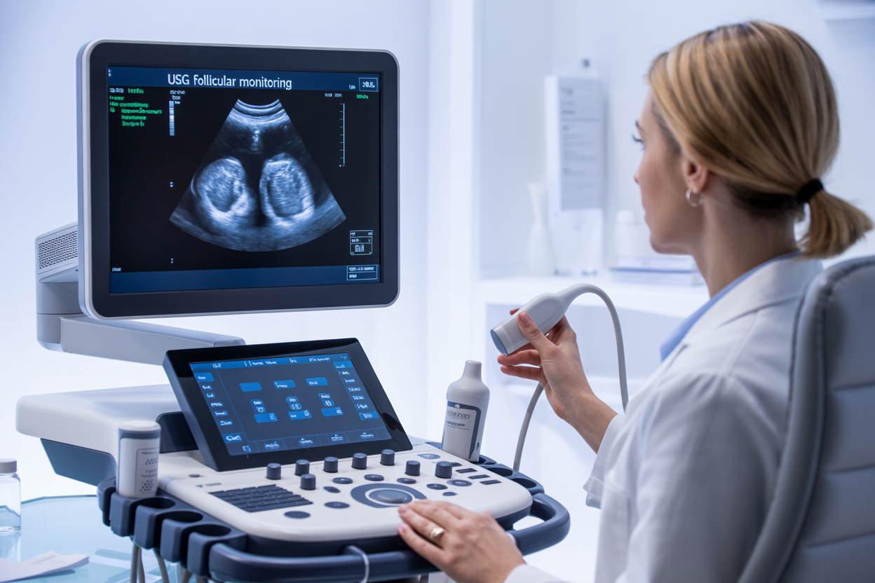
Follicular monitoring ultrasound gives you a clear window into your reproductive cycle, tracking the development of egg-containing follicles in your ovaries throughout your menstrual cycle. This specialized USG follicular monitoring technique captures real-time images of follicular growth, measuring their size, shape, and maturation progress with remarkable precision.
During a single day follicular study, the ultrasound reveals crucial details about your ovarian function. You’ll see exactly how many follicles are developing, which ones are dominant, and whether they’re progressing toward ovulation at the expected pace. The scan measures follicle diameter down to the millimeter, helping predict when ovulation might occur – typically when dominant follicles reach 18-24mm in size.
This ovarian follicle tracking also uncovers important information about your endometrial lining thickness and pattern, which directly affects implantation chances. The ultrasound shows whether your uterine lining is developing synchronously with follicular growth, creating optimal conditions for conception.
For women undergoing fertility treatments, follicular monitoring ultrasound becomes even more valuable. It reveals how your ovaries respond to fertility medications, whether follicles are developing uniformly, and if dosage adjustments are needed. The scan can identify potential issues like ovarian hyperstimulation or poor follicular response before they become problematic.
Your fertility specialist can also assess blood flow patterns around developing follicles using color Doppler technology, providing additional insights into follicular health and viability. This comprehensive follicular development assessment helps create personalized treatment strategies based on your unique ovarian response patterns.
Key advantages of ultrasound over other monitoring methods
Ultrasound stands out as the gold standard for fertility ultrasound monitoring because it provides immediate, visual confirmation of what’s happening inside your ovaries. Unlike blood hormone tests that only give you numbers, reproductive ultrasound scan shows you the actual follicles developing in real-time, making treatment decisions more accurate and timely.
The non-invasive nature of ovulation monitoring USG makes it comfortable for repeated use throughout your cycle. You can have multiple scans without any radiation exposure or side effects, unlike other imaging methods. This safety profile makes ultrasound ideal for fertility treatment monitoring, where frequent assessments are often necessary.
Precision sets ultrasound apart from other monitoring approaches. While ovulation predictor kits can miss your fertile window and basal body temperature tracking only confirms ovulation after it happens, follicular maturation tracking through ultrasound predicts ovulation 24-36 hours before it occurs. This timing advantage is crucial for both natural conception attempts and assisted reproductive procedures.
Cost-effectiveness makes ultrasound monitoring accessible to more patients, especially at CGHS empanelled facilities where covered patients can access quality care without financial barriers. The immediate results eliminate waiting periods associated with laboratory hormone testing, allowing for real-time treatment adjustments.
Ultrasound also provides comprehensive information in a single session. One scan reveals follicle count, size, endometrial thickness, ovarian positioning, and potential abnormalities like cysts or masses. This holistic view helps fertility specialists make informed decisions quickly, improving your chances of successful treatment outcomes while reducing the need for multiple separate tests.
Preparing for Your Single-Day USG Follicular Study

Essential pre-appointment preparations for accurate results
Getting ready for your USG follicular monitoring study doesn’t require major lifestyle changes, but a few simple steps can make a big difference in the quality of your results. Think of it like preparing for any important medical test – the better you prepare, the clearer the picture your doctor gets of what’s happening with your reproductive health.
Timing Your Appointment
The timing of your follicular monitoring ultrasound matters more than you might think. Your healthcare provider will schedule this single day follicular study based on your menstrual cycle, typically between days 10-14 for a standard 28-day cycle. If your cycles are irregular, don’t worry – your doctor will work with you to find the optimal window for ovarian follicle tracking. Keep a detailed record of your cycle patterns for at least two months before scheduling, as this information helps pinpoint the best timing for accurate follicular development assessment.
Physical Preparation Steps
On the day of your fertility ultrasound monitoring, wear comfortable, loose-fitting clothing that allows easy access to your abdomen. Avoid wearing jewelry around your waist or lower torso, as metal can interfere with the ultrasound equipment. You don’t need to fast before the procedure, but eating a light meal about an hour before can help you feel more comfortable during the scan.
Your bladder preparation depends on the type of ultrasound approach your clinic uses. For transabdominal scans, you’ll typically need a moderately full bladder – drink about 32 ounces of water one hour before your appointment, then avoid urinating until after the scan. For transvaginal reproductive ultrasound scans, an empty bladder works better, so you can use the restroom right before the procedure.
Medication and Supplement Considerations
If you’re taking fertility medications as part of your treatment plan, continue following your prescribed schedule unless your doctor specifically tells you otherwise. Medications like Clomid, letrozole, or injectable gonadotropins are designed to work in conjunction with ovulation monitoring USG sessions, so stopping them could affect your results and treatment timeline.
Be upfront about any over-the-counter supplements you’re taking, especially those that might affect hormone levels. While most vitamins and minerals won’t interfere with the ultrasound itself, some herbal supplements can impact follicular development, and your doctor needs this information for accurate interpretation.
Managing Stress and Expectations
Your mental state can actually influence your body’s hormonal responses, which in turn affects follicular development. Try to get a good night’s sleep before your appointment and avoid scheduling stressful activities on the same day. Simple relaxation techniques like deep breathing or gentle stretching can help you feel more at ease.
Remember that follicular maturation tracking is a process, not a one-time snapshot. Your results from this single study will be part of a larger picture that helps guide your fertility treatment monitoring decisions. Don’t put pressure on yourself for “perfect” results – your medical team is there to work with whatever your body shows them.
Interpreting Your Follicular Monitoring Results
Normal follicle size measurements and growth patterns
Understanding what constitutes normal follicular development helps you make sense of your USG follicular monitoring results. During a natural menstrual cycle, multiple follicles begin developing, but typically only one becomes dominant and continues to mature.
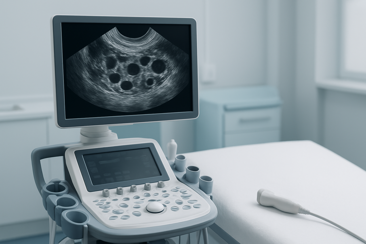
In the early follicular phase, antral follicles measure between 2-9mm in diameter. These small structures appear as dark, fluid-filled circles on your fertility ultrasound monitoring scan. As the cycle progresses, follicles that will continue developing grow at approximately 1-3mm per day during the mid to late follicular phase.
The dominant follicle usually emerges around cycle day 7-10, measuring roughly 10mm in diameter. From this point forward, it grows consistently at about 2mm per day until it reaches pre-ovulatory size. Healthy follicular development assessment shows this leading follicle reaching 18-25mm before ovulation occurs.
Your ovarian follicle tracking results will also reveal the follicle wall thickness, which becomes more prominent as maturation progresses. The follicular fluid should appear echo-free (completely dark) on the ultrasound, indicating proper development. Any internal echoes or irregular shapes may suggest developmental issues that your healthcare provider will discuss with you.
Temperature, stress levels, and underlying health conditions can affect these growth patterns, which is why single day follicular study results are often interpreted alongside your medical history and symptoms.
Identifying dominant follicles and ovulation readiness
Recognizing signs of ovulation readiness through your reproductive ultrasound scan requires looking at several key indicators beyond just follicle size. The dominant follicle stands out clearly from surrounding smaller follicles, typically measuring at least 18mm when ovulation approaches.
Your follicular maturation tracking will show specific characteristics that signal readiness. The follicle wall often becomes thinner and more defined, while the follicular fluid maintains its clear, echo-free appearance. Some women develop a small cumulus oophorus – a cluster of cells visible within the follicle that indicates final maturation stages.
Multiple dominant follicles can develop, particularly in women undergoing fertility treatments. Your fertility treatment monitoring will identify each mature follicle, as this affects timing for procedures like intrauterine insemination or timed intercourse protocols. Generally, follicles measuring 16-18mm are considered nearly mature, while those over 18mm are ready for ovulation.
The ovulation monitoring USG also evaluates endometrial thickness and pattern, which should complement follicular development. A trilaminar (three-layer) endometrial pattern with thickness of 7-10mm typically accompanies a mature follicle, creating optimal conditions for conception.
Your doctor will correlate these ultrasound findings with hormone levels and clinical symptoms to determine the best timing for your specific treatment plan. Each woman’s response varies, making personalized interpretation of these measurements essential for successful outcomes.
Clinical Applications and Treatment Planning
Using results to optimize natural conception timing
Your USG follicular monitoring results provide precise insights into your reproductive cycle, helping you pinpoint the optimal window for natural conception. When follicles reach 18-22mm in diameter, ovulation typically occurs within 12-48 hours. This critical information transforms guesswork into strategic timing.
The fertility ultrasound monitoring reveals dominant follicle development patterns unique to your cycle. Some women ovulate when their leading follicle reaches 18mm, while others need 24mm for mature egg release. Understanding your personal ovulation threshold through follicular development assessment eliminates the uncertainty that couples often face when trying to conceive.
Endometrial thickness measurements complement follicle tracking data. A triple-line endometrial pattern measuring 8-12mm indicates optimal uterine receptivity. When both follicular maturation and endometrial preparation align, conception chances increase significantly.
Your single day follicular study results help identify the best 2-3 day window for conception attempts. Digital reports delivered via WhatsApp allow you to share findings instantly with your partner, ensuring coordinated timing. Many couples report successful pregnancies within 3-6 months of implementing targeted timing based on their ovarian follicle tracking data.
Irregular cycles benefit tremendously from this approach. Rather than relying on calendar calculations that may be inaccurate, reproductive ultrasound scan results provide real-time cycle information, adapting recommendations to your body’s actual hormonal patterns.
Supporting IVF and fertility treatment protocols
Fertility treatment monitoring through USG forms the backbone of successful IVF protocols. Your reproductive specialist uses follicular monitoring ultrasound data to make critical decisions about medication dosing, trigger shot timing, and egg retrieval scheduling. Each scan provides vital information that directly impacts treatment success rates.
During controlled ovarian stimulation, follicular maturation tracking occurs every 2-3 days. Your fertility centre monitors multiple follicles simultaneously, adjusting hormone medications based on growth patterns. When 60-70% of follicles reach 17-20mm diameter, human chorionic gonadotropin (hCG) triggers final egg maturation.
The ovulation monitoring USG reveals response variations to fertility medications. Some patients respond rapidly to lower doses, while others require protocol adjustments. Your medical team uses this data to prevent ovarian hyperstimulation syndrome while maximizing egg yield for retrieval procedures.
Frozen embryo transfer cycles rely heavily on endometrial monitoring. Your fertility treatment monitoring includes measuring endometrial thickness and pattern development. Transfer scheduling depends on achieving optimal uterine lining conditions, typically 8mm thickness with appropriate hormonal support.
CGHS empanelled facilities provide comprehensive fertility ultrasound monitoring as part of approved treatment protocols. Patients can collect physical scan films from the centre at their convenience while receiving immediate digital reports via WhatsApp for treatment planning discussions with their fertility specialists.
Maximizing the Value of Your Follicular Study
Questions to Ask Your Healthcare Provider About Results
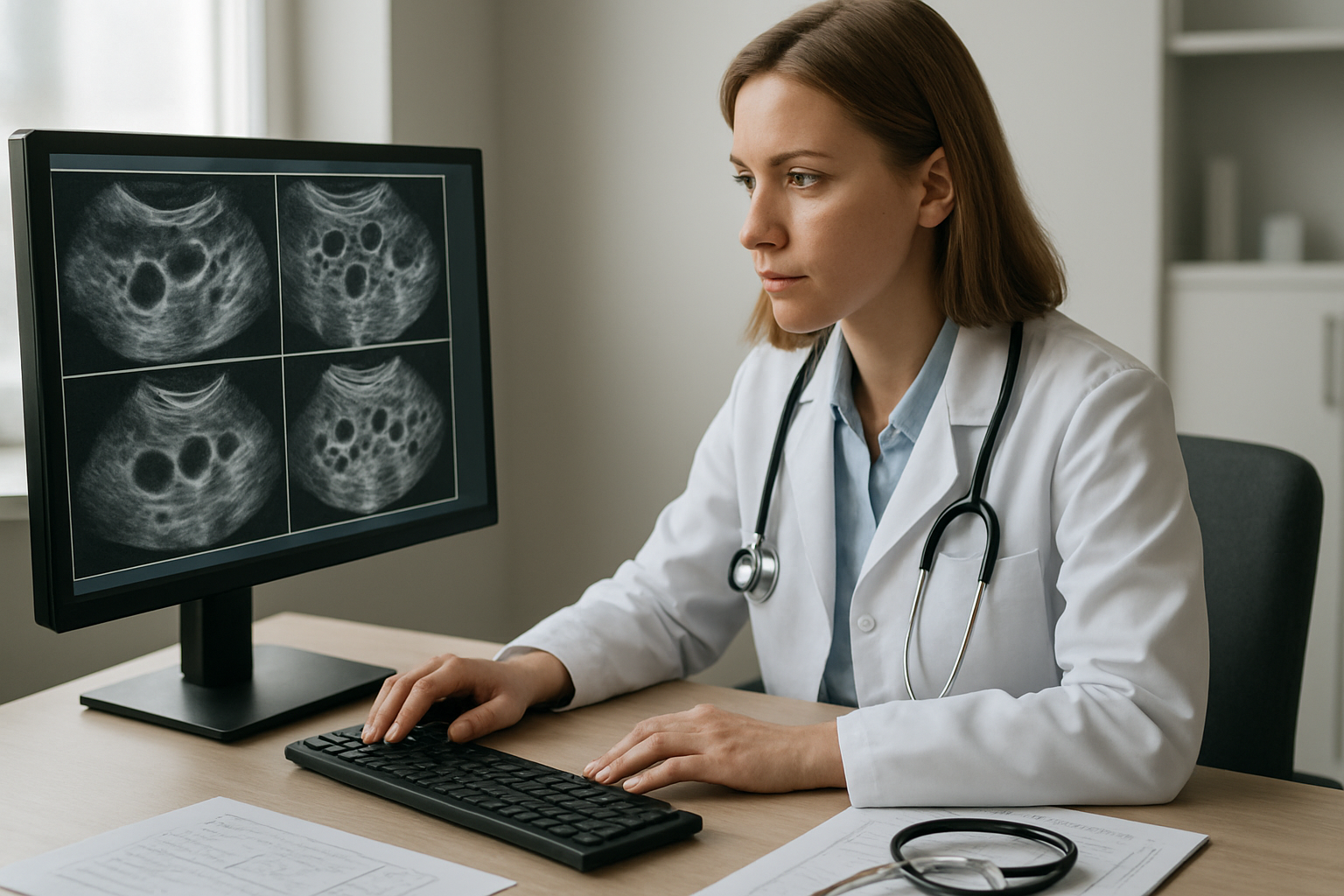
Getting the most from your USG follicular monitoring study means asking the right questions during your consultation. Start by understanding what your specific follicle measurements indicate about your ovarian response. Ask your doctor to explain the size and number of developing follicles, as these numbers directly impact your fertility treatment timeline and medication adjustments.
Request clarification on your endometrial thickness measurements and what they mean for implantation readiness. Your healthcare provider should explain how your current cycle compares to previous monitoring sessions and whether any adjustments to your treatment protocol are needed based on these follicular development assessment results.
Don’t hesitate to inquire about the timing of your next monitoring appointment or trigger shot administration. Understanding the correlation between your hormone levels and ultrasound findings helps you grasp the complete picture of your reproductive cycle. Ask about any concerning findings and what steps might be taken if follicular maturation tracking shows suboptimal development.
If you’re working with a CGHS empanelled facility, confirm that all necessary documentation is properly processed for your benefits coverage. Request information about accessing your digital reports on WhatsApp and when you can collect physical films from the centre at your convenience.
Tracking Patterns Across Multiple Cycles for Better Outcomes
Consistent ovarian follicle tracking across multiple cycles reveals valuable patterns that single-day studies cannot capture. Maintaining detailed records of your follicular monitoring ultrasound results helps identify your unique response patterns to fertility medications and natural hormonal fluctuations.
Create a cycle log that includes follicle counts, sizes, and growth rates from each monitoring session. Track your endometrial thickness progression alongside follicular development to understand your body’s coordinated response. This comprehensive fertility ultrasound monitoring data becomes invaluable for optimizing future treatment protocols.
Pay attention to how your ovaries respond to different medication dosages or timing adjustments. Some patients show consistent patterns in follicle recruitment, while others demonstrate cycle-to-cycle variations that require protocol modifications. Your reproductive specialist can use this accumulated data to predict optimal stimulation parameters for subsequent cycles.
Document any side effects or symptoms alongside your ultrasound results. Correlating physical sensations with follicular development helps you recognize signs of ovarian hyperstimulation or inadequate response early in future cycles. This proactive approach to fertility treatment monitoring significantly improves your chances of successful outcomes.
Regular pattern analysis also helps identify the best candidates for single day follicular study timing, ensuring maximum diagnostic value from each monitoring session while minimizing unnecessary visits to the centre.
Ultrasound follicular monitoring gives you clear insights into your fertility journey by tracking egg development in real-time. This single-day study helps your doctor time treatments perfectly, whether you’re planning natural conception or undergoing fertility procedures. The detailed images and measurements from your scan provide the roadmap your healthcare team needs to make informed decisions about your care.
Getting the most from your follicular study means choosing a facility that combines advanced technology with expert interpretation. Our CGHS empanelled centre in Sector 65 ensures accurate results while keeping costs manageable for government employees. We make the process convenient with digital reports delivered directly to your WhatsApp, and you can pick up your scan films whenever it works for your schedule. Your fertility journey deserves precise monitoring – this simple ultrasound study gives you the clarity and confidence to move forward with your treatment plan.