

“Echocardiography explained: A safe, accurate way to see your heart in motion and detect problems early.”
Understanding Echocardiography: Your Complete Guide to Heart Ultrasound
Echocardiography is a non-invasive cardiac imaging technique that uses sound waves to create detailed pictures of your heart. This heart ultrasound technology helps doctors diagnose heart conditions, monitor treatment progress, and assess overall cardiac health without any discomfort or radiation exposure.
This guide is designed for healthcare professionals, medical students, and patients who want to learn more about the echocardiogram procedure and what to expect during cardiac ultrasound examinations.
We’ll walk you through the fundamental technology behind echocardiographic examination, covering how sound waves create real-time images of your heart’s structure and function. You’ll discover the different types of echo tests available, from standard transthoracic exams to specialized stress echocardiograms, and learn which approach works best for specific situations.
We’ll also break down the patient preparation process and examination steps, so you know exactly what happens during your visit. Finally, you’ll gain practical insights into echo results interpretation, helping you understand how doctors use these cardiac imaging findings to identify heart problems and develop treatment plans.
Master the Fundamentals of Echocardiography Technology
Discover How Sound Waves Create Detailed Heart Images
Echocardiography works on a simple but brilliant principle – sound waves bounce off your heart structures to create moving pictures. Think of it like sonar that submarines use, but instead of finding objects underwater, we’re mapping your heart’s chambers, valves, and blood flow patterns.
The cardiac ultrasound technology sends high-frequency sound waves (typically 2-10 MHz) through a special probe called a transducer. When these waves hit different heart tissues, they reflect back at varying speeds and intensities. Dense structures like heart valves reflect more sound waves, appearing brighter on the screen, while blood-filled chambers appear darker.
The magic happens when sophisticated computer algorithms process these returning sound waves in real-time. The system calculates the time delay between sending and receiving each wave, then translates this information into detailed moving images. You can actually watch your heart beating, see blood flowing through chambers, and observe valve movements frame by frame.
Different frequencies penetrate tissues at various depths. Lower frequencies travel deeper but provide less detail, while higher frequencies offer crisp images of surface structures. Modern echocardiogram procedure technology automatically adjusts these settings based on the patient’s body type and the specific heart structures being examined.
The Doppler effect adds another layer of information by detecting moving blood cells. As red blood cells move toward or away from the transducer, they slightly change the frequency of reflected sound waves. This creates color-coded maps showing blood flow direction and velocity throughout your heart.
Learn the Key Components of Echocardiography Equipment
The transducer serves as the heart of any echo test system. These sophisticated devices contain piezoelectric crystals that convert electrical energy into sound waves and vice versa. Different transducer types are designed for specific applications – phased array transducers create sector-shaped images perfect for cardiac imaging, while linear transducers produce rectangular images better suited for vascular studies.
| Component | Function | Key Features |
|---|---|---|
| Transducer | Sound wave generation and reception | Multiple frequencies, beam focusing |
| Console | Image processing and display | Real-time processing, measurement tools |
| Monitor | Image visualization | High-resolution display, adjustable brightness |
| Storage System | Data archiving | Digital storage, network connectivity |
The main console houses powerful processors that handle millions of calculations per second. These systems filter noise, enhance image quality, and provide measurement tools for cardiac assessment. Modern machines offer preset optimization settings for different types of echocardiographic examination, automatically adjusting gain, time gain compensation, and image depth.
Cardiac imaging systems include sophisticated software packages for quantitative analysis. These programs can automatically trace heart chamber borders, calculate ejection fractions, and measure valve areas. Advanced features like strain imaging and 3D reconstruction provide detailed information about heart muscle function and structural abnormalities.
Storage capabilities have evolved from analog videotapes to digital systems supporting DICOM standards. This allows seamless integration with hospital networks, enabling physicians to review heart ultrasound studies remotely and share images with specialists worldwide. Cloud-based storage solutions now offer virtually unlimited capacity for archiving patient studies while maintaining strict security protocols.
The ergonomic design of modern equipment reduces operator fatigue during lengthy examinations. Adjustable monitor arms, keyboard positioning, and transducer cable management systems help sonographers maintain proper posture while delivering high-quality diagnostic images.
Explore Essential Types of Echocardiographic Examinations
Maximize Diagnostic Value with Transthoracic Echocardiography
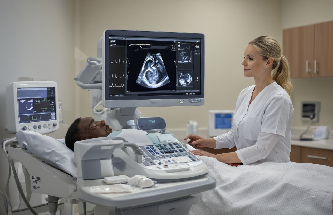
Transthoracic echocardiography stands as the most common and accessible form of cardiac imaging, offering a non-invasive window into heart function. This standard echocardiogram procedure involves placing an ultrasound transducer directly on the chest wall, capturing real-time images of the beating heart through sound waves.
The beauty of transthoracic heart ultrasound lies in its versatility and safety profile. Patients experience no radiation exposure, making it suitable for repeated monitoring and safe for pregnant women. The examination typically takes 30-45 minutes and provides comprehensive information about:
- Heart chamber sizes and wall thickness
- Valve function and blood flow patterns
- Ejection fraction and overall pump function
- Presence of blood clots or masses
- Pericardial abnormalities
Cardiac imaging specialists can assess multiple heart conditions using this technique, from valve disease to heart failure. The Doppler capabilities allow measurement of blood flow velocities, helping detect stenosis or regurgitation. Color Doppler imaging adds another layer of diagnostic power by visualizing blood flow direction and turbulence.
Modern transthoracic systems offer exceptional image quality with advanced features like tissue harmonics and compound imaging. These technologies improve visualization even in challenging patients with obesity or lung disease. The real-time nature of echocardiographic examination enables dynamic assessment during stress testing or with position changes, providing crucial diagnostic information that static imaging cannot deliver.
Access Advanced Imaging Through Transesophageal Echocardiography
Transesophageal echocardiography represents the gold standard for detailed cardiac visualization when transthoracic imaging proves insufficient. This semi-invasive procedure involves inserting a specialized ultrasound probe through the mouth into the esophagus, positioning it directly behind the heart for superior image quality.
The proximity advantage cannot be overstated. With no intervening lung tissue or chest wall structures, transesophageal echo test delivers crystal-clear images of posterior cardiac structures. This technique excels in visualizing:
| Structure | Diagnostic Advantage |
|---|---|
| Left atrial appendage | Clot detection before cardioversion |
| Mitral valve apparatus | Detailed leaflet and chordal assessment |
| Aortic root | Dissection evaluation and measurements |
| Prosthetic valves | Function assessment and complications |
| Endocarditis vegetations | Small vegetation detection |
Cardiac ultrasound technology reaches its peak sophistication with 3D transesophageal imaging. Three-dimensional reconstruction provides surgeons with precise anatomical details, enabling better surgical planning for complex valve repairs. Real-time 3D imaging during interventional procedures guides catheter placement and device deployment with remarkable precision.
Patient preparation requires conscious sedation and throat numbing, making this procedure more involved than standard transthoracic imaging. The examination duration ranges from 15-30 minutes, depending on clinical indications. Despite the semi-invasive nature, complications remain rare when performed by experienced practitioners. The superior diagnostic yield makes transesophageal echocardiography essential for evaluating stroke sources, guiding complex interventions, and assessing surgical candidates where precise anatomical detail determines treatment success.
Identify Critical Heart Conditions Through Echo Analysis

Detect Valve Abnormalities and Assess Their Severity
Heart valves act like one-way doors, controlling blood flow through your heart’s four chambers. When these valves don’t work properly, echocardiography becomes the gold standard for spotting problems and measuring how serious they are.
The heart ultrasound can reveal two main valve issues: stenosis (when valves don’t open wide enough) and regurgitation (when valves don’t close completely). Your technologist will use Doppler technology to measure blood flow velocities across each valve, creating colorful maps that show turbulent flow patterns.
For aortic stenosis, the echo measures the valve area and pressure gradients. A normal aortic valve opens to about 3-4 cm², but severe stenosis shows areas less than 1 cm². The echocardiogram procedure also calculates peak velocities – readings above 4 m/s typically indicate severe narrowing.
Mitral valve problems show up differently on your echo test. Mitral regurgitation appears as a colorful jet shooting backward into the left atrium during heart contraction. The size and intensity of this jet helps doctors grade severity from mild to severe.
| Valve Condition | Key Echo Measurements | Severe Criteria |
|---|---|---|
| Aortic Stenosis | Valve area, peak velocity | <1.0 cm², >4 m/s |
| Mitral Regurgitation | Jet area, vena contracta | >40% LA area, >7mm |
| Tricuspid Regurgitation | Peak velocity, PA pressure | >3.6 m/s velocity |
Modern cardiac imaging technology also measures valve replacement function, checking for proper leaflet motion and detecting any complications like paravalvular leaks or valve thrombosis.
Evaluate Heart Muscle Function and Wall Motion
Your heart’s pumping ability depends on how well the muscle contracts and relaxes. Echocardiographic examination provides detailed pictures of these movements, helping doctors spot areas where the heart isn’t working as it should.
The ejection fraction (EF) serves as the primary measurement of heart strength. This percentage shows how much blood your left ventricle pumps out with each beat. Normal EF ranges from 55-70%, while readings below 40% suggest significant heart muscle weakness.
Wall motion analysis breaks your heart into 17 segments, each scored based on how well it moves:
- Normal (score 1): Good inward movement during contraction
- Hypokinetic (score 2): Reduced movement
- Akinetic (score 3): No movement
- Dyskinetic (score 4): Outward movement during contraction
Cardiac ultrasound technology uses strain imaging to catch subtle muscle problems before they become obvious. This advanced technique measures how much each heart segment stretches and contracts, often revealing damage from heart attacks, infections, or other conditions before standard measurements show problems.
The echo results interpretation includes diastolic function assessment – how well your heart fills with blood between beats. Problems here often appear before pumping issues develop, making early detection possible.
Stress echocardiography takes this evaluation further by recording heart function during exercise or medication-induced stress. Areas with poor blood supply may look normal at rest but show movement problems when the heart works harder, revealing hidden coronary artery disease.
Navigate Patient Preparation and Examination Process

Prepare Patients for Optimal Image Quality
Getting the best possible images during an echocardiogram procedure starts with proper echo patient preparation. Clear communication with patients helps reduce anxiety and ensures they understand what to expect during their cardiac imaging session. Patients should wear comfortable, loose-fitting clothing that allows easy access to the chest area, as they’ll need to remove clothing from the waist up and change into a hospital gown.
Food and medication restrictions are minimal for standard echocardiographic examinations. Patients can eat normally before their heart ultrasound appointment and should continue taking prescribed medications unless specifically instructed otherwise by their physician. However, certain specialized echo studies may require specific preparations, such as fasting before stress echocardiograms or contrast-enhanced examinations.
Skin preparation plays a crucial role in image quality. Patients should avoid using lotions, oils, or powders on their chest area on the day of the examination, as these substances can interfere with ultrasound gel transmission and reduce image clarity. Hair removal may be necessary for patients with excessive chest hair, as it can create air pockets that impair acoustic coupling between the transducer and skin.
Creating a comfortable environment helps patients relax, which improves their cooperation during the echo test. Explaining each step of the process, including the need to hold their breath occasionally and remain still during imaging, ensures better diagnostic quality. Room temperature should be comfortable, as shivering can create artifacts that compromise image interpretation.
Position Patients Correctly for Different Echo Views
Proper patient positioning is essential for obtaining comprehensive cardiac imaging views during the echocardiogram procedure. The standard examination begins with the patient lying on their left side in the left lateral decubitus position, which brings the heart closer to the chest wall and improves acoustic windows for imaging.
The left lateral decubitus position allows optimal visualization of the apical views, which are critical for assessing left ventricular function and wall motion abnormalities. Patients should lie with their left arm extended above their head or tucked under their head for comfort, while the right arm remains free for the sonographer to access the chest area. A pillow or wedge behind the patient’s back helps maintain stable positioning throughout the examination.
Different echocardiographic examination views require specific positioning adjustments:
| Echo View | Patient Position | Key Benefits |
|---|---|---|
| Parasternal | Left lateral decubitus | Best acoustic window for left heart structures |
| Apical | Left lateral decubitus, rotated toward left | Optimal left ventricular assessment |
| Subcostal | Supine with knees bent | Clear view of all four chambers |
| Suprasternal | Supine with neck extended | Aortic arch and great vessel imaging |
For subcostal views, patients transition to a supine position with knees bent and feet flat on the examination table. This positioning relaxes the abdominal muscles and allows better transducer placement below the rib cage. The sonographer may ask patients to take deep breaths and hold them briefly to move the diaphragm and improve visualization of cardiac structures.
Suprasternal notch imaging requires patients to lie flat with their neck slightly extended, often with a small pillow under their shoulders. This position provides access to the suprasternal notch for imaging the aortic arch and assessing for abnormalities in the great vessels. Throughout all positioning changes, clear communication ensures patient comfort and cooperation for optimal cardiac ultrasound technology results.
Interpret Echo Results for Better Patient Outcomes
Read Normal vs Abnormal Echo Measurements
Echo results interpretation starts with understanding the key measurements that define healthy heart function. Normal echocardiogram values serve as benchmarks for detecting heart conditions and making accurate diagnoses.
Key Normal Echo Parameters:
| Parameter | Normal Range | What It Measures |
|---|---|---|
| Ejection Fraction (EF) | 55-70% | Heart’s pumping strength |
| Left Ventricular End-Diastolic Dimension | 3.5-5.6 cm | Heart chamber size at rest |
| Interventricular Septum Thickness | 0.6-1.1 cm | Wall thickness between chambers |
| Mitral Valve E/A Ratio | 1.0-2.0 | Filling pattern of left ventricle |
Abnormal echo measurements reveal specific patterns that point to different cardiac conditions. An ejection fraction below 40% suggests heart failure, while values above 70% might indicate hypertrophic cardiomyopathy. Wall thickness measurements exceeding 1.2 cm often signal high blood pressure damage or genetic heart muscle disease.
Valve function appears clearly in echo results through velocity measurements and visual assessment. Normal mitral valve flow velocities range from 0.6 to 1.3 meters per second, while aortic valve velocities should stay under 2.0 meters per second. Values outside these ranges suggest valve stenosis or regurgitation requiring medical attention.
Red Flag Measurements:
- EF less than 35%: Severe heart failure
- Wall motion abnormalities: Previous heart attack
- Pericardial effusion: Fluid around the heart
- Enlarged chambers: Various heart diseases
Correlate Echo Findings with Clinical Symptoms
Matching echo results with patient symptoms creates a complete picture for accurate heart condition diagnosis. This correlation process helps distinguish between similar-looking conditions and guides treatment decisions.
Shortness of breath paired with reduced ejection fraction on echocardiographic examination clearly indicates heart failure. However, the same symptom combined with normal pumping function but thickened walls suggests diastolic dysfunction or heart failure with preserved ejection fraction.
Common Symptom-Echo Correlations:
Chest pain symptoms take on different meanings based on echo findings. Normal echo results with chest pain might suggest coronary artery disease not yet affecting heart muscle, while wall motion abnormalities during stress echo testing confirm blocked arteries causing symptoms.
Palpitations become more concerning when echo shows structural abnormalities. Normal heart ultrasound results with palpitations often indicate benign rhythm issues, but enlarged chambers or valve problems suggest underlying cardiac disease requiring closer monitoring.
Critical Correlation Patterns:
- Fatigue + low EF = Systolic heart failure
- Ankle swelling + enlarged right ventricle = Right heart failure
- Dizziness + aortic stenosis = Critical valve disease
- Syncope + thick walls = Hypertrophic cardiomyopathy risk
Cardiac imaging specialists look for patterns rather than isolated findings. A patient with diabetes showing early wall motion changes might need aggressive preventive treatment even with mild symptoms, while someone with longstanding high blood pressure might have significant structural changes requiring immediate intervention.
The timing of symptoms relative to echo findings also matters. Recent onset shortness of breath with new wall motion abnormalities suggests acute heart attack, while gradual symptom progression with slowly enlarging chambers indicates chronic heart disease development.

Echocardiography opens a window into the heart’s inner workings, giving doctors the power to see what’s happening in real time. From understanding the basic technology and different types of scans to spotting serious heart conditions, this imaging technique has become a game-changer in cardiac care. The ability to prepare patients properly and guide them through the examination process makes all the difference in getting clear, reliable results.
Getting comfortable with echo interpretation can transform how you approach patient care. Whether you’re reviewing those wavy lines on the monitor or explaining results to worried patients, remember that each echo tells a unique story about someone’s heart health. Take the time to really understand what you’re seeing – your patients are counting on your expertise to help them make the best decisions about their cardiac health moving forward.