

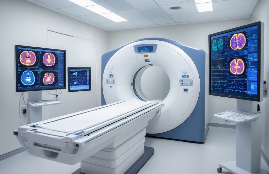
PSMA PET-CT has become a game-changer for men facing prostate cancer diagnosis and monitoring. This advanced imaging technology offers doctors and patients a clearer, more accurate picture of cancer spread than traditional scans.
This guide is designed for prostate cancer patients, their families, and anyone wanting to understand how PSMA PET-CT works and why it matters. We’ll break down the technology behind PSMA PET-CT for prostate cancer and explain why doctors are calling it a breakthrough in cancer detection.
You’ll learn about the superior accuracy rates that make this scan stand out from older imaging methods. We’ll also cover the real clinical benefits for both patients and healthcare teams, and how early detection capabilities are improving treatment outcomes.
Understanding PSMA PET-CT Technology for Prostate Cancer Detection

How PSMA targeting enhances prostate cancer cell visualization
PSMA (Prostate-Specific Membrane Antigen) acts like a beacon on prostate cancer cells, making them light up on imaging scans. This protein is present on the surface of cancer cells at much higher concentrations than in healthy tissue, creating a clear target for radioactive tracers to bind.
Key differences between PSMA PET-CT and traditional imaging methods
| Feature | PSMA PET-CT | Traditional Imaging |
| Detection sensitivity | 95-98% accuracy | 70-80% accuracy |
| Small lesion detection | Spots 2-3mm tumors | Misses lesions under 1cm |
| Bone metastases | Shows early spread | Detects advanced changes only |
Technical advantages of combining PET and CT scanning technologies
PET scanning captures the metabolic activity of cancer cells while CT provides detailed anatomical structure. Together, they create a complete picture that pinpoints exactly where cancer lurks in the body, giving doctors precise locations for treatment planning and monitoring disease progression with unmatched clarity.
Superior Accuracy Rates of PSMA PET-CT Imaging
Detection precision for primary prostate tumors
PSMA PET-CT achieves remarkable detection rates for primary prostate cancer, with sensitivity levels reaching 85-95% for clinically significant tumors. The radiotracer binds explicitly to prostate-specific membrane antigen, allowing precise tumor localization within the prostate gland. This high-resolution imaging helps urologists identify tumor locations, size, and aggressiveness levels that conventional MRI might miss.
Enhanced identification of lymph node metastases
Traditional imaging methods struggle to detect lymph node involvement smaller than 10mm, but PSMA PET-CT can identify metastatic nodes as small as 2-4mm. Studies show detection sensitivity rates of 75-85% for nodal metastases, significantly outperforming conventional CT scans, which typically achieve only 25-35% accuracy for lymph node assessment.
Improved bone metastasis detection capabilities
| Imaging Method | Bone Metastasis Detection Rate |
| Bone Scan | 70-85% |
| CT Scan | 60-75% |
| PSMA PET-CT | 90-98% |
PSMA PET-CT demonstrates superior detection of bone metastases compared to traditional bone scintigraphy. The technology identifies both osteoblastic and osteolytic lesions with exceptional clarity, detecting bone involvement months earlier than conventional methods. This early detection capability proves crucial for treatment planning and patient prognosis.
Reduced false-positive and false-negative results
The specificity of PSMA targeting dramatically reduces false-positive results compared to other imaging modalities. False-positive rates drop to less than 10% with PSMA PET-CT, while conventional imaging methods often produce 20-30% false-positive results. The technology’s ability to distinguish between benign conditions and malignant tissue provides clinicians with greater diagnostic confidence and reduces unnecessary biopsies or treatments.
Clinical Benefits for Patients and Physicians

More precise staging for treatment planning optimization
PSMA PET-CT revolutionizes cancer staging by pinpointing exact tumor locations and spread patterns with remarkable detail. This precision helps doctors create personalized treatment plans that target specific cancer sites while avoiding unnecessary interventions in unaffected areas.
Reduced need for multiple diagnostic procedures
Traditional prostate cancer diagnosis often requires several separate scans and tests, creating stress and delays for patients. PSMA PET-CT combines multiple imaging modalities into a single comprehensive scan, streamlining the diagnostic process, reducing patient anxiety, and providing a complete cancer assessment in a single session.
Earlier intervention opportunities leading to better outcomes
The enhanced sensitivity of PSMA PET-CT detects cancer recurrence months before conventional imaging methods. Early identification allows doctors to begin treatment when tumors are smaller and more responsive to therapy, thereby significantly improving patient survival rates and quality of life.
Cost-effective approach to comprehensive cancer assessment
While PSMA PET-CT may have higher upfront costs, it eliminates the need for multiple diagnostic procedures and reduces treatment delays. This efficiency translates to lower overall healthcare costs and faster patient care delivery, making it a valuable investment for healthcare systems seeking optimal cancer management strategies.
Early Detection Capabilities Transforming Patient Outcomes

Identifying recurrent cancer at lower PSA levels
PSMA PET-CT revolutionizes prostate cancer monitoring by detecting recurrence when PSA levels remain relatively low, often below 1.0 ng/mL. Traditional imaging methods struggle at these levels, leaving patients and doctors in a diagnostic gray area. This advanced technology can pinpoint cancer cells that produce low PSA levels, enabling targeted treatment before the disease progresses significantly.
Detecting microscopic disease spread before symptoms appear
The imaging technique captures microscopic cancer deposits in lymph nodes, bones, and soft tissues long before patients experience pain or other symptoms. This early visualization prevents the anxiety of rising PSA without clear answers. Doctors can now map disease spread with remarkable precision, identifying lesions as small as 4-6 millimeters that would remain invisible on conventional scans for months or years.
Enabling personalized treatment strategies based on disease extent
Treatment decisions become more precise when physicians can see precisely where cancer has spread. Local recurrence may warrant targeted radiation, whereas distant metastases may require systemic therapy. PSMA PET-CT findings directly influence surgical planning, radiation field design, and medication choices. Patients receive treatments tailored to their specific disease pattern rather than generic approaches based on incomplete information.
Optimal Timing and Patient Selection for PSMA PET-CT

Ideal PSA levels for maximum diagnostic benefit
PSMA PET-CT shows excellent detection rates when PSA levels exceed 2.0 ng/mL, with sensitivity reaching 95% above this threshold. The scan performs best in patients with PSA doubling times of less than 10 months, as rapid PSA kinetics often indicate aggressive disease requiring immediate intervention.
Risk stratification criteria for scan recommendation
High-risk patients with Gleason scores ≥7, initial PSA >20 ng/mL, or clinical stage T3-T4 disease benefit most from PSMA imaging. Intermediate-risk patients should undergo scanning when conventional imaging fails to locate recurrent disease or when treatment planning requires precise lesion mapping.
Pre-scan preparation requirements for accurate results
Patients must discontinue antiandrogen therapy 48-72 hours before scanning to prevent false-negative results. Adequate hydration and frequent urination help clear the radiotracer from the urinary system, reducing bladder interference.
Contraindications and patient safety considerations
| Contraindication | Reason |
| Pregnancy | Radiation exposure risk |
| Active UTI | Increased background uptake |
| Recent biopsy | Inflammatory uptake patterns |
Patients with claustrophobia may require mild sedation, while those with diabetes need blood glucose monitoring during the procedure.

PSMA PET-CT has emerged as a game-changing technology that’s revolutionizing how doctors detect and monitor prostate cancer. With its exceptional accuracy and ability to detect cancer cells that other imaging methods might miss, this advanced scan provides patients and their medical teams with the detailed information they need to make better treatment decisions. The technology’s precision helps doctors create more targeted treatment plans, reducing unnecessary procedures and patient anxiety.
The real power of PSMA PET-CT lies in its ability to catch prostate cancer early and track how well treatments are working. When doctors can see exactly where cancer cells are hiding in the body, they can act faster and more effectively. If you or someone you know is dealing with prostate cancer, talk to your physician about whether PSMA PET-CT could be right for your situation. This technology represents a significant step forward in prostate cancer care, offering hope of better outcomes and more personalized treatment.