

“PSMA PET CT explained: A powerful scan that shows where prostate cancer is and how far it has spread.”
PSMA PET CT represents a breakthrough in cancer imaging that’s changing how doctors detect and track certain types of cancer. This advanced scanning technology combines the precision of PET scans with detailed CT imaging to spot cancer cells that other tests might miss.
This guide is designed for cancer patients, their families, and anyone wanting to understand how PSMA PET CT works and why doctors are increasingly recommending it. You’ll also find valuable information if you’re scheduled for a PSMA scan and want to know what to expect.
We’ll explore how PSMA imaging technology works and why it’s revolutionizing cancer detection. You’ll learn about the clinical benefits that make PSMA PET CT scan benefits so significant for treatment planning. Finally, we’ll walk through the patient experience, including PSMA scan preparation requirements and how to understand your results.
Understanding PSMA PET CT Technology and Its Revolutionary Impact
How PSMA PET CT Works to Detect Cancer Cells
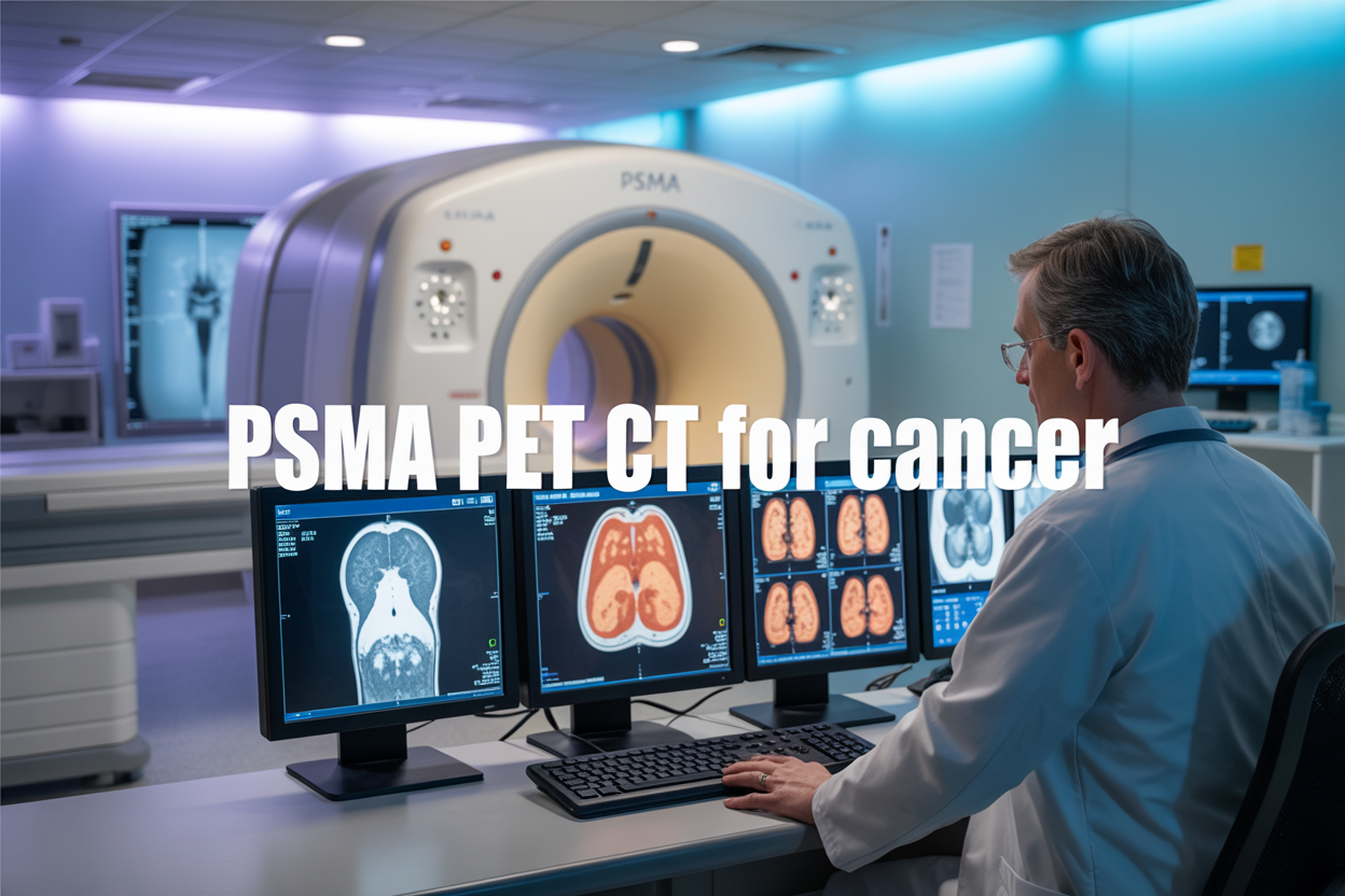
PSMA PET CT technology represents a breakthrough in medical imaging by targeting a specific protein found on prostate cancer cells called prostate-specific membrane antigen (PSMA). The scan combines two powerful imaging techniques: positron emission tomography (PET) and computed tomography (CT), creating detailed pictures that reveal both the metabolic activity and anatomical structure of tissues.
The process begins with injecting a small amount of radioactive tracer that binds specifically to PSMA proteins. Cancer cells typically produce much higher levels of PSMA compared to healthy cells, making them light up brightly on the scan. The radioactive tracer emits positrons that collide with electrons in nearby tissues, creating gamma rays that the PET scanner detects. Meanwhile, the CT component provides precise anatomical details, allowing doctors to pinpoint exactly where the cancer cells are located within the body.
This targeted approach makes PSMA PET CT incredibly effective at finding even tiny cancer deposits that other imaging methods might miss. The technology works particularly well for prostate cancer imaging because PSMA expression increases dramatically in cancerous prostate tissue. The scan can detect cancer recurrence, identify metastases in lymph nodes, bones, and other organs, and help track how well treatments are working.
The entire scanning process typically takes about 60-90 minutes, with patients lying still on a table that moves through the scanner. The radioactive tracer has a short half-life and poses minimal risk to patients.
Key Advantages Over Traditional Imaging Methods
PSMA PET CT scan benefits far exceed those of conventional imaging techniques like bone scans, CT scans, or MRI when it comes to detecting and staging prostate cancer. Traditional imaging methods often struggle to identify small cancer deposits or distinguish between cancerous and non-cancerous tissue, especially when PSA levels are rising but conventional scans appear normal.
The superior sensitivity of PSMA imaging technology allows doctors to detect cancer recurrence at much lower PSA levels compared to traditional methods. While conventional imaging typically requires PSA levels above 10-20 ng/mL to reliably detect metastases, PSMA PET CT can identify cancer spread when PSA levels are as low as 0.2 ng/mL. This early detection capability completely changes treatment planning and patient outcomes.
Another major advantage lies in the scan’s ability to provide whole-body imaging in a single session. Instead of requiring multiple separate scans to check different body regions, PSMA PET CT delivers comprehensive information about cancer spread throughout the entire body. This reduces patient inconvenience, lowers healthcare costs, and speeds up diagnosis.
The precision of PSMA PET CT also significantly improves cancer staging PSMA protocols. Doctors can more accurately determine whether cancer is confined to the prostate, has spread to nearby lymph nodes, or has metastasized to distant organs. This detailed staging information directly impacts treatment decisions, helping oncologists choose between surgery, radiation, hormone therapy, or combination approaches.
Unlike some traditional imaging methods that can produce false positives from inflammation or benign conditions, PSMA PET CT offers much higher specificity for cancer detection, reducing unnecessary anxiety and additional testing for patients.
Specific Cancer Types That Benefit Most from PSMA PET CT Scanning

Prostate Cancer Detection and Monitoring Excellence
PSMA PET CT stands as the gold standard for prostate cancer imaging, offering unmatched precision that traditional imaging methods simply can’t match. This advanced prostate cancer imaging technology targets PSMA proteins that appear in abundance on prostate cancer cells, creating a beacon that lights up even the smallest tumors.
The PSMA PET scan cancer approach excels particularly when PSA levels rise after initial treatment but conventional scans come up empty. Many men find themselves in this frustrating situation where their blood work suggests cancer activity, yet standard imaging shows nothing. PSMA imaging fills this critical gap, detecting cancer cells with remarkable sensitivity even when they’re microscopic.
Advanced prostate cancer detection becomes especially powerful during biochemical recurrence scenarios. When PSA levels climb above 0.2 ng/mL following radical prostatectomy or increase during radiation therapy, PSMA PET CT can pinpoint exactly where cancer cells are hiding. This precision proves invaluable for urologists and oncologists who need to make informed decisions about targeted treatments.
The technology’s ability to detect prostate cancer extends beyond initial diagnosis. For men with intermediate to high-risk prostate cancer, PSMA imaging technology provides detailed staging information that directly influences treatment planning. Unlike conventional bone scans or CT scans that might miss small lesions, PSMA PET CT can identify cancer deposits as small as 4-5 millimeters.
PET CT scan prostate cancer applications also shine in monitoring treatment response. After hormone therapy, radiation, or surgical interventions, this imaging method accurately assesses whether treatments are working effectively, allowing doctors to adjust strategies quickly when needed.
Effectiveness in Identifying Metastatic Disease
Metastatic prostate cancer detection reaches new heights with PSMA PET CT technology. This sophisticated imaging method revolutionizes how doctors identify cancer spread throughout the body, providing clarity that conventional imaging often misses entirely.
Cancer staging PSMA capabilities prove most impressive when evaluating lymph node involvement. Traditional CT scans rely on size criteria, often missing small but cancerous lymph nodes while flagging enlarged but benign ones. PSMA PET CT bypasses this limitation by targeting the actual cancer cells regardless of node size, dramatically improving staging accuracy.
Bone metastases, which affect roughly 90% of men with advanced prostate cancer, become clearly visible with PSMA imaging. While bone scans can show areas of increased activity, they can’t distinguish between cancer, arthritis, or old fractures. PSMA PET CT scan benefits include precise identification of actual cancer deposits in bones, eliminating the guesswork that plagues traditional bone scans.
The technology excels at detecting cancer in unexpected locations. Prostate cancer can spread to organs like the liver, lungs, or even brain tissue. PSMA PET CT’s whole-body scanning approach catches these unusual metastases that might go unnoticed with organ-specific imaging protocols.
Oligometastatic disease, where cancer has spread to just a few sites, represents another area where PSMA shines. Identifying these limited metastases opens doors for targeted treatments like stereotactic radiation therapy, potentially extending survival while maintaining quality of life. This precision transforms what might be palliative care scenarios into opportunities for aggressive, curative treatments.
Clinical Benefits and Treatment Planning Improvements

Precise Tumor Localization for Surgical Planning
PSMA PET CT scan technology transforms surgical decision-making by providing unprecedented detail about tumor location and extent. Unlike traditional imaging methods, PSMA PET CT can detect cancer cells at the molecular level, revealing tiny lesions that might otherwise go unnoticed. This level of precision helps surgeons map out exactly where cancer has spread, allowing them to plan the most effective surgical approach.
When planning prostate cancer surgery, knowing the exact boundaries of tumor involvement makes the difference between a successful procedure and one that leaves cancer behind. PSMA imaging technology shows surgeons which lymph nodes contain cancer cells, which areas of the prostate are affected, and whether the cancer has extended beyond the prostate capsule. This detailed roadmap allows for more targeted surgical techniques while preserving healthy tissue whenever possible.
For patients with recurrent prostate cancer, PSMA PET CT benefits become even more apparent. The scan can pinpoint exactly where cancer has returned, whether in the prostate bed, nearby lymph nodes, or distant organs. This precision enables surgeons to perform focused salvage procedures rather than extensive exploratory operations, reducing complications and improving recovery times.
The technology also helps determine whether surgery is the best treatment option at all. If PSMA PET CT reveals widespread disease, oncologists can redirect patients toward systemic therapies that would be more appropriate than surgical intervention.
Better Assessment of Treatment Response
Monitoring how well cancer treatments are working becomes much more accurate with PSMA PET CT scanning. Traditional methods of tracking treatment response often rely on changes in tumor size, which can take months to become apparent. PSMA scans can detect changes in cancer cell activity much earlier, sometimes within weeks of starting treatment.
This early detection of treatment response helps doctors make critical decisions about continuing, modifying, or switching therapies. If a PSMA PET scan shows that cancer cells are still highly active despite treatment, oncologists can quickly pivot to more aggressive approaches or try different medications. Conversely, when scans show dramatic reductions in PSMA expression, it confirms that the current treatment is working effectively.
For patients receiving hormone therapy, chemotherapy, or newer targeted treatments, PSMA PET CT results interpretation provides objective evidence of treatment success. The scan measures not just tumor size but also the biological activity of cancer cells, offering a more complete picture of how the disease is responding.
Advanced prostate cancer detection through PSMA scanning also helps identify patients who might benefit from newer therapies like PSMA-targeted radioligand treatments. These specialized therapies work specifically on cells that express high levels of PSMA, making the PET scan both a diagnostic tool and a predictor of treatment success.
The ability to track treatment response so precisely means fewer patients receive ineffective treatments for extended periods, reducing side effects and improving quality of life while cancer remains controlled.
Patient Experience and Preparation Requirements

Simple Pre-Scan Preparation Steps
Getting ready for your PSMA PET CT scan is straightforward, with just a few key steps to follow. Your medical team will provide specific instructions, but most patients can eat normally on the day of their scan. Unlike some other imaging procedures, PSMA scan preparation doesn’t require fasting or special dietary restrictions.
You’ll need to arrive at the imaging center about 60-90 minutes before your actual scan time. This allows for the preparation process, including the injection of the PSMA radiotracer. The radiotracer needs time to circulate through your body and bind to PSMA-positive cells before imaging begins.
Wear comfortable, loose-fitting clothing without metal objects like zippers, buttons, or jewelry. Metal can interfere with image quality, so you might need to change into a hospital gown. Remove watches, rings, and any removable dental work before the procedure.
Stay well-hydrated by drinking plenty of water before your appointment, unless your doctor advises otherwise. Good hydration helps with the elimination of the radiotracer from your system after the scan. Bring a list of all medications you’re currently taking, as some might need temporary adjustment.
Tell your medical team about any recent medical procedures, especially if you’ve had other imaging studies or treatments within the past few weeks. This information helps them interpret your PSMA PET CT results more accurately.
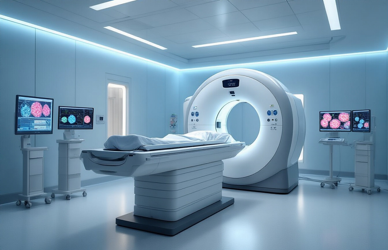
What to Expect During the PSMA PET CT Procedure
The PSMA PET CT procedure typically takes 2-3 hours from start to finish, though the actual scanning time is much shorter. When you arrive, a technologist will explain the process and answer any questions you might have.
First, you’ll receive an intravenous injection of the PSMA radiotracer through a small needle, usually in your arm. This injection contains a tiny amount of radioactive material that’s designed to seek out and bind to PSMA proteins on cancer cells. The injection itself feels like any routine blood draw and takes just a few seconds.
After the injection, you’ll wait in a quiet, comfortable room for about 60-90 minutes. During this uptake period, the radiotracer travels through your bloodstream and accumulates in areas with PSMA-positive cells. You can read, listen to music, or simply relax during this waiting time. Drinking water during this period helps improve image quality and reduces radiation exposure to your bladder.
The actual scanning process involves lying still on a padded table that slides slowly through the PET CT scanner. The machine looks like a large donut and makes some noise, but it’s not as loud as an MRI. You’ll be able to breathe normally, and the technologist can see and hear you throughout the scan.
The scanning portion usually takes 20-30 minutes, depending on the areas being imaged. Some patients may need additional focused images of specific body regions. The technologist will let you know how long to expect and will communicate with you throughout the procedure to ensure your comfort.
Interpreting Results and Next Steps in Cancer Care

How Medical Teams Analyze PSMA PET CT Images
Medical teams use a systematic approach when analyzing PSMA PET CT scan results to identify prostate cancer cells throughout the body. Radiologists and nuclear medicine specialists look for areas where the radioactive tracer has accumulated, appearing as bright spots or “hot spots” on the imaging. These areas indicate high concentrations of PSMA receptors, which are typically found on prostate cancer cells.
The analysis process involves examining multiple factors including the intensity of tracer uptake, the size and location of suspicious areas, and how these findings compare to surrounding normal tissue. Specialists measure standardized uptake values (SUVs) to quantify tracer concentration in different body regions. Higher SUV readings often suggest more aggressive cancer activity.
Medical teams also correlate PSMA PET CT results with other diagnostic information, including PSA blood levels, previous biopsy results, and conventional imaging studies. This comprehensive approach helps distinguish between cancer recurrence, scar tissue, and other benign conditions that might appear similar on standard scans.
The multidisciplinary team typically includes urologists, oncologists, radiologists, and nuclear medicine physicians who collaborate to ensure accurate interpretation. They examine lymph nodes, bones, soft tissues, and the prostate bed area for signs of cancer spread or recurrence. Advanced software tools help create detailed 3D reconstructions that allow doctors to pinpoint exact locations of cancerous tissue.
Treatment Decision Making Based on Scan Results
PSMA PET CT scan results directly influence treatment strategies for prostate cancer patients. When scans reveal localized disease recurrence, doctors might recommend targeted radiation therapy or surgical intervention to remove specific areas of concern. The precise location information helps radiation oncologists design focused treatment plans that minimize damage to healthy surrounding tissues.
For patients with metastatic disease detected through PSMA imaging technology, treatment decisions become more complex. Oncologists consider the number of metastatic sites, their locations, and the intensity of PSMA uptake to determine whether systemic treatments like hormone therapy, chemotherapy, or newer targeted therapies would be most effective.
The scan results also help determine eligibility for emerging treatments like PSMA-targeted radioligand therapy. This innovative approach uses radioactive compounds that specifically bind to PSMA receptors, delivering targeted radiation directly to cancer cells while sparing healthy tissue.
Doctors use PSMA PET CT results interpretation to monitor treatment response over time. Follow-up scans help assess whether chosen therapies are working effectively or if treatment modifications are needed. This real-time feedback allows medical teams to adjust approaches quickly, potentially improving patient outcomes.
The detailed staging information from PSMA scans helps clinicians discuss prognosis more accurately with patients and their families. This information becomes valuable for making informed decisions about quality of life considerations, treatment intensity, and long-term care planning.

PSMA PET CT technology has truly changed how doctors approach cancer detection and treatment planning. This advanced scanning method offers incredibly detailed views of specific cancer types, especially prostate cancer, allowing medical teams to spot problems earlier and track treatment progress more accurately. The benefits go far beyond just better images – patients get more personalized treatment plans, and doctors can make faster, more confident decisions about next steps in care.
If you or a loved one is facing a cancer diagnosis, don’t hesitate to ask your doctor about PSMA PET CT scanning. While the preparation process requires some simple steps and the scan itself takes time, the detailed information it provides can make a real difference in your treatment journey. Understanding what the results mean and how they guide your care plan puts you in a better position to work with your medical team and feel more confident about the path ahead.