
PET-CT in Lung Cancer: Your Complete Guide to Advanced Imaging
PET-CT imaging has changed how doctors detect, stage, and monitor lung cancer treatment. This powerful scanning technology combines two imaging methods to give doctors a detailed picture of what’s happening inside your body.
This guide is written for patients recently diagnosed with lung cancer, their families, and healthcare professionals who want to understand how PET-CT fits into cancer care. We’ll break down the medical jargon and explain what you can expect from these scans.
We’ll cover how PET-CT screening helps catch lung cancer early when treatment works best. You’ll also learn how doctors use these scans to determine your cancer’s exact stage and location, which helps them create your personalized treatment plan. Finally, we’ll explore how PET-CT monitoring shows whether your treatment is working and helps your medical team adjust your care as needed.
Understanding PET-CT Technology for Lung Cancer Detection
How PET-CT Combines Metabolic and Anatomical Imaging
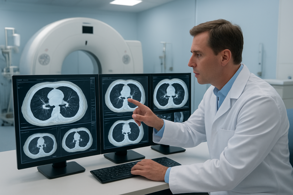
PET-CT technology merges the power of two imaging techniques, creating a comprehensive view of lung cancer that neither method achieves alone. The PET component uses radioactive glucose to identify areas where cells consume high amounts of energy – a hallmark of rapidly dividing cancer cells. Meanwhile, the CT portion captures detailed anatomical structures, showing exact tumor locations and surrounding tissue relationships.
This dual approach gives doctors a complete picture of both what’s happening metabolically and where it’s occurring physically. Cancer cells light up on a PET scan due to increased glucose uptake, while a CT scan provides a roadmap showing precise tumor boundaries, lymph node involvement, and potential spread to other organs.
Superior Accuracy Over Traditional CT Scans
PET-CT dramatically improves diagnostic accuracy compared to CT scans alone, reducing both missed cancers and unnecessary procedures. Traditional CT imaging relies solely on structural changes, often missing small tumors or confusing benign inflammation with malignancy. Studies show PET-CT achieves up to 95% accuracy in lung cancer detection, compared to 70-80% with CT alone.
Enhanced Detection of Small Malignant Lesions
The metabolic sensitivity of PET imaging catches tiny cancerous lesions before they become visible on standard imaging. Even tumors smaller than 1 centimeter can be detected when they show increased glucose metabolism, allowing for earlier intervention and better patient outcomes.
Reduced False Positive Results
PET-CT significantly cuts down on false alarms that plague other imaging methods. By combining metabolic activity with anatomical detail, radiologists can better distinguish between active cancer and benign conditions like scarring, infection, or inflammation that might appear suspicious on CT alone.
PET-CT Screening Benefits for Early Lung Cancer Detection

Identifying Cancer Before Symptoms Appear
PET-CT scans detect metabolic changes in lung tissue months or even years before physical symptoms develop. Cancer cells consume glucose at higher rates than normal cells, creating bright spots on PET images that reveal tumors as small as 4-6 millimeters. This early detection window allows doctors to catch lung cancer in Stage I, when five-year survival rates exceed 90% compared to just 15% for advanced stages.
Distinguishing Between Benign and Malignant Nodules
Standard CT scans often identify lung nodules but cannot determine if they’re cancerous. PET-CT imaging measures the metabolic activity of these nodules, with malignant tumors showing significantly higher glucose uptake. This capability reduces unnecessary biopsies and surgeries while providing accurate diagnosis. Studies show PET-CT achieves 95% accuracy in differentiating between benign and malignant nodules, compared to 65% with CT alone.
Cost-Effective Alternative to Multiple Diagnostic Tests
A single PET-CT scan replaces multiple separate procedures including chest CT, bone scans, and brain MRI. This comprehensive imaging approach reduces total healthcare costs by 30-40% while providing faster results. Patients avoid repeated appointments and radiation exposure from multiple tests, streamlining the diagnostic process from weeks to days.
Precise Staging Advantages with PET-CT Imaging
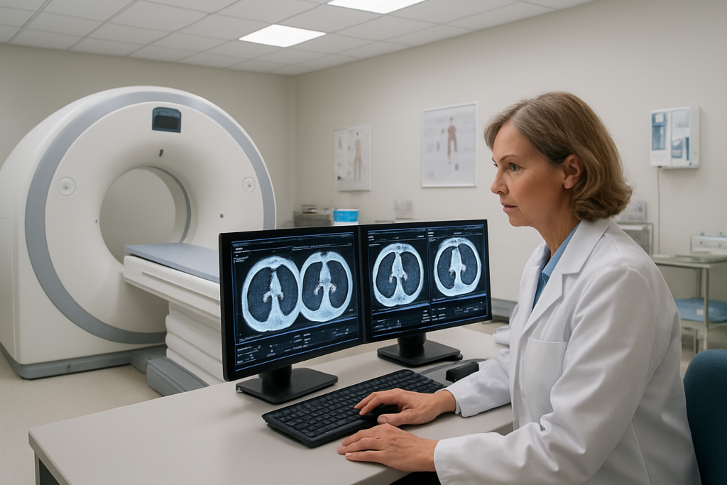
Accurate Assessment of Primary Tumor Size and Location
PET-CT imaging provides unmatched precision in measuring lung tumors and pinpointing their exact location within the chest cavity. This dual-modality approach combines the metabolic activity detection of PET with the anatomical detail of CT scans, giving doctors a complete picture of tumor characteristics. The technology can distinguish between active cancer tissue and surrounding inflammation or scar tissue, which is crucial for accurate staging decisions.
Comprehensive Detection of Lymph Node Involvement
The ability to identify cancer spread to nearby lymph nodes dramatically impacts staging accuracy. PET-CT detects metabolically active lymph nodes that may appear normal on CT alone, revealing hidden metastases that could change treatment strategies. This comprehensive lymph node assessment helps determine whether surgery is viable or if systemic therapy should be the primary approach.
Early Identification of Distant Metastases
PET-CT excels at detecting cancer spread to distant organs before symptoms appear or other imaging methods can identify them. The scan evaluates the entire body in one session, uncovering metastases in bones, liver, brain, and other organs that might otherwise go unnoticed. Early detection of distant spread prevents unnecessary surgeries and redirects patients toward more appropriate systemic treatments.
Improved Treatment Planning and Prognosis Prediction
Accurate staging through PET-CT imaging directly translates to better treatment decisions and more reliable prognosis estimates. Oncologists can tailor therapy protocols based on precise disease extent, avoiding over-treatment in early-stage cases and ensuring aggressive approaches for advanced disease. The detailed staging information helps patients and families understand their situation and make informed decisions about treatment options and life planning.
Treatment Monitoring Capabilities Through PET-CT

Real-Time Assessment of Therapy Response
PET-CT scans provide oncologists with immediate insights into how lung cancer responds to treatment. The technology tracks metabolic changes in tumor cells within weeks of starting therapy, revealing whether cancer cells are dying or continuing to grow. This rapid feedback allows doctors to make quick decisions about treatment effectiveness.
Early Detection of Treatment Resistance
When standard treatments stop working, PET-CT imaging catches resistance patterns before they become visible on regular CT scans. The enhanced sensitivity helps identify areas where cancer cells have adapted to current medications, enabling swift switches to alternative treatment approaches that might work better.
Optimization of Chemotherapy and Radiation Protocols
Treatment protocols get fine-tuned based on PET-CT findings throughout the cancer journey. Doctors can adjust chemotherapy doses, modify radiation targeting, or combine different therapies based on how tumors respond. This personalized approach maximizes treatment benefits while minimizing unnecessary side effects for each patient’s unique situation.
Reduction in Unnecessary Treatment Cycles
PET-CT monitoring prevents patients from enduring ineffective treatment cycles that cause side effects without benefits. When scans show poor response early in treatment, doctors can stop unsuccessful protocols and explore different options. This approach saves patients from months of ineffective therapy while preserving their quality of life and overall health.
Patient Safety and Comfort During PET-CT Procedures
Minimal Radiation Exposure with Maximum Diagnostic Value
PET-CT scans deliver remarkably low radiation doses while providing comprehensive imaging of lung tumors. Modern equipment optimizes radiation levels to stay well below safety thresholds, making the procedure safe for repeated monitoring. The dual imaging approach captures both metabolic activity and anatomical details, giving doctors complete diagnostic information without requiring multiple separate scans.
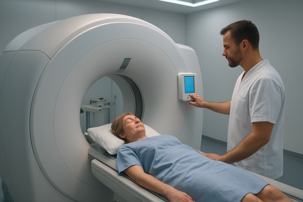
Non-Invasive Outpatient Procedure Benefits
The entire PET-CT process happens on an outpatient basis, requiring no surgery or invasive procedures. Patients receive a simple injection of radioactive glucose and rest comfortably during the 60-90 minute scan. Most people return to normal activities immediately afterward, with no recovery time needed or restrictions on daily routines.
Faster Results Leading to Quicker Treatment Decisions
PET-CT results are typically available within 24-48 hours, dramatically accelerating treatment planning. This rapid turnaround allows oncologists to begin appropriate therapies sooner, whether that’s surgery, chemotherapy, or radiation. Quick diagnostic clarity reduces patient anxiety and prevents delays that could impact treatment effectiveness.
PET-CT has changed the game for lung cancer patients by offering unmatched accuracy in catching cancer early, staging it properly, and tracking how well treatments work. This advanced imaging combines the best of both worlds – metabolic activity detection and detailed anatomical pictures – giving doctors the clear information they need to create personalized treatment plans. The technology’s ability to detect even small tumors and monitor treatment response in real time means patients receive more targeted care and better outcomes.
If you or someone you know is at risk for lung cancer or is currently battling the disease, talk to your healthcare team about whether PET-CT imaging could be right for your situation. The procedure is designed with patient comfort in mind, and the detailed insights it provides can make all the difference in your cancer journey. Don’t wait to explore this powerful diagnostic tool that’s helping save lives every day.