
“PET CT FDG Whole Body Scan: A Complete Guide to Cancer Detection and Body Imaging”
A PET CT FDG whole body scan combines two powerful imaging technologies to create detailed pictures of your body’s metabolic activity and structure. This advanced diagnostic tool is designed for patients who need comprehensive cancer screening, those monitoring treatment progress, or individuals with unexplained symptoms that require thorough investigation.
Your doctor might recommend this scan to detect cancer cells, stage existing tumors, or monitor how well your treatment is working. The scan can also identify other medical conditions affecting your organs and tissues throughout your body.
We’ll walk you through the essential preparation steps you need to follow before your appointment, including dietary restrictions and medication guidelines. You’ll also learn exactly what happens during the scanning process, from the injection of the radioactive tracer to the actual imaging procedure. Finally, we’ll explain how doctors interpret your results and what those findings mean for your next steps in treatment or follow-up care.
Understanding PET CT FDG Technology and Its Medical Applications
How PET CT combines metabolic and anatomical imaging for superior diagnostic accuracy
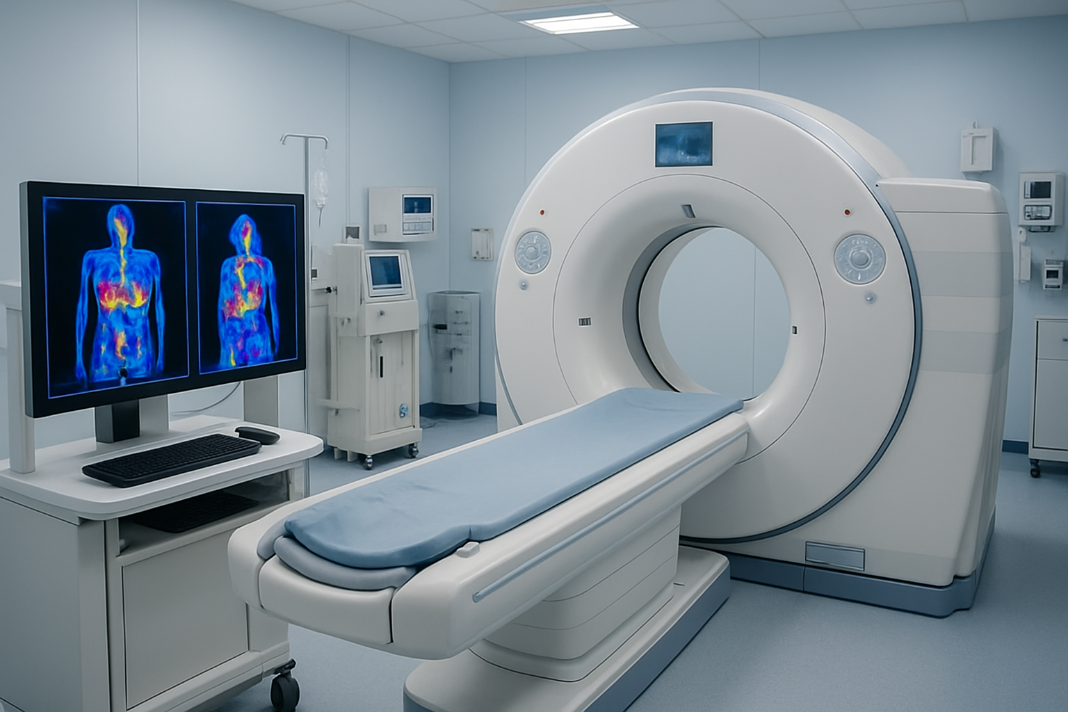
PET CT technology brings together two powerful imaging methods to create a comprehensive picture of what’s happening inside your body. Think of it like combining a road map with a heat sensor – you get both the exact location and the activity level at each spot.
The PET (Positron Emission Tomography) component tracks metabolic activity by detecting radioactive tracers as they move through your body. Meanwhile, the CT (Computed Tomography) scan captures detailed structural images of your organs, bones, and tissues. When doctors merge these two sets of images, they can pinpoint exactly where abnormal activity is occurring and see the precise anatomical context.
This dual approach eliminates much of the guesswork that comes with using either technology alone. A CT scan might show a small mass, but it can’t tell doctors whether that mass is actively growing or metabolically quiet. A PET scan reveals hotspots of activity but lacks the crisp anatomical detail needed for precise localization. Together, they provide a complete diagnostic picture.
The combination proves especially valuable in cancer detection and monitoring. Cancer cells typically consume glucose at much higher rates than normal cells, creating bright spots on PET images. When overlaid on CT’s anatomical framework, doctors can determine not just that cancer is present, but exactly where it’s located, how extensive it is, and whether it has spread to lymph nodes or other organs.
The role of FDG tracer in detecting cellular activity and glucose metabolism
FDG (fluorodeoxyglucose) acts as a molecular spy, mimicking glucose to reveal how cells consume energy throughout your body. This radioactive sugar analog gets injected into your bloodstream about an hour before your scan, giving it time to circulate and accumulate in metabolically active tissues.
Your cells can’t distinguish FDG from regular glucose, so they absorb it eagerly through their normal glucose uptake mechanisms. However, once inside the cell, FDG gets trapped because it can’t complete the usual glucose breakdown process. This trapping effect causes FDG to build up in areas where cells are consuming glucose rapidly.
The radioactive fluorine in FDG emits positrons that the PET scanner detects, creating bright signals wherever glucose consumption is high. Normal organs like your brain, heart, and kidneys naturally show some FDG uptake because they require constant energy. Your brain alone uses about 20% of your body’s glucose supply.
Cancer cells often display dramatically increased glucose consumption – sometimes 10 to 100 times higher than surrounding healthy tissue. This metabolic difference makes FDG particularly effective at spotting malignant tumors, even when they’re quite small. The tracer also helps identify inflammatory processes, infections, and other conditions that increase cellular metabolic demands.
The amount of FDG uptake, measured as a standardized uptake value (SUV), helps doctors assess how aggressive a tumor might be and monitor treatment response over time.
Essential Pre-Scan Preparation for Optimal Results

Dietary restrictions and fasting requirements 12-24 hours before your appointment
Your body needs to be in a specific metabolic state for the FDG tracer to work properly. Think of it like preparing for a blood test, but more involved. You’ll need to fast for at least 12 hours before your scan, though some facilities prefer 24 hours for the clearest results.
During your fasting period, you can only drink plain water. No coffee, tea, juice, or anything with calories. Even sugar-free gum and mints are off-limits because they can trigger insulin responses that interfere with glucose uptake patterns. Your cells need to be hungry for glucose so the FDG tracer concentrates where it should.
The day before your scan, stick to a low-carb, high-protein diet. Skip the pasta, bread, rice, and sugary foods. Instead, focus on lean meats, eggs, and non-starchy vegetables. This helps deplete your normal glucose stores and makes the tracer more effective at highlighting abnormal tissue activity.
Avoid strenuous exercise for 24 hours before your appointment. Working out can cause your muscles to take up glucose, creating confusing bright spots on your scan that aren’t related to any medical condition. Light walking is fine, but save the gym session for after your scan.
If you’re diabetic, your preparation will be different and more complex. Your medical team will give you specific instructions about timing your medications and meals to keep your blood sugar stable while still allowing the scan to work properly.
Medication adjustments and supplements to avoid before scanning
Several medications can interfere with your scan results, so your doctor will review your current prescriptions and supplements well before your appointment. Don’t stop any prescribed medications without explicit instructions from your healthcare team.
If you take metformin for diabetes, you’ll typically need to stop it 48 hours before your scan and wait another 48 hours after before restarting. Metformin can affect how your kidneys handle the contrast agent, though not all PET scans require contrast.
Blood thinners like warfarin usually don’t need adjustment, but newer medications might require timing changes. Your doctor will coordinate with whoever manages your anticoagulation therapy to ensure your safety.
Some supplements can affect scan quality. High-dose vitamin C can interfere with glucose metabolism, so you might need to skip it for a few days. Iron supplements can sometimes create artifacts on images, depending on the timing and dosage.
Steroids like prednisone can suppress the uptake of FDG in certain tissues, potentially masking inflammation or infection. If you’re on steroids for a chronic condition, your doctor will weigh the risks and benefits of temporary adjustment.
Anxiety medications won’t typically interfere with the scan itself, but you might want to take your usual dose to help you stay relaxed during the procedure. Being tense can cause muscle uptake of the tracer, which can complicate interpretation of your results.
Always bring a complete list of everything you take, including over-the-counter medications, vitamins, and herbal supplements. Even seemingly harmless items can sometimes affect scan quality or interact with the radiotracer in unexpected ways.
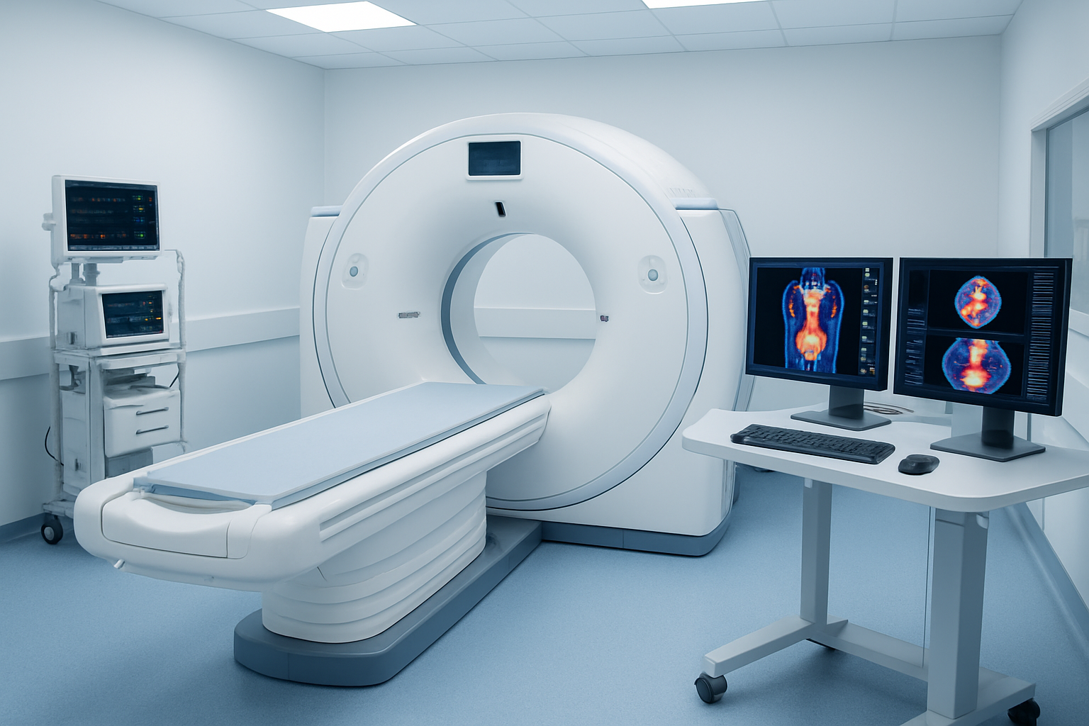
Step-by-Step Scan Process and What to Expect
Initial consultation and medical history review with technologist
When you arrive at the imaging center, you’ll first meet with a certified nuclear medicine technologist who will guide you through the entire process. This consultation typically takes 15-20 minutes and covers several important aspects of your medical background that could affect the scan results.
The technologist will review your current medications, especially diabetes medications, blood thinners, and any recent chemotherapy treatments. They’ll ask about your fasting status since proper preparation requires at least 6 hours without food. Your blood glucose levels will be checked using a simple finger stick test, as elevated glucose can interfere with FDG uptake and compromise image quality.
You’ll discuss any recent surgeries, radiation treatments, or other medical procedures within the past few months, as these can cause inflammation that might appear on the scan. The technologist will also review your medical history for conditions like diabetes, kidney disease, or claustrophobia that might require special accommodations.
Pregnancy screening is mandatory for women of childbearing age since radioactive tracers pose risks to developing fetuses. You’ll be asked to remove jewelry, dental appliances, and any metal objects that could interfere with imaging quality. The technologist will explain what you can expect during the waiting period and scan itself, addressing any concerns or questions you might have about the procedure.
FDG injection procedure and mandatory waiting period
The FDG injection marks the beginning of the actual scanning process. The technologist will start an IV line, usually in your arm, and inject a small amount of radioactive glucose (FDG). The injection itself feels similar to any standard IV procedure and takes just a few seconds. The amount of radiation exposure is comparable to what you’d receive from a CT scan.
After the injection, you’ll enter a mandatory waiting period of 45-60 minutes. This waiting time is critical because it allows the FDG to circulate throughout your body and be absorbed by your cells. Cancer cells and areas of infection or inflammation typically absorb more FDG than normal tissues, which is what makes them visible on the scan.
During this waiting period, you’ll rest in a quiet, dimly lit room to minimize muscle activity that could affect tracer uptake. Physical activity, talking, chewing gum, or even fidgeting can cause muscles to absorb more FDG, potentially creating false positive results. You’ll be encouraged to lie still and relax, though light reading is usually acceptable.
The technologist will monitor you periodically and may offer blankets for comfort. You can use the restroom during this time, and drinking water is encouraged to help flush the tracer through your kidneys after the scan. Some facilities provide comfortable recliners or beds to help you stay relaxed during this crucial uptake period.
Comprehensive Cancer Detection and Staging Benefits
Early-stage tumor identification before symptoms appear
PET CT FDG scans excel at catching cancer in its earliest stages, often years before patients notice any symptoms. The radioactive glucose tracer highlights metabolically active cells, and cancer cells typically consume glucose at much higher rates than normal tissue. This means even tiny tumors, sometimes as small as 4-6 millimeters, can show up as bright spots on the scan.
The real game-changer here is timing. Traditional methods like physical exams, blood tests, or standard CT scans might miss these microscopic cancers entirely. By the time symptoms appear – whether it’s unexplained weight loss, persistent fatigue, or localized pain – cancer may have already progressed significantly. PET CT FDG scanning flips this timeline, allowing doctors to spot trouble long before the body sends out distress signals.
This early detection capability proves especially valuable for high-risk patients, including those with strong family histories of cancer, previous cancer survivors, or individuals with genetic predispositions. Regular PET CT FDG surveillance can catch recurrences or new primary tumors at their most treatable stages.
The scan’s sensitivity also extends to detecting pre-cancerous lesions in certain organs. These abnormal tissue changes haven’t yet become malignant but show increased metabolic activity that warrants close monitoring or immediate intervention.
Accurate cancer staging and metastasis detection throughout the body
Once cancer is confirmed, PET CT FDG scanning becomes the gold standard for staging – determining how far the disease has spread. Unlike other imaging methods that examine specific body regions, this scan provides a comprehensive whole-body view in a single session, typically lasting 45-90 minutes.
The scan’s ability to detect metastases changes everything about treatment planning. Cancer staging follows the TNM system – Tumor size, Node involvement, and Metastasis presence. PET CT FDG excels at all three components, but its metastasis detection capabilities truly set it apart. The scan can identify cancer spread to lymph nodes, bones, liver, lungs, brain, and other organs with remarkable precision.
This whole-body approach prevents treatment mistakes that could occur with limited imaging. For example, a patient might appear to have localized breast cancer on mammography, but PET CT FDG could reveal liver metastases that completely change the treatment strategy from surgery to systemic therapy.
The scan also distinguishes between active cancer and scar tissue or inflammation from previous treatments. This proves crucial for monitoring treatment response and detecting recurrence. Standard CT or MRI scans might show suspicious masses that could be either cancer or benign tissue changes, while PET CT FDG reveals the metabolic activity that indicates living cancer cells.
For treatment monitoring, serial PET CT FDG scans track how tumors respond to chemotherapy, radiation, or targeted therapies. Shrinking metabolic activity often appears before tumors physically shrink, giving doctors early feedback about treatment effectiveness.
Beyond Cancer: Additional Medical Conditions Diagnosed

Heart Disease Assessment and Cardiac Viability Evaluation
PET CT FDG scans reveal critical information about heart muscle function and blood flow patterns that traditional cardiac tests might miss. When heart muscle cells are healthy and functioning properly, they actively consume glucose and show bright uptake on FDG imaging. Areas of the heart that have suffered damage from heart attacks or other cardiac events may show reduced or absent FDG uptake, helping cardiologists distinguish between dead tissue and hibernating muscle that could potentially recover with proper treatment.
The scan excels at identifying viable heart tissue in patients with severe coronary artery disease. This information becomes crucial when deciding whether a patient would benefit from bypass surgery or angioplasty procedures. If large areas of heart muscle show no metabolic activity, invasive procedures may not improve the patient’s condition. However, when hibernating muscle tissue is detected, revascularization procedures often restore normal heart function and significantly improve quality of life.
Cardiac PET imaging also helps evaluate inflammatory heart conditions like sarcoidosis and myocarditis. These conditions cause abnormal glucose uptake patterns that appear distinctly different from typical heart attack damage. Early detection of cardiac inflammation allows doctors to start anti-inflammatory treatments before permanent damage occurs.
The test proves particularly valuable for patients with heart failure symptoms but unclear underlying causes. By mapping metabolic activity throughout the heart muscle, doctors can pinpoint specific areas of dysfunction and tailor treatment approaches accordingly.
Brain Disorders Including Dementia and Seizure Localization
Brain PET CT scans provide detailed maps of glucose metabolism throughout different brain regions, revealing functional abnormalities that structural imaging like MRI cannot detect. In healthy brains, glucose uptake follows predictable patterns, with higher activity in areas like the cerebral cortex and lower activity in white matter regions. When disease processes affect brain function, these metabolic patterns change in characteristic ways.
Dementia diagnosis benefits tremendously from PET imaging, as different types of dementia create distinct metabolic fingerprints. Alzheimer’s disease typically shows reduced glucose uptake in the parietal and temporal lobes, often appearing years before symptoms become severe. Frontotemporal dementia affects the frontal and anterior temporal regions, while Lewy body dementia creates a different pattern of metabolic changes. These metabolic signatures help doctors distinguish between various dementia types and plan appropriate treatments.
For epilepsy patients, PET scans locate seizure focus areas with remarkable precision. Between seizures, the brain regions where seizures originate often show decreased glucose metabolism. During active seizure periods, these same areas may show dramatically increased uptake. This information guides neurosurgeons when considering epilepsy surgery, helping them remove seizure-causing tissue while preserving normal brain function.
The technology also detects brain tumors that may not appear clearly on other imaging studies. Malignant brain tumors typically show intense glucose uptake, while benign tumors often have lower metabolic activity. This metabolic information helps distinguish tumor types and guides treatment decisions.
Interpreting Your Scan Results and Next Steps
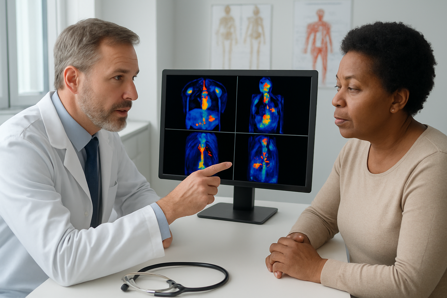
Understanding SUV values and their clinical significance
SUV stands for Standardized Uptake Value, and it’s basically a measurement that shows how much glucose your tissues are absorbing during the scan. Think of it as a numerical score that helps doctors understand what they’re seeing on your images.
Here’s how it works: when you receive the FDG injection before your scan, the radioactive glucose travels through your bloodstream and gets taken up by your cells. Normal tissues use glucose at predictable rates, but abnormal tissues – like cancer cells – often gobble up glucose much faster than they should. The SUV number reflects this uptake intensity.
Most normal tissues have SUV values between 1.0 and 2.5. When doctors see areas with higher SUV values, especially above 2.5 or 3.0, they pay closer attention. However, don’t panic if you hear about elevated SUV numbers – they don’t automatically mean cancer. Infections, inflammation, recent injuries, or even muscle tension can cause increased glucose uptake.
The tricky part is that SUV values aren’t black and white. A reading of 4.0 might be concerning in one part of your body but completely normal in another. Your brain naturally has high SUV values because it constantly burns glucose for energy. Same goes for your heart and liver.
Your doctor considers SUV values alongside other factors like the size, shape, and location of any abnormal areas. They also compare your results to previous scans if you’ve had them, looking for changes over time rather than focusing solely on absolute numbers.
Timeline for receiving comprehensive results from your physician
Getting your PET CT results involves several steps, and the timing can vary depending on your specific situation and healthcare facility. Most patients can expect to hear from their doctor within 2-7 business days after their scan.
The process starts immediately after your scan when a nuclear medicine technologist reviews the images for technical quality. Within 24-48 hours, a radiologist specializing in nuclear medicine interprets your scans, comparing them to your medical history and previous imaging studies. This specialist writes a detailed report describing their findings.
Your referring physician then receives this report and needs time to review it carefully. They’ll compare the results to your symptoms, physical exam findings, lab work, and other tests you may have had. This comprehensive review helps them determine what the results mean for your specific case and what steps to take next.
If your scan shows urgent findings that need immediate attention, most facilities have protocols to contact your doctor within hours rather than days. Emergency situations might prompt same-day communication between the radiologist and your physician.
Some healthcare systems now offer patient portals where you can access preliminary results before your doctor visit. While this provides faster access to information, always wait for your physician’s interpretation before drawing conclusions. They understand your complete medical picture and can explain what the findings mean for your treatment plan.
Complex cases sometimes require additional consultation with specialists, which can extend the timeline by a few extra days. Your doctor might want to discuss your results with oncologists, surgeons, or other experts before meeting with you.
PET CT FDG whole body scans have revolutionized how doctors detect and monitor cancer, offering a powerful combination of metabolic and anatomical imaging in one comprehensive test. This advanced technology goes beyond traditional imaging by showing how your body’s cells are actually functioning, making it incredibly valuable for cancer staging, treatment planning, and monitoring response to therapy. The scan’s ability to detect other conditions like infections and inflammatory diseases makes it an even more versatile diagnostic tool.
Getting the best results from your PET CT FDG scan comes down to proper preparation – following fasting guidelines, staying hydrated, and avoiding strenuous activity beforehand. While the actual scan process takes about 90 minutes and involves minimal discomfort, the detailed images it produces can provide life-changing information about your health. If your doctor has recommended this scan, remember that it’s designed to give them the clearest possible picture of what’s happening inside your body, helping them create the most effective treatment plan for your specific situation.