

“A CT PNS scan gives a detailed view of your sinuses, helping detect infections, blockages, or inflammation—here’s how it works.”
A CT scan of the paranasal sinuses uses specialized X-ray technology to create detailed cross-sectional images of your nasal passages and surrounding air-filled spaces. This diagnostic tool helps doctors see inside your sinuses when symptoms like persistent facial pain, chronic congestion, or recurring infections won’t go away.
This guide is designed for patients scheduled for a sinus CT scan, their families, and anyone wanting to understand this common imaging procedure. You’ll learn what happens during the scan and how to prepare for the best results. We’ll also explain how doctors use these images to spot conditions like sinusitis, polyps, and structural problems that might be causing your symptoms.
Understanding CT Scans for Paranasal Sinus Evaluation
What CT imaging reveals about your sinus anatomy
CT scans provide an incredibly detailed three-dimensional view of your paranasal sinuses, showing structures that other imaging methods simply can’t capture with the same clarity. The scan reveals the four main sinus cavities – the frontal sinuses above your eyebrows, the ethmoid sinuses between your eyes, the sphenoid sinuses deep behind your nose, and the maxillary sinuses in your cheekbones.
What makes CT imaging so valuable is its ability to show the thickness and condition of the sinus walls, the presence of any fluid or mucus buildup, and the health of the tiny drainage pathways called ostia. These drainage openings are crucial for proper sinus function, and even small blockages can lead to significant problems. The scan also reveals the nasal septum’s position and any deviation that might affect breathing or sinus drainage.
CT images can detect polyps, cysts, tumors, and infections with remarkable precision. They show whether inflammation has spread to surrounding tissues and can identify anatomical variations that might predispose you to chronic sinus problems. The bone detail is particularly impressive – CT can spot even hairline fractures or subtle changes in bone density that might indicate chronic infection or other conditions.
How CT scans differ from other sinus imaging methods
Regular X-rays have been largely replaced by CT for sinus evaluation because they only provide a flat, two-dimensional image that often misses important details. X-rays might show fluid levels in the maxillary sinuses, but they can’t reveal the complex anatomy of the ethmoid air cells or detect subtle changes in the smaller sinus cavities.
MRI scans, while excellent for soft tissue detail, don’t show bone structures as clearly as CT. MRI is better for distinguishing between different types of soft tissue masses and can help differentiate between inflammatory tissue and tumors. However, MRI takes longer to perform and costs more than CT scanning.
Nasal endoscopy allows direct visualization of the nasal passages and sinus openings, but only shows surfaces that can be reached with the scope. CT provides the complete picture, showing what’s happening inside the sinus cavities themselves, behind the walls that endoscopy can’t penetrate.
The speed of CT scanning is another major advantage – most sinus CT scans take just a few minutes to complete, while MRI can take 30-45 minutes. CT also works well for patients who have metal implants or pacemakers, which can be problematic with MRI.
Preparing for Your Paranasal Sinus CT Scan

Essential pre-scan instructions to follow
Before your paranasal sinus CT scan, there are several important steps to ensure the best possible imaging results. Unlike some medical procedures, sinus CT scans require minimal preparation, making them convenient for most patients.
You don’t need to fast or avoid eating before your scan. Your regular medications can be taken as prescribed, though you should inform your healthcare team about any medications you’re currently using, especially blood thinners or diabetes medications.
Remove all metal objects from your head and neck area before entering the scanning room. This includes jewelry, earrings, hairpins, dentures with metal components, and hearing aids. These items can create artifacts on the images, potentially obscuring important details your doctor needs to see.
Wear comfortable, loose-fitting clothing without metal zippers, buttons, or decorative elements around the chest and head area. Many imaging centers provide hospital gowns to ensure optimal scanning conditions.
If you have a history of claustrophobia or anxiety, discuss this with your healthcare provider beforehand. While CT scanners are much more open than MRI machines, some patients still feel uncomfortable. Your doctor might prescribe mild sedation if needed.
Inform the technologist if you’re pregnant or suspect you might be pregnant. CT scans use X-rays, and while the radiation exposure is minimal, special precautions may be necessary during pregnancy.
Bring your insurance cards, identification, and any previous sinus imaging studies. Having prior scans available allows radiologists to compare current findings with previous results, providing valuable insights into changes over time.
What to expect during the scanning process
The actual CT scanning process for paranasal sinuses is straightforward and typically takes only 10-15 minutes from start to finish. Most patients find the experience much easier than anticipated.
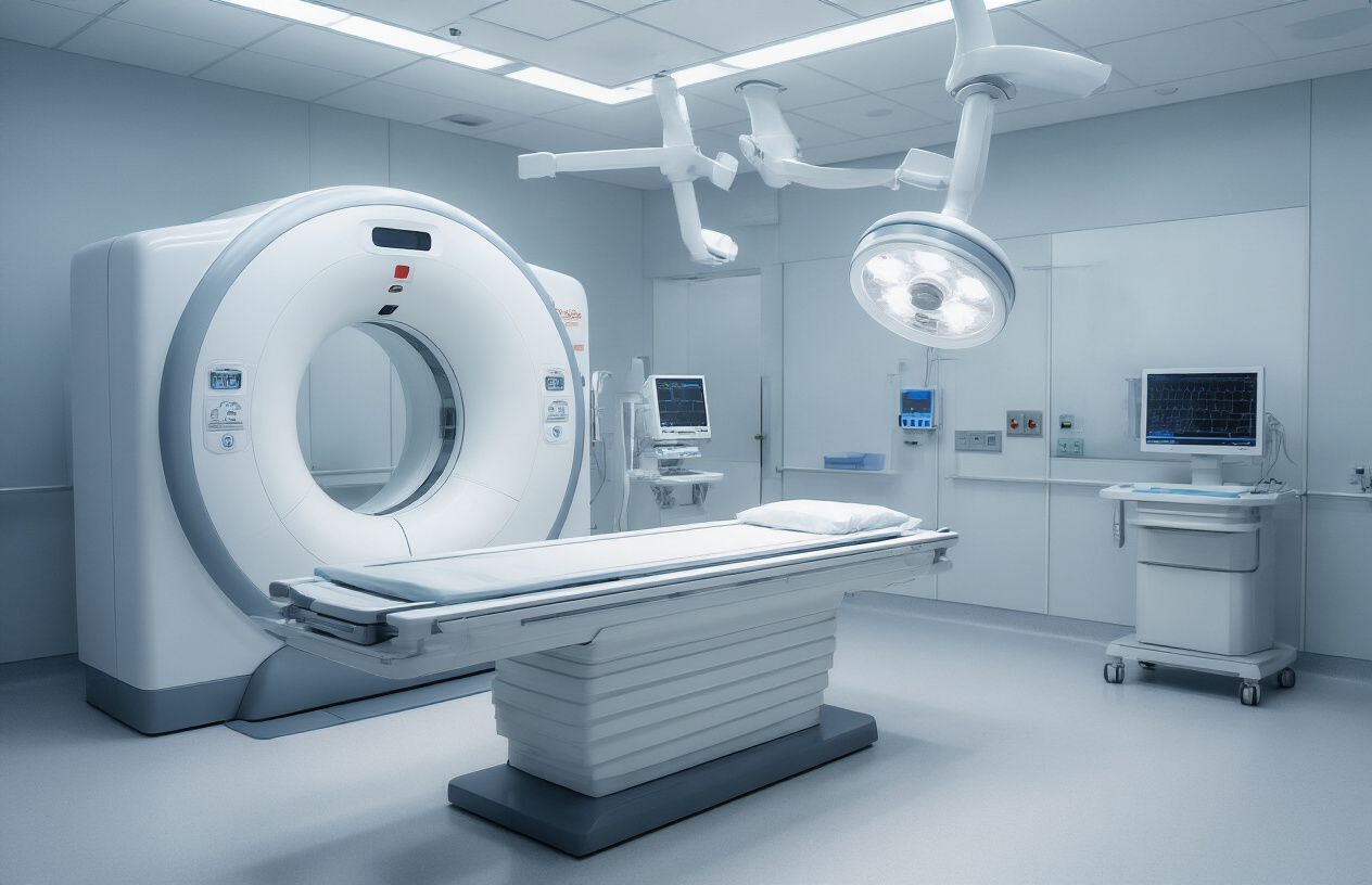
When you arrive at the imaging center, you’ll check in and complete any remaining paperwork. A technologist will escort you to the CT room and explain the procedure. The CT scanner looks like a large donut-shaped machine with a narrow table that slides through the center opening.
You’ll lie on your back on the padded table, and the technologist will position your head carefully to ensure optimal imaging angles. Small cushions or straps might be used to help you maintain the correct position throughout the scan. The table will slowly move through the scanner opening while the X-ray tube rotates around your head.
During the scan, you’ll hear mechanical whirring and clicking sounds as the machine operates. These noises are completely normal and indicate the scanner is working properly. The technologist will communicate with you through an intercom system and can see you at all times through a window.
You’ll need to remain very still during the actual imaging, which typically lasts only a few minutes. The technologist may ask you to hold your breath briefly for certain images to reduce motion blur, though this isn’t always necessary for sinus scans.
The procedure is painless, and most patients experience no discomfort beyond lying still on a firm surface. Some people feel slightly anxious about the enclosed space, but the scanner opening is much larger and less confining than other imaging equipment.
After the scan completes, you can immediately return to normal activities. There are no restrictions on driving, eating, or taking medications following a sinus CT scan.
Conditions Diagnosed Through Sinus CT Imaging
Chronic sinusitis detection and severity assessment
CT scans provide the clearest picture of chronic sinusitis, showing exactly where inflammation has taken hold and how severe the problem has become. When sinuses become chronically inflamed, the scan reveals thickened mucous membranes that line the sinus cavities, creating a distinctive “ground glass” appearance on the images. Doctors can see blocked drainage pathways, fluid buildup, and areas where healthy air-filled spaces have been replaced by inflamed tissue.
The scan helps distinguish between different types of sinusitis patterns. Some patients show pan-sinusitis, where all sinus cavities are affected, while others have isolated involvement of specific areas like the maxillary or ethmoid sinuses. The degree of mucosal thickening tells doctors how long the inflammation has persisted and helps predict which treatments might work best.
Chronic sinusitis often creates changes in the bony structures surrounding the sinuses. The CT scan can detect these subtle bone changes, including sclerosis (thickening) or erosion, which indicates long-standing disease. These findings help doctors understand whether the condition is responding to medical treatment or if surgical intervention might be necessary.
The scan also reveals complications that can develop with chronic sinusitis, such as osteomyelitis (bone infection) or the formation of retention cysts. These complications require different treatment approaches and monitoring strategies.
Identifying nasal polyps and structural abnormalities
Nasal polyps appear as soft tissue masses that grow from the inflamed sinus linings, and CT scans can spot them even when they’re too small to see during a physical exam. These grape-like growths typically develop in the ethmoid sinuses first, then extend into the nasal passages, where they can block breathing and reduce the sense of smell. The scan shows their exact location, size, and how much they’re obstructing normal airflow.
Structural abnormalities become clearly visible on CT imaging, helping explain why some people are more prone to sinus problems. A deviated septum shows up as an obvious curve or bend in the wall separating the nostrils, potentially blocking proper drainage. Concha bullosa, where the normally thin turbinate bones contain air pockets, can crowd the nasal passages and interfere with normal sinus function.
The scan reveals variations in sinus anatomy that might predispose someone to recurring infections. Some people have narrower drainage openings, while others might have additional septae (thin walls) dividing their sinuses into smaller compartments. These variations aren’t necessarily problems by themselves, but they can make it harder for mucus to drain properly.
CT imaging also detects more serious structural issues like previous surgical changes, fractures, or developmental abnormalities that might require specialized treatment approaches. The three-dimensional view helps surgeons plan procedures by showing the exact relationship between different anatomical structures.
Interpreting Your CT Scan Results

Understanding Normal Sinus Anatomy on CT Images
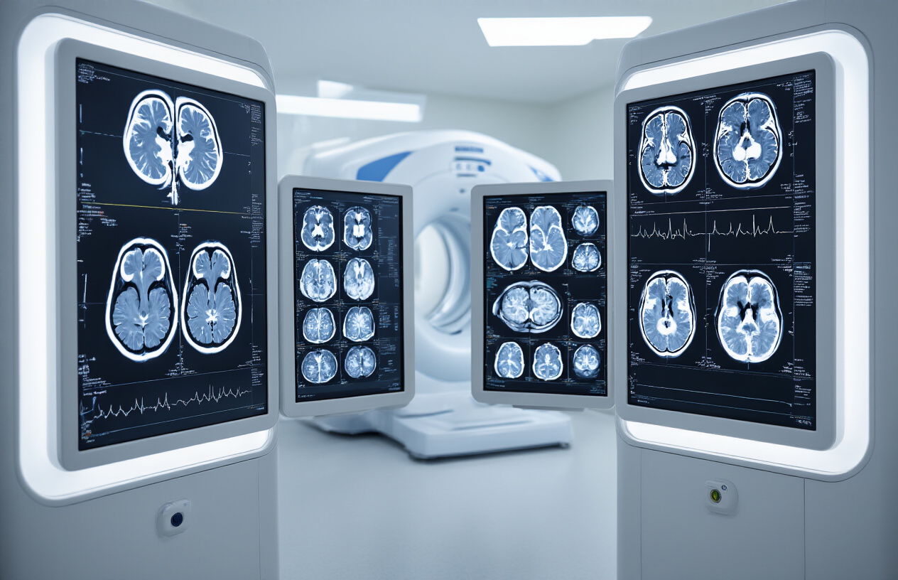
When you look at your CT scan, you’ll see several air-filled spaces around your nose and eyes. These are your paranasal sinuses, and they should appear black or dark gray on the images. The maxillary sinuses are the largest ones, sitting in your cheekbones on both sides of your nose. You’ll also see the ethmoid sinuses, which look like a honeycomb structure between your eyes, the frontal sinuses above your eyebrows, and the sphenoid sinuses deeper in your skull.
Healthy sinuses have thin, smooth walls that create clear boundaries on the scan. The openings between sinuses, called ostia, should be visible and unobstructed. You might notice some natural variations in size and shape between your left and right sides – this is completely normal and doesn’t indicate a problem.
The nasal septum, which divides your nose down the middle, appears as a thin line on the scan. Many people have a slightly deviated septum, which shows up as a curved rather than straight line. The turbinates, small structures that help warm and humidify air, appear as curved ridges along the nasal walls.
Recognizing Signs of Inflammation and Infection
Inflamed or infected sinuses look dramatically different from healthy ones on CT scans. Instead of appearing black with air, affected sinuses show up as gray or white areas filled with mucus, pus, or swollen tissue. This change in density is one of the clearest indicators that something’s wrong.
Acute sinusitis typically shows up as fluid levels or complete opacification of the affected sinuses. You might see a horizontal line where fluid meets air, creating what doctors call an “air-fluid level.” The sinus walls may appear thicker than normal due to inflammation.
Chronic sinusitis presents differently, often showing patchy areas of thickening along the sinus walls or polyps that appear as soft tissue masses. The bone around chronically inflamed sinuses may become thicker and more irregular over time.
Blockages at the sinus openings are another key finding. These obstructions prevent proper drainage and ventilation, leading to the buildup of secretions. On your scan, blocked areas will appear as narrowed or completely closed passageways where you should see open spaces.
Your radiologist will also look for complications like bone erosion, which appears as irregular or missing bone borders, or extension of infection into surrounding areas.
Treatment Planning Based on CT Findings

Surgical options revealed through CT imaging
CT scans provide surgeons with detailed roadmaps that help determine the best surgical approach for paranasal sinus conditions. The images reveal the exact location and extent of blockages, polyps, or structural abnormalities that may require surgical intervention. When conservative treatments fail to provide relief, these scans become the foundation for planning endoscopic sinus surgery.
The CT scan shows surgeons which specific sinuses are affected and how severely. For instance, if the scan reveals extensive polyposis throughout the ethmoid sinuses with involvement of the maxillary and frontal sinuses, the surgeon can plan a comprehensive approach targeting all affected areas. The images also highlight anatomical variations that might complicate surgery, such as a deviated septum or concha bullosa, allowing surgeons to modify their techniques accordingly.
Functional endoscopic sinus surgery (FESS) planning relies heavily on CT findings to identify the ostiomeatal complex – the critical drainage pathway for the frontal, maxillary, and anterior ethmoid sinuses. When this area shows significant narrowing or blockage on the scan, targeted widening procedures can restore proper drainage. The CT also helps surgeons avoid critical structures like the orbit, brain, and major blood vessels by clearly showing their proximity to the surgical site.
For patients with chronic rhinosinusitis who haven’t responded to medical therapy, CT scans help determine if they’re good candidates for balloon sinuplasty, a less invasive option than traditional surgery.
Medical therapy guidance from scan results
CT scan findings directly influence medication choices and treatment intensity for sinus conditions. When scans show mild mucosal thickening without significant blockage, doctors typically start with conservative medical management including nasal corticosteroids and saline irrigation. The degree of inflammation visible on CT helps determine whether oral steroids might be necessary for initial treatment.
Patients with extensive sinus involvement shown on CT often require more aggressive medical therapy. Scans revealing complete opacification of multiple sinuses usually prompt doctors to prescribe longer courses of antibiotics, sometimes guided by cultures obtained during endoscopy. The CT findings also help predict which patients might need systemic steroids to reduce severe inflammation before other treatments can be effective.
For fungal sinusitis, CT characteristics like hyperdense material or bone erosion guide antifungal therapy decisions. Allergic fungal sinusitis typically shows a characteristic “cerebriform” pattern on CT, which helps doctors know to focus on anti-inflammatory treatments and allergy management rather than antibiotics alone.
The scan results also help monitor treatment effectiveness over time. Follow-up CTs after medical therapy can show whether inflammation has decreased, helping doctors decide if current treatments should continue or if surgical intervention becomes necessary. This approach prevents unnecessary procedures while ensuring patients receive appropriate escalation of care when medical therapy proves insufficient.
A CT scan of your paranasal sinuses gives doctors a clear picture of what’s happening inside your head when you’re dealing with persistent sinus problems. This imaging test helps identify everything from chronic sinusitis and nasal polyps to structural issues that might be causing your symptoms. The scan itself is quick and painless, and with minimal preparation needed, you can get answers about your sinus health without much hassle.
Your CT results become the roadmap for your treatment plan. Whether you need medication, surgery, or other interventions, having detailed images of your sinuses helps your doctor make the best decisions for your specific situation. If you’re struggling with ongoing sinus issues that won’t go away, talk to your healthcare provider about whether a sinus CT scan might be right for you. Getting to the bottom of what’s causing your symptoms is the first step toward feeling better and breathing easier.