

“Confused about NCCT PNS Coronal? Let’s simplify what this sinus scan is all about.”
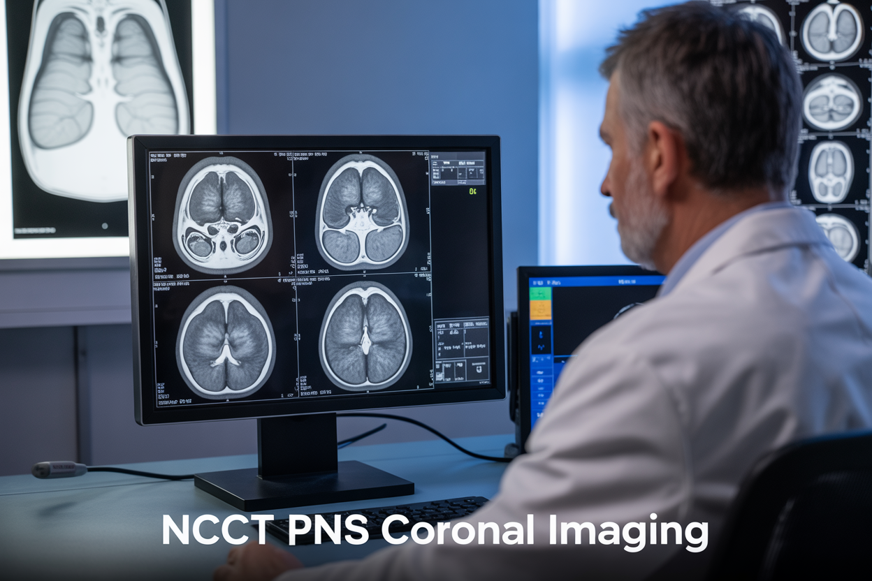
NCCT PNS Coronal: Your Guide to Paranasal Sinus CT Imaging
NCCT PNS coronal scans are the go-to imaging method for diagnosing sinus problems, giving doctors a clear view of your nasal passages and surrounding structures. This comprehensive guide is designed for radiology residents, medical students, and healthcare professionals who want to master non-contrast CT paranasal sinuses interpretation and improve their diagnostic skills.
You’ll learn the essential fundamentals of coronal CT sinus imaging, including proper positioning and scan protocols that produce high-quality results every time. We’ll walk you through PNS CT scan interpretation techniques, showing you how to spot normal anatomical variants and distinguish them from actual disease. Finally, you’ll discover how to recognize common sinus pathology patterns, from simple infections to complex inflammatory conditions that require immediate attention.
Whether you’re just starting out or looking to sharpen your skills, this guide covers everything you need to confidently read CT paranasal sinus studies and provide accurate diagnoses for your patients.
Understanding NCCT PNS Coronal Imaging Fundamentals
Key anatomical structures visualized in coronal plane
The coronal plane offers exceptional visualization of paranasal sinus anatomy, cutting through structures from front to back like slicing bread. This perspective reveals the complete vertical relationship between sinuses and surrounding structures that axial views simply can’t match.
NCCT PNS coronal imaging displays the maxillary sinuses prominently, showing their pyramid-shaped configuration with the apex pointing toward the zygomatic process. The ethmoid air cells appear as intricate honeycomb structures between the orbits, with the middle meatus clearly visible as the critical drainage pathway. The frontal sinuses sit above the orbits, though their size varies dramatically between individuals.
The ostiomeatal complex becomes beautifully apparent in coronal views, revealing the uncinate process, middle turbinate, and infundibulum. This region serves as the primary drainage route for the frontal, maxillary, and anterior ethmoid sinuses, making its assessment crucial for CT sinus pathology recognition.
Bony landmarks stand out clearly, including the nasal septum’s deviation patterns, the cribriform plate’s delicate structure, and the orbital floor’s integrity. The sphenoid sinus appears posteriorly, often extending into the sphenoid body with variable pneumatization patterns.
Paranasal sinus anatomy CT reveals soft tissue structures equally well, showing the nasal turbinates’ normal configuration, mucosal thickening patterns, and any polyps or masses. The relationship between sinuses and critical structures like the optic nerve, carotid arteries, and brain becomes immediately apparent, essential for surgical planning.
Optimal scan parameters for diagnostic quality
Achieving diagnostic excellence in non-contrast CT paranasal sinuses requires precise technical parameters tailored to sinus imaging demands. The coronal plane acquisition typically uses thin-section technique with slice thickness between 0.625-1.25mm, providing excellent spatial resolution for detecting subtle mucosal changes and small anatomical variants.
Coronal CT sinus imaging benefits from specific positioning protocols. Patients lie supine with the head positioned to achieve true coronal sections perpendicular to the hard palate. Direct coronal acquisition remains superior to reformatted images, though modern multidetector CT scanners produce acceptable reformats when direct positioning proves difficult.
Radiation dose optimization balances image quality with patient safety. Tube voltage settings typically range from 100-120 kVp, while tube current modulation adjusts automatically based on patient size. Modern PNS imaging protocols incorporate iterative reconstruction algorithms, allowing dose reduction of 30-50% while maintaining diagnostic quality.
Window settings play a crucial role in CT paranasal sinus diagnosis. Bone algorithm reconstruction with wide window settings (bone window: 4000 HU width, 700 HU level) optimizes visualization of bony structures and subtle fractures. Soft tissue windows (350 HU width, 50 HU level) enhance mucosal detail and fluid levels within sinuses.
Field of view should encompass the entire sinus region from frontal sinuses to sphenoid sinus floor, typically requiring 18-22cm coverage. Matrix size of 512×512 ensures adequate spatial resolution for detecting small pathological changes while keeping scan times reasonable for patient comfort.
Clinical Applications and Diagnostic Benefits
Sinusitis Detection and Severity Assessment
NCCT PNS coronal imaging serves as the gold standard for diagnosing and evaluating sinusitis across all paranasal sinus compartments. The coronal plane provides excellent visualization of the ostiomeatal complex, which plays a crucial role in sinus drainage and ventilation. Radiologists can easily identify mucosal thickening, fluid levels, and complete opacification patterns that characterize different stages of inflammatory disease.
Acute sinusitis typically presents with mucosal thickening exceeding 4mm, often accompanied by air-fluid levels within affected sinuses. The coronal CT sinus imaging allows precise measurement of mucosal changes and helps differentiate between viral and bacterial infections based on severity patterns. Chronic sinusitis demonstrates more extensive changes, including polypoid mucosal thickening, sclerotic bony changes, and potential osteoneogenesis around sinus walls.
The coronal plane excels at evaluating maxillary, ethmoid, and frontal sinus involvement simultaneously. Ethmoid air cells appear particularly well-defined in coronal sections, making it easier to assess the extent of inflammatory changes and their impact on neighboring structures. This comprehensive view helps clinicians determine appropriate treatment strategies, from conservative medical management to surgical intervention.
CT paranasal sinus diagnosis becomes more accurate when radiologists can correlate findings with clinical symptoms. The ability to assess both soft tissue and bony changes in a single examination makes NCCT PNS coronal the preferred imaging modality for sinusitis evaluation.
Nasal Polyp Identification and Characterization
Coronal CT sinus imaging provides exceptional detail for detecting and characterizing nasal polyps throughout the sinonasal cavity. These benign inflammatory masses appear as soft tissue densities that conform to the shape of surrounding anatomical structures, creating characteristic “cerebriform” or convoluted patterns easily recognizable on coronal sections.
The coronal plane allows optimal visualization of polyp origins, most commonly arising from the middle meatus and ethmoid region. Small polyps may appear as focal mucosal thickening, while larger lesions can completely fill sinus cavities and extend into the nasal cavity. The imaging helps distinguish between inflammatory polyps and other masses such as inverted papillomas or malignant tumors.
PNS CT scan interpretation for polyp assessment includes evaluating associated findings like bone erosion, which may suggest more aggressive pathology. Inflammatory polyps typically cause smooth bone remodeling or expansion without destructive changes. The presence of cerebriform pattern, bilateral involvement, and association with chronic rhinosinusitis supports the diagnosis of benign inflammatory polyps.
Non-contrast CT paranasal sinuses effectively demonstrates the relationship between polyps and critical anatomical landmarks, including the lamina papyracea, skull base, and sphenoid sinus. This information proves essential for surgical planning, helping surgeons anticipate potential complications and determine the extent of endoscopic resection required for optimal patient outcomes.
Image Interpretation Techniques and Normal Variants
Systematic Approach to Coronal PNS Analysis
Reading NCCT PNS coronal images requires a methodical approach that ensures no critical findings are missed. Start by examining the bony architecture of the skull base and orbital walls, looking for fractures or erosions that might indicate aggressive pathology. The ethmoid air cells deserve particular attention since their complex anatomy can harbor subtle inflammatory changes that appear as soft tissue opacification or air-fluid levels.
Move systematically through each sinus cavity, beginning with the maxillary sinuses and their dependent drainage patterns. Check the ostiomeatal complex carefully, as this narrow drainage pathway frequently becomes obstructed in sinusitis. The frontal sinuses should be evaluated for symmetry and drainage through their respective frontonasal ducts.
Pay close attention to soft tissue windows when reviewing coronal CT sinus imaging. Mucosal thickening greater than 3-4mm typically indicates inflammatory changes, while complete opacification suggests more advanced disease. The nasal septum position and any associated deviation should be noted, as this can impact drainage patterns and contribute to recurrent infections.
Bone window settings reveal subtle osteitic changes along sinus walls that might be missed on soft tissue windows. These reactive bone changes often accompany chronic inflammatory conditions and can help differentiate acute from chronic sinusitis patterns.
Recognizing Normal Anatomical Variations
CT paranasal sinus anatomy displays remarkable variation between individuals, making familiarity with common variants essential for accurate interpretation. Concha bullosa, where the middle turbinate contains an air cell, occurs in approximately 35% of the population and can contribute to ostiomeatal complex narrowing when significantly enlarged.
Asymmetric frontal sinus development is extremely common, with complete aplasia occurring in about 5% of individuals. Don’t mistake this normal variant for pathology, especially when bilateral. The frontal sinuses may also demonstrate intersinus septations that create complex drainage patterns.
Ethmoid air cell variants include agger nasi cells, which pneumatize anteriorly to the middle meatus attachment, and Haller cells that extend into the maxillary sinus floor. These variants can predispose to drainage obstruction and should be specifically mentioned when present, as they may influence surgical planning.
Sphenoid sinus pneumatization varies dramatically, ranging from minimal aeration to extensive pneumatization that extends into the pterygoid processes or anterior clinoid processes. Sinus CT normal variants like these require recognition to avoid misinterpretation as pathological findings.
Dehiscence of the lamina papyracea occurs in up to 15% of individuals and represents a normal developmental variant rather than traumatic injury when seen in isolation.
Common Pathological Findings and Recognition Patterns
Acute versus chronic sinusitis appearances
Distinguishing between acute and chronic sinusitis on NCCT PNS coronal imaging requires careful attention to specific imaging characteristics and patterns. Acute sinusitis typically presents with complete opacification of affected sinuses, creating homogeneous soft tissue density that fills the entire sinus cavity. The mucosal surfaces appear smooth and uniform, often showing air-fluid levels when the patient is scanned in an upright position. These fluid levels create characteristic horizontal interfaces between dependent fluid and overlying air, appearing as straight lines across the sinus floor.
Chronic sinusitis displays markedly different features on coronal CT sinus imaging. The hallmark finding is mucosal thickening that appears as irregular, nodular soft tissue lining the sinus walls. This thickened mucosa often creates a “cobblestone” or “cerebriform” pattern, particularly evident in the ethmoid air cells. Sclerosis of the surrounding bone becomes apparent as increased density and thickening of the sinus walls, especially noticeable in the maxillary and frontal sinuses.
PNS CT scan interpretation reveals additional chronic changes including osteomeatal complex obstruction, polypoid tissue formation, and retention cysts. The maxillary sinuses often show dependent layering of inspissated secretions, appearing as areas of varying density within the same sinus. Chronic changes may also include calcifications within retained secretions, appearing as small hyperattenuating foci scattered throughout the affected sinuses.
Fungal sinusitis characteristic features
Fungal sinusitis presents unique imaging patterns on non-contrast CT paranasal sinuses that help differentiate it from bacterial infections. Allergic fungal sinusitis (AFS) creates a distinctive “cerebriform” or convoluted pattern within the affected sinuses, resembling the folds of brain tissue. This appearance results from alternating areas of fungal debris and inspissated mucin, creating a characteristic heterogeneous density pattern.
The presence of hyperattenuating areas within the sinus contents serves as a key diagnostic clue for fungal involvement. These areas of increased density represent fungal concretions, mycetomas, or calcified debris that appear brighter than typical mucosal thickening or fluid. The maxillary and sphenoid sinuses are commonly affected, with the hyperattenuating material often settling in dependent portions.
CT sinus pathology recognition becomes critical when evaluating for invasive fungal sinusitis, which shows more aggressive features. Look for bone erosion, particularly of the lamina papyracea, cribriform plate, or skull base. The affected sinuses may show complete opacification with areas of fat stranding extending into adjacent soft tissues. Unlike AFS, invasive fungal disease often demonstrates irregular bone destruction rather than the smooth remodeling seen in chronic bacterial sinusitis.
Fungal balls or mycetomas appear as discrete, rounded masses of mixed density within otherwise clear sinuses, most commonly in the maxillary or sphenoid sinuses. These lesions often show internal calcifications and may be surrounded by a rim of mucosal thickening, creating a characteristic “halo” sign on coronal images.
Technical Optimization Strategies for Enhanced Results
Window and Level Adjustments for Optimal Visualization
Getting the most out of NCCT PNS coronal imaging starts with mastering window and level settings. The bone window settings (typically 2000-4000 HU window width with a center around 400-600 HU) provide the best visualization of sinus anatomy, septal deviations, and bony structures like the ethmoid air cells and sphenoid sinus walls. These settings make subtle bone erosions and osteomeatal complex abnormalities pop off the screen.
Soft tissue windows (300-400 HU window width with center around 40-60 HU) become essential when evaluating fluid levels, mucosal thickening, and distinguishing between different tissue densities within the paranasal sinus anatomy CT. Many radiologists switch between these windows during interpretation to catch pathology that might be missed with a single setting.
The lung window settings can be surprisingly useful for coronal CT sinus imaging, especially when looking for air-fluid levels in the maxillary sinuses or evaluating pneumatization patterns. A window width of 1500-2000 HU with a center around -500 HU helps identify even minimal fluid collections that standard soft tissue windows might miss.
Brain window settings (80-100 HU window width, center around 35-45 HU) prove valuable when the scan includes intracranial structures, allowing assessment of potential complications like intracranial extension of sinus disease or orbital involvement.
Slice Thickness Selection for Detailed Assessment
Choosing the right slice thickness dramatically impacts diagnostic confidence in PNS CT scan interpretation. Ultra-thin sections of 0.5-0.75mm provide exceptional detail for CT sinus pathology recognition, especially when evaluating the osteomeatal complex, ethmoid air cells, and subtle septal abnormalities. These thin slices excel at detecting small polyps, minimal mucosal thickening, and early erosive changes.
Standard 1-2mm slice thickness strikes an excellent balance for routine coronal sinus CT technique, offering good spatial resolution while maintaining reasonable file sizes and faster reconstruction times. This thickness works well for most diagnostic scenarios and provides adequate detail for surgical planning.
Thicker sections of 3-5mm might seem outdated, but they still have value when screening for gross pathology or when radiation dose reduction is paramount. However, these thicker slices can miss subtle findings and should be reserved for specific clinical scenarios.
The key lies in matching slice thickness to clinical indication. Presurgical planning demands the thinnest possible sections, while follow-up studies might tolerate slightly thicker cuts. Modern PNS imaging protocols increasingly favor thin-section acquisition with multiplanar reformatting capabilities, giving clinicians the flexibility to review cases in multiple planes while maintaining optimal diagnostic quality.

NCCT PNS coronal imaging stands out as a powerful diagnostic tool that gives doctors clear, detailed views of the paranasal sinuses without using contrast. From understanding the basic imaging setup to recognizing complex pathological patterns, this technique offers reliable insights for diagnosing everything from simple sinusitis to more serious conditions. The ability to spot normal variants and distinguish them from actual disease makes this imaging method particularly valuable in everyday clinical practice.
Getting the most out of NCCT PNS coronal scans comes down to proper technique and knowing what to look for. When radiologists and technicians work together to optimize image quality and apply solid interpretation skills, patients get faster, more accurate diagnoses. If you’re working with sinus imaging, take time to master these fundamentals – your patients will benefit from the clearer answers and more targeted treatments that come from truly understanding what these scans reveal.