

Wrist and hand pain can be frustrating when you can’t pinpoint the cause. MRI wrist & hand imaging helps doctors see exactly what’s happening inside your joints, tendons, and bones when X-rays don’t tell the whole story.
This guide is for patients with persistent wrist or hand pain, athletes with sports injuries, and anyone whose doctor has recommended an MRI. You’ll learn what to expect during the process and how these detailed images help your healthcare team create the right treatment plan.
We’ll walk you through the most common wrist injuries that show up on MRI scans, from ligament tears to stress fractures that might be invisible on regular X-rays. You’ll also discover what hand injuries look like on MRI images and when your doctor might suggest this type of scan over other imaging options.
By the end, you’ll understand why MRI technology is so valuable for diagnosing tricky wrist and hand problems, plus get practical tips for preparing for your appointment.
Understanding MRI Technology for Wrist and Hand Imaging

How MRI Works to Visualize Soft Tissues and Bones
MRI technology uses powerful magnetic fields and radio waves to create detailed images of your wrist and hand structures. Unlike other imaging methods that rely on radiation, MRI uses a strong magnetic field to align hydrogen atoms in your body’s tissues. When radio frequency pulses are applied, these atoms temporarily move out of alignment, then snap back into place, releasing energy signals that the machine captures and converts into detailed cross-sectional images.
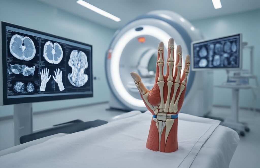
The wrist and hand contain intricate networks of bones, ligaments, tendons, cartilage, and muscles packed into a relatively small space. MRI excels at distinguishing between these different tissue types because each responds differently to the magnetic field. Soft tissues such as ligaments and cartilage appear clearly defined on MRI scans, allowing detection of tears, inflammation, or other damage that might be invisible on different imaging modalities.
The scanning process typically takes 30-60 minutes for wrist and hand imaging, during which multiple sequences are performed to capture different views and highlight specific structures. These sequences can be adjusted to emphasize particular tissues – some sequences highlight fluid and inflammation, while others focus on bones or blood vessels.
Superior Imaging Capabilities Compared to X-rays and CT Scans
While X-rays remain the first-line imaging for suspected fractures, they have significant limitations when evaluating wrist and hand injuries. X-rays clearly show bones but miss soft-tissue problems entirely. Many common wrist conditions, such as ligament tears or cartilage damage, simply don’t appear on X-ray films.
CT scans provide better bone detail than X-rays and can create cross-sectional images, but they still struggle with soft tissue visualization. CT scans are excellent for complex fractures but fall short when doctors need to evaluate the intricate soft tissue structures that make your wrist and hand function properly.
| Imaging Method | Bone Visualization | Soft Tissue Detail | Radiation Exposure | Scan Time |
| X-ray | Good for fractures | Poor | Low dose | Seconds |
| CT Scan | Excellent | Limited | Moderate dose | 5-10 minutes |
| MRI | Good | Excellent | None | 30-60 minutes |
MRI provides unmatched soft tissue contrast, making it the gold standard for diagnosing conditions like:
- Triangular fibrocartilage complex (TFCC) tears
- Scaphoid bone blood supply problems
- Tendon and ligament injuries
- Cartilage damage
- Nerve compression syndromes
The ability to see these structures in multiple planes (axial, sagittal, and coronal) gives doctors a complete three-dimensional understanding of your injury.
Safety Benefits of Radiation-Free Diagnostic Method
One of MRI’s most significant advantages is its complete lack of ionizing radiation. X-rays and CT scans expose patients to radiation, which carries a small but cumulative cancer risk over time. This radiation exposure becomes a particular concern for younger patients or those requiring multiple imaging studies.
MRI uses only magnetic fields and radio waves, both of which are considered safe for human exposure at the levels used in medical imaging. This safety profile makes MRI the preferred choice when detailed imaging is needed, especially for:
- Pediatric patients whose developing tissues are more sensitive to radiation
- Pregnant women requiring wrist or hand imaging
- Patients who need repeated scans to monitor healing
- Athletes undergoing preventive screening
The magnetic field strength in most clinical MRI machines ranges from 1.5 to 3 Tesla, which is strong enough to attract ferromagnetic objects but doesn’t cause biological harm to human tissues. The radio frequency energy used is similar to FM radio waves, but at different frequencies.
Safety considerations do exist – patients with certain metal implants, pacemakers, or claustrophobia may not be suitable candidates for MRI. However, newer MRI-safe implants and open MRI designs have expanded access to this valuable diagnostic tool for patients who previously couldn’t undergo scanning.
Most Common Wrist Injuries Detected by MRI

TFCC Tears and Triangular Fibrocartilage Complex Damage
The triangular fibrocartilage complex sits on the pinky side of your wrist, acting like a shock absorber between your forearm bones and the small wrist bones. When this structure is damaged, it causes a nagging pain that often worsens when you rotate your wrist or grip something tightly.
MRI excels at detecting TFCC tears because it can visualize soft-tissue details that X-rays miss. The scan reveals different types of tears – some happen right in the center of the cartilage disc, while others occur where the TFCC attaches to the bones. Athletes who play tennis, gymnastics, or baseball frequently develop these injuries from repetitive twisting motions.
What makes TFCC injuries tricky is that they often feel similar to other wrist problems. You might experience a clicking sensation, weakness when gripping, or pain that shoots up your forearm. MRI images help doctors precisely locate the tear and assess its severity, which directly affects treatment decisions.
Scaphoid Fractures and Hidden Bone Injuries
The scaphoid bone sits right below your thumb and plays a crucial role in wrist movement. This boat-shaped bone has a reputation for being problematic due to its poor blood supply, especially in its middle section. When you fall and catch yourself with your hand, the scaphoid often takes the brunt of the impact.
Regular X-rays miss scaphoid fractures about 15% of the time, especially hairline cracks that are just beginning to develop. MRI detects these hidden fractures by showing bone swelling and subtle stress changes that occur even before a complete break. Early detection can prevent a small crack from becoming a complete fracture that requires surgery.
The location of a scaphoid fracture matters enormously for healing. Breaks closer to your thumb typically heal well, while fractures in the middle or upper portions often struggle to mend properly due to limited blood flow. MRI shows doctors exactly where the fracture sits and whether the bone is getting adequate blood supply for natural healing.
Carpal Tunnel Syndrome and Nerve Compression
Carpal tunnel syndrome happens when the median nerve gets squeezed as it travels through the narrow carpal tunnel in your wrist. While doctors can often diagnose this condition through physical examination and nerve conduction studies, MRI provides valuable visual confirmation and helps rule out other causes of similar symptoms.
MRI images clearly show the median nerve, revealing when it’s swollen, flattened, or compressed by surrounding tissues. The scan also displays the carpal tunnel itself – the narrow passageway formed by wrist bones and a tough ligament. Sometimes the tunnel appears smaller than usual, or the ligament seems thicker and more prominent than it should.
Beyond just confirming carpal tunnel syndrome, MRI helps identify other conditions that mimic its symptoms. These include tendon inflammation, cysts pressing on the nerve, or unusual muscle formations that crowd the tunnel space. The detailed images also show whether conservative treatments like splinting and injections might work, or if surgical release of the carpal tunnel becomes necessary.
Ligament Tears and Joint Instability
Your wrist contains multiple small ligaments that work together to keep the joint stable during movement. These tough fibrous bands connect the various wrist bones and prevent them from shifting out of position when you lift, twist, or apply pressure through your hand.
MRI reveals ligament injuries with remarkable clarity, showing partial tears, complete ruptures, and areas where ligaments have pulled away from their bone attachments. The scapholunate ligament, which connects two essential wrist bones, tears frequently in sports injuries and falls. When this ligament fails, the wrist bones can shift abnormally, creating long-term arthritis if left untreated.
Another commonly injured structure is the lunotriquetral ligament, which connects bones on the pinky side of the wrist. Damage here often causes pain and clicking with wrist rotation. MRI images help surgeons determine whether these ligaments can heal naturally or need surgical repair to restore proper wrist function and prevent future joint problems.
Frequent Hand Injuries Revealed Through MRI Scanning
Tendon Ruptures and Inflammatory Conditions
Hand tendons work incredibly hard every day, making them vulnerable to both acute injuries and chronic inflammation. MRI scans excel at detecting tendon ruptures that might not be evident on X-rays or physical examination alone. Complete ruptures appear as gaps in the tendon structure, while partial tears show up as areas of increased signal intensity with thinning or irregularity.
Flexor tendon injuries are common in the fingers, often resulting from deep cuts or forceful gripping. The MRI can differentiate between injury zones, helping surgeons plan the most effective repair approach. Extensor tendon ruptures, though less frequent, can occur from seemingly minor trauma and may go undiagnosed without proper imaging.
Tenosynovitis, inflammation of the tendon sheath, creates a distinctive MRI appearance with fluid accumulation around the tendon. This condition frequently affects people who perform repetitive hand movements, from assembly-line workers to musicians. De Quervain’s tenosynovitis, affecting the thumb-side tendons, shows characteristic thickening and inflammation that MRI captures beautifully.
Trigger finger involves inflammation and thickening of the tendon sheath, creating a nodule that catches as the finger moves. MRI can reveal this thickening and help determine whether conservative treatment or surgical release would be most beneficial.
Ganglion Cysts and Soft Tissue Masses
Ganglion cysts rank among the most common hand masses, appearing as fluid-filled sacs that typically arise from joints or tendon sheaths. These benign growths appear brightly on MRI as well-defined, fluid-signal masses that are usually attached to nearby joint capsules or tendons via a small stalk.
The dorsal wrist is the most common site for ganglion cysts, which often arise from the scapholunate joint. Volar wrist ganglions typically occur from the radiocarpal or midcarpal joints and can sometimes compress the nearby radial artery. MRI helps distinguish these cysts from other masses and can identify their exact origin, which is valuable for surgical planning.
Occult ganglion cysts present a diagnostic challenge because they’re too small to feel but large enough to cause pain. MRI frequently uncovers these hidden culprits in patients with unexplained wrist discomfort.
Other soft tissue masses that MRI can identify include lipomas, which appear as fatty-signal masses, and giant cell tumors of the tendon sheath, which show characteristic low signal intensity. Nerve sheath tumors, though rare, display specific imaging features that help differentiate them from other masses.
Enchondromas, benign cartilage tumors in the hand bones, have distinctive appearances, with lobulated cartilage signal intensity and sometimes calcifications that help confirm the diagnosis.
Arthritis and Joint Degeneration
Hand arthritis takes many forms, and MRI provides detailed information about joint damage that X-rays might miss in early stages. Osteoarthritis commonly affects the thumb base (CMC joint), finger joints, and wrist, creating characteristic changes in cartilage, bone, and surrounding soft tissues.
Rheumatoid arthritis has a predilection for the wrist and finger joints, causing synovial inflammation that appears as thickened, enhancing tissue around joints. MRI can detect erosions and bone edema months before they become visible on conventional radiographs. This early detection capability enables more timely treatment interventions.
Psoriatic arthritis often affects the finger joints in a distinctive pattern, and MRI can show the characteristic sausage-like swelling of entire fingers along with joint inflammation. The imaging also reveals enthesitis, inflammation at the sites where tendons and ligaments attach to bone.
Post-traumatic arthritis develops after fractures or ligament injuries, and an MRI shows both the cartilage damage and any underlying bone changes. The scans can also identify loose bodies within joints that may require removal.
Crystalline arthropathies, such as gout, can affect hand joints, and MRI may show characteristic tophi (crystal deposits) as masses with specific signal characteristics.
Sports-Related Finger and Thumb Injuries
Athletes put tremendous stress on their hands, leading to specific injury patterns that MRI can accurately diagnose. Gamekeeper’s thumb, an injury to the ulnar collateral ligament of the thumb, is standard in skiing and other ball sports. MRI shows the extent of ligament damage and whether the torn ligament end has flipped over (Stener lesion), which requires surgical repair.
Mallet finger injuries involve rupture of the extensor tendon at the fingertip, often from ball sports. While the clinical diagnosis is usually straightforward, MRI can show associated fractures and help determine the best treatment approach.
Climber’s finger, involving pulley ruptures in the flexor tendon system, has become increasingly familiar with the popularity of rock climbing and bouldering. MRI clearly shows which pulleys are damaged and the extent of tendon bowstringing, information that guides both treatment and return-to-sport decisions.
Jersey finger occurs when the flexor digitorum profundus tendon avulses from its insertion, typically during football when a player grabs another player’s jersey. MRI locates the retracted tendon end and identifies any associated bone fragments.
Boxer’s fractures affecting the fifth metacarpal neck are common. Still, MRI can also reveal associated soft-tissue injuries, such as extensor tendon damage or joint capsule tears, which may influence treatment decisions.
What MRI Images Actually Show Healthcare Professionals

Detailed Soft Tissue Contrast and Abnormality Detection
MRI excels at revealing the intricate details of soft tissues that other imaging methods simply can’t match. When radiologists examine your wrist and hand MRI, they’re looking at incredibly detailed pictures that show the difference between tendons, ligaments, cartilage, muscles, and nerves with stunning clarity. This high-resolution view allows them to spot even tiny tears in ligaments, such as the scapholunate ligament or the triangular fibrocartilage complex (TFCC), that might be completely invisible on X-rays.
The magic happens because different tissues respond uniquely to the magnetic fields, creating distinct signals that translate into varying shades of gray on the images. Healthy cartilage appears smooth and uniform, while damaged areas show up as irregular bright spots or dark gaps. Inflamed tendons become swollen and exhibit characteristic signal changes that immediately catch a trained eye.
Healthcare professionals can also identify fluid collections, cysts, and masses with precision. Ganglion cysts, for example, appear as well-defined, bright areas filled with fluid, while more concerning masses exhibit characteristics that help distinguish between benign and potentially problematic growths. The ability to detect nerve compression, such as median nerve entrapment in carpal tunnel syndrome, provides doctors with crucial information about the severity and exact location of the problem.
Bone Marrow Changes and Stress Fractures
While bones might seem solid, MRI reveals the hidden world inside them that X-rays often miss. Bone marrow produces specific signals that change dramatically in response to injury or disease. Fresh stress fractures create a telltale pattern of bone marrow edema – essentially swelling inside the bone – that shows up as bright white areas on specific MRI sequences.
These stress fractures are prevalent in athletes and people who perform repetitive motions. The scaphoid bone, notorious for its poor blood supply and tendency to develop complications, frequently shows early changes on MRI long before they become visible on standard X-rays. This early detection can be the difference between successful conservative treatment and the need for surgery.
Bone marrow changes can also reveal other conditions, such as avascular necrosis, in which parts of bone tissue die due to poor blood supply. In the wrist, this commonly affects the lunate (Kienböck’s disease) or the scaphoid. MRI shows these changes as distinct patterns of signal alteration, helping doctors stage the disease and plan appropriate treatment strategies.
Blood Flow Patterns and Vascular Issues
MRI provides unique insights into blood flow and vascular health throughout the wrist and hand. Memorable sequences can track how blood moves through vessels, revealing blockages, abnormal connections, or areas with compromised circulation. This capability proves invaluable when assessing conditions like hypothenar hammer syndrome, where repeated trauma damages the ulnar artery.
Contrast-enhanced MRI takes vascular imaging even further by injecting a special dye that highlights blood vessels and areas with increased blood flow. This technique helps identify active inflammation, tumors, and infection by showing which areas are receiving more blood than usual. Infected tissues typically exhibit enhanced blood flow patterns that help doctors distinguish between different types of soft-tissue problems.
The vascular information also helps surgeons plan procedures by mapping out the blood supply to different structures. Before complex reconstructive surgeries, knowing exactly where major vessels run and how they branch can prevent complications and improve outcomes. This detailed vascular roadmap becomes especially important in trauma cases where normal anatomy might be disrupted.
When Your Doctor Will Recommend an MRI for Hand or Wrist Pain

Persistent Pain Despite Normal X-ray Results
X-rays show bones clearly but miss the soft tissues that often cause wrist and hand pain. When you’re dealing with ongoing discomfort that won’t go away, but your X-ray comes back normal, your doctor faces a puzzle that MRI can solve.
Many patients experience this frustrating scenario. You know something’s wrong—your wrist aches during daily activities or your hand feels weak when gripping objects—but traditional imaging doesn’t reveal the culprit. MRI becomes the detective tool that uncovers hidden injuries to ligaments, tendons, cartilage, and other soft tissues that X-rays simply can’t detect.
This situation commonly occurs with:
- Ligament tears that aren’t visible on standard films
- Early-stage arthritis affecting cartilage
- Tendon inflammation or partial tears
- Nerve compression syndromes
- Minor fractures in areas poorly visualized by X-rays
Your doctor will likely recommend an MRI when pain persists for several weeks despite conservative treatment, especially if the pain interferes with your daily activities or work performance.
Suspected Soft Tissue Injuries Requiring Detailed Imaging
Soft-tissue injuries account for the majority of hand and wrist problems, yet they remain invisible to conventional imaging methods. MRI excels at revealing these hidden injuries with remarkable detail.
Sports-related injuries often involve complex soft tissue damage. A fall on an outstretched hand might cause multiple ligament injuries simultaneously, creating instability that’s difficult to diagnose without detailed imaging. Rock climbers, gymnasts, and racquet-sport athletes frequently develop overuse injuries affecting tendons and ligaments, which require MRI evaluation.
Common soft tissue injuries that need MRI include:
- Triangular fibrocartilage complex (TFCC) tears
- Scapholunate ligament injuries
- Tendon ruptures or partial tears
- Joint capsule injuries
- Muscle strains affecting hand function
The ability to detect inflammation, swelling, and tissue changes makes MRI invaluable for diagnosing conditions such as tendinitis, bursitis, and early degenerative changes. Your doctor can also assess blood flow to tissues, helping identify areas with poor healing potential.
Pre-surgical Planning for Complex Cases
Surgeons rely heavily on MRI when planning intricate procedures involving the hand and wrist. These areas contain densely packed structures where millimeter precision matters, making precise preoperative planning essential for successful outcomes.
MRI provides surgeons with a detailed roadmap before entering the operating room. The technology reveals the exact location and extent of injuries, helping determine the best surgical approach and techniques needed. This planning reduces surgery time, minimizes tissue damage, and improves patient outcomes.
Complex cases requiring MRI-guided planning include:
- Multi-ligament reconstructions
- Tendon transfer procedures
- Carpal tunnel release with suspected complications
- Mass or tumor removal
- Fracture repairs involving joint surfaces
- Revision surgeries after previous procedures
Surgeons can also identify anatomical variations that might complicate surgery. Some people have unusual tendon or blood vessel arrangements that could affect surgical planning. MRI reveals these variations before the procedure begins, allowing surgeons to adapt their approach accordingly.
Monitoring Treatment Progress and Healing
MRI is an excellent tool for tracking how well treatments are working and whether tissues are healing correctly. This monitoring capability proves especially valuable for complex injuries or when healing seems slower than expected.
During the healing process, an MRI can show:
- Reduction in tissue inflammation and swelling
- New tissue formation and repair
- Blood flow improvements to injured areas
- Scar tissue development that might affect function
- Complications like infection or poor healing
Athletes returning to sports benefit significantly from MRI monitoring. The images help determine when tissues have healed sufficiently to handle increased stress and activity levels. This prevents re-injury and ensures safe return to competition.
Your doctor might order follow-up MRI scans if:
- Symptoms aren’t improving with treatment
- New symptoms develop during recovery.
- Physical therapy progress stalls unexpectedly
- You’re considering returning to high-demand activities.s
- Previous treatments haven’t worked as expected.
Detailed information from follow-up MRI scans helps doctors adjust treatment plans, modify therapy approaches, or consider alternative interventions when initial treatments aren’t producing the desired results.
Preparing for Your MRI Appointment and What to Expect
Pre-scan Instructions and Metal Object Removal
Before your MRI appointment, you’ll receive specific instructions. The most critical step involves removing all metal objects from your body and clothing. This includes jewelry, watches, hairpins, belts with metal buckles, underwire bras, and apparel with metallic threads or zippers. Even small items, like coin-sized metal buttons, can degrade image quality.
Your medical team will provide you with a comprehensive checklist days before your scan. Some facilities offer metal-free gowns or scrubs to ensure complete compliance. If you have any implanted medical devices, such as pacemakers, cochlear implants, or metal plates from previous surgeries, inform your technologist immediately. These require special consideration and may affect your ability to safely undergo the procedure.
Remove all makeup, nail polish, and hair products that might contain metallic particles. Credit cards, phones, and keys must stay in the changing area locker. The magnetic field is powerful enough to erase data from cards and potentially damage electronic devices.
Positioning Techniques for Optimal Image Quality
Proper positioning makes the difference between transparent, diagnostic images and scans that need repeating. For wrist and hand MRI, you’ll typically lie on your back with your arm extended above your head in what’s called the “Superman position.” Your hand and wrist will be placed in a specialized coil – a device that looks like a small cage or tube, explicitly designed for extremity imaging.
The technologist will use cushions and supports to keep your arm comfortable and perfectly still throughout the scan. Your fingers might be gently secured with soft straps or positioned in a specific way, depending on what structures your doctor wants to examine. Some facilities use dedicated extremity MRI units where you can sit upright with just your hand and wrist inside the machine while the rest of your body remains outside.
The positioning process takes several minutes as the technologist fine-tunes everything for optimal image quality. They’ll check that you’re comfortable because staying completely still is essential for clear pictures. Even small movements can blur the images and require additional scanning time.
Scan Duration and Comfort Measures During Procedure
A typical wrist or hand MRI takes 30 to 45 minutes to complete. The machine creates loud knocking, tapping, and buzzing sounds during different sequences. You’ll receive earplugs or noise-canceling headphones to reduce the noise level and make the experience more comfortable.
Most facilities offer music options through headphones to help you relax. The technologist maintains constant communication through an intercom system and can hear you if you need anything. They’ll explain each sequence before it starts and let you know how long each one will take.
You can expect multiple scanning sequences, each focusing on different tissue types or injury patterns. Some sequences last just two minutes, while others might run for eight to ten minutes. The technologist will give you breaks between more extended sequences if needed.
Temperature control keeps the scanning room comfortable, though some people feel cool lying still for extended periods. Blankets are available upon request. If you experience claustrophobia, discuss this with your doctor beforehand – they might prescribe mild sedation or recommend an open MRI unit if available in your area.

MRI scans give doctors an incredibly detailed look at what’s happening inside your wrist and hand when you’re dealing with pain or injury. From torn ligaments and fractured bones to damaged tendons and compressed nerves, these scans can spot problems that might be missed with regular X-rays. The technology provides clear images of soft tissues, bones, and joints, helping your healthcare team determine precisely what’s causing your discomfort and how best to treat it.
If you’re experiencing persistent wrist or hand pain that isn’t getting better with basic treatment, don’t hesitate to talk with your doctor about whether an MRI might be right for you. Getting an early diagnosis can make a huge difference in your recovery and help prevent long-term complications. Remember that the scan itself is painless and straightforward – the most challenging part is often just staying still for 30-45 minutes while the machine does its work.