
MRI Liver With Contrast: Complete Guide to Enhanced Diagnostic Imaging
Looking for an MRI scan near you to evaluate liver health? Contrast-enhanced liver MRI offers doctors the clearest view of liver tissue, blood vessels, and potential abnormalities that standard imaging might miss.
This comprehensive guide is written for patients scheduled for liver MRI scans, their families, and anyone seeking to understand this advanced diagnostic tool. Whether you’re researching local MRI imaging centers near me or preparing for an upcoming appointment, we’ll walk you through everything you need to know.
We’ll cover the essential medical conditions that require contrast-enhanced liver MRI, including tumor detection and monitoring of treatment progress. You’ll also learn about the complete procedure process, including what happens during your MRI test appointment and how the contrast agent works to highlight liver structures.
Finally, we’ll explore the clinical benefits that make this imaging technique so valuable for accurate diagnosis and treatment planning, helping you understand why your doctor may have recommended this specific type of scan at your local MRI centre.
Understanding MRI Liver with Contrast Technology
How contrast-enhanced MRI differs from standard liver imaging
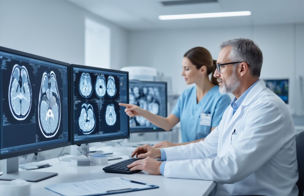
Contrast-enhanced liver MRI provides dramatically more precise visualization of liver structures compared to standard imaging techniques. The contrast agent highlights blood vessels, tissue boundaries, and abnormalities that might otherwise go undetected on routine scans. This enhanced clarity allows radiologists to detect smaller lesions and better characterize tissue properties, making it significantly more accurate than standard MRI or CT scans for liver evaluation.
Types of contrast agents used for liver evaluation
Gadolinium-based contrast agents are the primary choice for liver MRI, offering excellent safety profiles and superior image enhancement. These agents flow through blood vessels and accumulate in different tissues at varying rates, creating distinct contrast patterns. Hepatobiliary-specific agents like gadoxetic acid provide additional benefits by being taken up by healthy liver cells, allowing radiologists to distinguish between normal and diseased tissue more effectively.
Superior soft tissue visualization capabilities
MRI technology excels at distinguishing among various soft-tissue types within the liver, providing unmatched detail of organ structure and pathology. The magnetic resonance signals reveal subtle differences in tissue composition, water content, and blood flow patterns that other imaging methods cannot detect.
Non-invasive nature compared to traditional diagnostic methods
Unlike liver biopsy procedures that require tissue sampling, contrast-enhanced MRI offers comprehensive liver assessment without surgical intervention. Patients can receive detailed diagnostic information through this painless procedure, avoiding the risks and recovery time associated with invasive techniques. When scheduling your MRI scan appointment at an MRI centre near you, you can expect a comfortable, non-surgical diagnostic experience.
Medical Conditions Requiring Contrast-Enhanced Liver MRI
Detection and characterization of liver tumors
Contrast-enhanced MRI excels at spotting both primary liver cancers like hepatocellular carcinoma and metastatic tumors that spread from other organs. The contrast agent highlights blood flow patterns, helping doctors distinguish between benign lesions like hemangiomas and malignant growths. This detailed imaging allows for precise tumor staging and surgical planning.
When searching for an ‘MRI scan near me’ or ‘MRI centre near me’, patients with suspected liver masses benefit from facilities equipped with advanced contrast protocols. The enhanced visualization helps determine tumor size, location, and relationship to blood vessels, making it easier to plan treatments like surgery, ablation, or chemotherapy.
Evaluation of chronic liver diseases and cirrhosis
Chronic conditions like hepatitis, fatty liver disease, and cirrhosis cause progressive liver scarring that shows up clearly on contrast MRI. The imaging reveals fibrosis patterns, portal hypertension signs, and early cirrhotic changes that might not appear on standard scans. This helps doctors track disease progression and adjust treatment plans.
Patients monitoring chronic liver conditions should schedule regular MRI scan appointments at specialized MRI imaging centers to track changes over time. The contrast enhancement provides detailed views of liver architecture, blood flow abnormalities, and complications like varices or ascites formation.
Assessment of bile duct abnormalities and blockages
Contrast MRIcreates detailed maps of bile duct anatomy using specialized sequences that highlight fluid-filled structures. This technique, called MRCP, detects strictures, stones, tumors, or inflammatory changes affecting bile flow. The contrast helps differentiate between benign and malignant causes of bile duct obstruction.
Jaundiced patients or those with elevated liver enzymes often need this specialized imaging available at comprehensive MRI labs. The non-invasive nature makes it safer than invasive procedures like ERCP while providing similar diagnostic information about bile duct pathology and drainage patterns.
Monitoring treatment response in liver cancer patients
Cancer patients undergoing chemotherapy, targeted therapy, or immunotherapy need regular imaging to assess treatment effectiveness. Contrast MRI shows changes in tumor vascularity, size, and metabolic activity that indicate whether treatments are working. This helps oncologists adjust treatment protocols or consider alternative approaches.
Regular follow-up scans at reliable MRI test centers locations allow for consistent monitoring using the same imaging protocols. The contrast enhancement reveals subtle changes in tumor characteristics that might indicate treatment response or disease progression, guiding clinical decision-making.
Investigation of unexplained abdominal pain or liver dysfunction
When patients present with unexplained right upper quadrant pain, abnormal liver function tests, or systemic symptoms, contrast MRIprovides comprehensive liver evaluation. The imaging can reveal inflammatory conditions, vascular abnormalities, or subtle lesions that explain the clinical symptoms.
Emergency departments and gastroenterology practices often refer patients to nearby MRI imaging centers for urgent liver evaluation. The detailed contrast-enhanced images help exclude severe conditions like liver abscesses, vascular thrombosis, or occult malignancies that require immediate treatment intervention.
Pre-Procedure Preparation and Safety Considerations

Essential fasting requirements before the examination
Patients need to avoid food and beverages for 4-6 hours before their MRI scan to reduce the risk of nausea when contrast material is administered. Clear liquids may be permitted up to 2 hours prior, but check with your MRIcentre near me for specific guidelines. This fasting period helps reduce the risk of complications during the procedure.
Screening for kidney function and contrast allergies
Blood tests measuring creatinine levels are required within 30 days of the exam to ensure the kidneys can safely process the contrast agent. Patients with prior allergic reactions to MRI contrast must inform their MRI imaging centers at the time of scheduling. Those with severe kidney disease may need alternative imaging approaches or special preparation protocols.
Medication adjustments and medical history review
| Medication Type | Adjustment Required |
| Blood thinners | May need temporary discontinuation |
| Diabetes medications | Timing adjustments for fasting |
| Blood pressure medications | Usually continued as normal |
Bring a complete list of current medications and medical conditions when visiting your MRI lab. Certain medications, such as metformin, may require temporary discontinuation in patients with kidney concerns.
Patient positioning and comfort measures
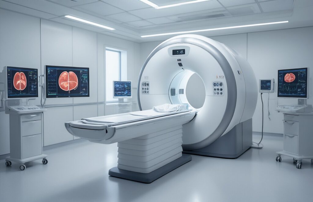
Comfortable clothing without metal components should be worn, though hospital gowns are typically provided. Patients lie on a padded table that slides into the MRI scanner, with positioning aids ensuring proper liver visualization. Communication systems allow constant contact with technologists during the 30-45 minute procedure at your chosen MRI test facility.
Step-by-Step MRILiver Procedure Process

Initial baseline imaging without contrast
Before any contrast agent enters your system, radiologists capture detailed baseline images of your liver using standard MRI sequences. These pre-contrast scans establish normal liver anatomy and identify abnormalities without contrast enhancement. The baseline imaging typically takes 10-15 minutes and serves as a crucial reference point for comparing subsequent contrast-enhanced phases.
Contrast injection timing and administration method
The contrast agent gets delivered through an IV line in your arm using an automated injector system that ensures precise timing and dosage. Most liver MRI protocols use gadolinium-based contrast agents administered at specific flow rates, typically 2-3 mL per second. The injection timing coordinates perfectly with imaging sequences to capture optimal enhancement patterns during different vascular phases.
Multiple imaging phases for optimal liver assessment
| Phase | Timing After Injection | Primary Purpose |
| Arterial Phase | 15-20 seconds | Detect hypervascular lesions |
| Portal Venous Phase | 60-70 seconds | Evaluate liver parenchyma |
| Delayed Phase | 3-5 minutes | Assess washout patterns |
| Hepatobiliary Phase | 10-20 minutes | Liver function evaluation |
Each phase captures different aspects of liver blood flow and cellular function, providing comprehensive diagnostic information that helps differentiate between various liver conditions.
Complete scan duration and patient monitoring
The entire MRI liver procedure typically spans 45-60 minutes from start to finish, including setup time and multiple imaging sequences. During the scan, technologists monitor your vital signs and comfort level through intercom communication. You’ll need to hold your breath for 15-20 seconds during certain sequences to minimize motion artifacts. When scheduling your MRI scan appointment at an MRI centre, expect this full duration for comprehensive liver imaging with contrast enhancement.
Clinical Benefits and Diagnostic Advantages
Enhanced Detection of Small Liver Lesions
Contrast-enhanced MRI liver scans excel at revealing tiny lesions that often remain hidden in standard imaging. The contrast agent brightens blood vessels and highlights abnormal tissue patterns, making even lesions smaller than 1 centimeter visible to radiologists.
Improved Differentiation Between Benign and Malignant Tumors
Different tumor types absorb and release contrast agents at distinct rates, creating unique enhancement patterns. This timing difference helps doctors distinguish between harmless cysts, benign adenomas, and dangerous cancers without requiring tissue samples.
Comprehensive Evaluation of Liver Blood Supply
The dual blood supply from hepatic arteries and portal veins becomes clearly mapped during contrast injection phases. This detailed vascular assessment is crucial for surgical planning and for identifying areas with compromised blood flow that may affect treatment decisions.
Reduced Need for Invasive Diagnostic Procedures
High-resolution contrast imaging often eliminates the need for liver biopsies or exploratory surgeries. Patients seeking an MRI scan appointment at their local MRI centre can receive definitive diagnoses through this non-invasive approach, reducing complications and recovery time.
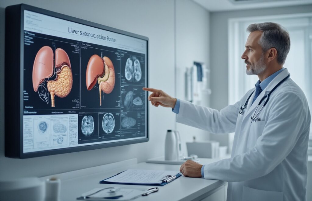
Interpreting Results and Treatment Planning
Key imaging findings that indicate liver pathology
Contrast-enhanced MRI reveals critical liver abnormalities through distinct imaging patterns. Hepatocellular carcinoma appears as hyperintense lesions in arterial phase with washout during delayed phases. Metastases typically show rim enhancement or heterogeneous patterns. Cirrhosis displays surface nodularity, altered liver contour, and regenerative nodules. Hemangiomas demonstrate characteristic peripheral nodular enhancement with centripetal fill-in.
How results influence surgical and medical decisions
MRI findings directly guide treatment strategies and surgical planning. Lesion size, number, and vascular involvement determine surgical candidacy for resection or transplantation. Tumor staging influences chemotherapy protocols and radiation therapy targeting. Portal vein invasion or extrahepatic spread alters surgical approach from curative to palliative care. Liver function assessment through contrast dynamics helps surgeons evaluate remaining healthy parenchyma before major hepatectomy procedures.
Follow-up imaging recommendations based on findings
| Finding | Initial Follow-up | Long-term Monitoring |
| Suspicious lesions | 3-6 months | Every 6 months |
| Post-surgical changes | 1-3 months | Annually |
| Treated tumors | 3 months | Every 6 months |
| Incidental findings | 6-12 months | Case-dependent |
High-risk patients require frequent surveillance at specialized MRI imaging centers to track disease progression. Scheduling your MRI scan appointment promptly after abnormal findings ensures timely intervention and optimal outcomes.

Getting an MRI liver scan with contrast gives doctors a powerful tool to spot problems that might otherwise go unnoticed. From detecting tumors and monitoring liver disease to evaluating blood flow issues, this imaging technique helps medical teams make accurate diagnoses and create effective treatment plans. The contrast agent acts like a spotlight, highlighting areas that need closer attention and providing the detailed information doctors need to help their patients.
If your doctor recommends this procedure, remember that proper preparation makes all the difference in getting precise, valuable results. Follow the pre-scan instructions carefully, tell your medical team about any health conditions or medications, and don’t hesitate to ask questions about what to expect. The temporary inconvenience of the scan often yields valuable insights into your liver health, giving you and your physician the information needed to move forward with confidence in your care plan.