

Getting an MRI of your knee can feel overwhelming, especially when you’re handed a report full of medical terms you’ve never heard before. This guide is for patients who want to understand what their MRI results for the knee joint actually mean and what those findings could mean for their health.
An MRI scan gives doctors a detailed look inside your knee without surgery, showing everything from cartilage and ligaments to bone and soft tissue. While the technology behind these scans is impressive, the real value comes from knowing how to read what the images reveal.
We’ll walk you through the most common problems that show up on knee MRI scans, like torn meniscus, ligament injuries, and cartilage damage. You’ll also learn how to decode the medical language in your report, turning confusing terms into plain English you can actually understand. Finally, we’ll cover what these findings typically mean for your treatment options and recovery.
Understanding MRI Technology for Knee Evaluation
How MRI Creates Detailed Images of Your Knee
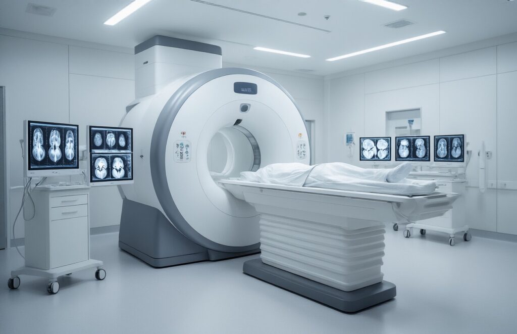
Magnetic Resonance Imaging works by using powerful magnets and radio waves to create detailed pictures of your knee’s internal structures. The MRI machine generates a strong magnetic field that aligns hydrogen atoms in your body, then sends radio frequency pulses that cause these atoms to emit signals. These signals are captured and processed by sophisticated computers to produce cross-sectional images showing bones, cartilage, ligaments, tendons, and fluid with remarkable clarity.
Different tissue types appear as varying shades of gray, white, or black on the images, allowing radiologists to distinguish between healthy and damaged structures. The scan captures multiple thin slices of your knee from different angles, creating a comprehensive three-dimensional view that reveals even subtle abnormalities that might be missed with other imaging methods.
Why Doctors Choose MRI Over Other Imaging Methods
MRI offers several advantages over X-rays and CT scans when evaluating knee problems. Unlike X-rays that only show bones clearly, MRI excels at visualizing soft tissues like cartilage, menisci, ligaments, and muscles. This makes it the gold standard for diagnosing torn ACLs, meniscal tears, cartilage damage, and other common knee injuries that don’t involve fractures.
The technology doesn’t use radiation, making it safer for repeated imaging when needed. MRI also provides superior contrast between different tissue types and can detect early degenerative changes, inflammation, and fluid accumulation that other imaging methods might miss.
What to Expect During Your Knee MRI Scan
Your knee MRI typically takes 30-45 minutes and requires you to lie still on a table that slides into the MRI machine. You’ll position your knee in a specialized coil designed to capture the clearest images possible. The machine makes loud knocking and tapping sounds during the scan, so you’ll receive earplugs or headphones for comfort.
Most knee MRIs don’t require contrast injection, though sometimes a gadolinium-based contrast agent may be administered through an IV to highlight certain structures or detect inflammation. The technologist will communicate with you throughout the procedure and can stop the scan if you experience any discomfort or claustrophobia.
Essential Knee Anatomy Visible on MRI Scans
Bones and Joint Structures Your Doctor Examines
MRI scans provide detailed views of your knee’s bony architecture, including the femur (thighbone), tibia (shinbone), and patella (kneecap). These bones form the primary joint surfaces where movement occurs. Radiologists examine bone marrow for signs of bruising, fractures, or degenerative changes that might not show up on X-rays.
The joint spaces between bones reveal crucial information about overall knee health. Your doctor looks for proper alignment, bone spurs, and any abnormal bone formations that could indicate arthritis or other conditions affecting your mobility.
Cartilage Components That Show Wear and Damage
Articular cartilage covers the ends of bones where they meet in the joint, acting like a smooth cushion during movement. MRI excels at showing cartilage thickness, surface irregularities, and areas where this protective layer has worn away. Different MRI sequences can highlight even small cartilage defects that cause pain and stiffness.
Cartilage damage appears as bright spots, thinning, or complete loss on MRI images. Your radiologist grades cartilage wear from mild surface roughening to full-thickness defects where bone rubs directly against bone.
Ligaments and Their Role in Knee Stability
The anterior cruciate ligament (ACL) and posterior cruciate ligament (PCL) cross inside your knee joint, preventing excessive forward and backward movement. MRI clearly shows tears, partial injuries, or complete ruptures in these critical stabilizing structures. The medial and lateral collateral ligaments on the sides of your knee also appear distinctly on MRI scans.
Ligament injuries show up as bright signals on certain MRI sequences, indicating swelling or tearing. Your doctor can determine whether surgery is needed based on the extent and location of ligament damage visible on your scan.
Meniscus Function and Common Problem Areas
The medial and lateral menisci are C-shaped cartilage pieces that act as shock absorbers between your thighbone and shinbone. These structures distribute weight evenly across the joint and provide stability during twisting movements. MRI shows meniscal tears as bright lines or irregular shapes within the normally dark meniscus tissue.
Common tear patterns include horizontal splits, vertical tears, and complex degenerative changes. The location and type of meniscal tear helps your doctor decide between conservative treatment and arthroscopic surgery to repair or remove damaged tissue.
Most Frequent Abnormal Findings in Knee MRI Reports
Meniscal Tears and Their Different Types
The meniscus acts like a shock absorber in your knee, and tears are among the most common findings on MRI scans. Radiologists classify these tears based on their shape and location – horizontal, vertical, radial, or complex patterns. Degenerative tears typically show up as irregular, frayed edges on MRI images, while acute traumatic tears appear as clean, sharp lines cutting through the meniscal tissue.
ACL and PCL Injuries That Affect Movement
Anterior cruciate ligament (ACL) tears create distinctive wavy or discontinuous signals on MRI scans, often accompanied by bone bruising patterns. Complete ACL ruptures show clear gaps in the ligament fibers, while partial tears display thickened, heterogeneous signals. Posterior cruciate ligament (PCL) injuries are less common but equally visible, appearing as increased signal intensity or complete discontinuity within the ligament structure.
Cartilage Degeneration and Arthritis Signs
Cartilage damage progresses through predictable stages that MRI can detect before symptoms become severe. Early degeneration shows as softening and surface irregularities, while advanced arthritis reveals full-thickness cartilage loss with exposed bone underneath. MRI sequences can differentiate between acute cartilage injuries and chronic wear patterns, helping doctors understand whether damage resulted from trauma or gradual deterioration over time.
Bone Bruises and Fractures Not Visible on X-rays
Bone bruises, technically called bone marrow edema, appear as bright white areas on certain MRI sequences where X-rays show nothing abnormal. These injuries represent microscopic fractures within the bone that haven’t created visible cracks yet. Stress fractures also show up as linear areas of increased signal intensity, often revealing overuse injuries in athletes weeks before they would appear on conventional imaging.
Fluid Accumulation and Swelling Patterns
Joint effusions and synovial thickening create characteristic patterns that help diagnose various knee conditions. Baker’s cysts form as fluid-filled sacs behind the knee, clearly visible on MRI as well-defined collections extending from the joint space. Inflammatory conditions like rheumatoid arthritis produce specific enhancement patterns when contrast is used, while infectious processes show different fluid characteristics and surrounding tissue changes that guide treatment decisions.
Decoding Medical Terminology in Your MRI Report

Understanding Signal Intensity Descriptions
MRI reports describe tissues using signal intensity terms that reflect how bright or dark structures appear on different scan sequences. “Hyperintense” means brighter than surrounding tissue, often indicating fluid or swelling. “Hypointense” appears darker and typically represents dense structures like bone or scar tissue. “Isointense” matches the brightness of nearby normal tissue. These descriptions help radiologists identify abnormal areas by comparing them to healthy tissue patterns.
Interpreting Tear Classifications and Grades
Tear grading follows a standardized system that describes the severity of structural damage. Grade 1 tears show minor signal changes without complete disruption, while Grade 2 tears involve partial thickness damage. Grade 3 represents complete tears with full-thickness disruption. Meniscal tears use specific classifications like horizontal, vertical, or complex patterns. Each grade helps your doctor understand the extent of injury and plan appropriate treatment strategies.
What Mild, Moderate, and Severe Really Mean
| Severity Level | Typical Characteristics | Treatment Implications |
| Mild | Minimal tissue changes, no functional limitation | Conservative management, physical therapy |
| Moderate | Noticeable damage with some symptoms | May require targeted treatment, monitoring |
| Severe | Significant structural damage, functional impairment | Often needs surgical intervention |
These severity descriptions directly correlate with your symptoms and activity limitations. Mild findings might explain occasional discomfort during sports, while severe findings usually match significant pain and mobility issues you’re experiencing daily.
Clinical Significance of Common MRI Findings

Which Abnormalities Require Immediate Treatment
Complete ACL ruptures, locked meniscus tears preventing knee extension, and fresh osteochondral fractures demand prompt surgical intervention. These injuries often cause significant pain, instability, and functional limitation that won’t improve with conservative care alone.
Age-Related Changes That May Not Need Surgery
Degenerative meniscal tears in patients over 40 frequently occur alongside normal wear and tear. Many respond well to physical therapy rather than arthroscopic surgery, especially when symptoms are mild and the tear pattern suggests age-related breakdown rather than acute injury.
How Findings Correlate With Your Symptoms
| MRI Finding | Typical Symptoms | Pain Location |
| Meniscal tear | Catching, locking | Joint line |
| ACL injury | Instability, giving way | Deep knee pain |
| Bone marrow edema | Aching, stiffness | Diffuse |
| Cartilage damage | Grinding, swelling | Activity-related |
Your pain pattern often matches specific anatomical damage. However, some patients with significant MRI abnormalities experience minimal symptoms, while others with minor findings report substantial discomfort.
When Conservative Treatment Is Most Effective
Non-surgical management works best for:
- Partial meniscus tears without mechanical symptoms
- Grade 1-2 ligament sprains
- Early degenerative changes
- Bone marrow edema from overuse
Physical therapy, activity modification, and anti-inflammatory treatments can resolve many knee problems within 6-12 weeks. Surgery becomes necessary when conservative approaches fail after adequate trial periods.
Next Steps After Receiving Your MRI Results

Questions to Ask Your Doctor About Your Report
Armed with your MRI results, prepare specific questions that will help you understand your diagnosis and treatment path. Ask about the severity of any findings, whether they explain your symptoms, and if conservative treatment might work before considering surgery. Request clarification on medical terms you don’t understand, and inquire about activity restrictions or modifications you should follow. Understanding your prognosis is crucial – ask whether your condition will worsen over time and what signs to watch for that might indicate progression.
Treatment Options Based on Specific Findings
| Finding | Conservative Options | Advanced Treatments |
| Meniscus Tears | Physical therapy, anti-inflammatories | Arthroscopic repair/removal |
| Ligament Injuries | Bracing, rehabilitation | Surgical reconstruction |
| Cartilage Damage | Injections, activity modification | Cartilage restoration procedures |
| Bone Marrow Edema | Rest, weight modification | Rarely requires surgery |
Your treatment plan depends heavily on your age, activity level, and specific MRI findings. Many knee problems respond well to non-surgical approaches including physical therapy, targeted exercises, and lifestyle changes.
Timeline Expectations for Recovery and Healing
Recovery timeframes vary dramatically based on your specific diagnosis and chosen treatment approach. Conservative treatments typically show improvement within 6-12 weeks, while surgical interventions may require 3-6 months for full recovery. Factors like your age, overall health, and commitment to rehabilitation significantly impact healing speed. Some conditions like bone marrow edema may resolve naturally over several months, while others like complete ligament tears often require surgical intervention for optimal outcomes.

Getting an MRI of your knee can feel overwhelming, especially when you’re handed a report filled with medical jargon. The good news is that most findings on knee MRIs are common and treatable. Whether your scan shows meniscal tears, ligament sprains, cartilage wear, or bone marrow changes, understanding what these terms mean puts you back in control of your health journey. Your radiologist and orthopedic doctor are looking for specific patterns that help explain your pain and guide your treatment plan.
Don’t let technical terms scare you – many knee MRI findings are part of normal aging or minor injuries that respond well to conservative treatment. The key is working with your healthcare team to connect your symptoms with what the scan reveals. Take your report to your doctor, ask questions about anything you don’t understand, and discuss realistic treatment options. Remember, an MRI is just one piece of the puzzle, and most knee problems can be managed effectively with the right approach and patience.