
“MRI Face With Neck With Contrast: How It Helps Diagnose Head and Neck Conditions Accurately”
MRI Face With Neck With Contrast: A Complete Guide for Patients
If your doctor has ordered an MRI face with neck with contrast, you probably have questions about what this scan involves and why you need it. This detailed imaging test helps doctors see soft tissues, blood vessels, and potential abnormalities in your head and neck area that might not show up on regular scans.
This guide is designed for patients scheduled for this procedure, their family members, and anyone wanting to understand this common diagnostic tool. We’ll walk through the medical conditions this scan can detect, from tumors and infections to vascular problems. You’ll also learn exactly how to prepare for your appointment, including what to eat (or not eat) beforehand and what to expect during the actual scanning process.
By the end, you’ll feel confident about your upcoming MRI and know what your results might mean for your health care moving forward.
Understanding MRI Face and Neck Imaging with Contrast
What contrast-enhanced MRI reveals about facial structures
Contrast-enhanced MRI provides incredibly detailed visualization of the complex anatomy within your face and neck region. When gadolinium-based contrast agents are injected into your bloodstream, they highlight blood vessels, inflammation, and abnormal tissue growth with remarkable clarity. This enhanced imaging allows radiologists to examine the intricate network of muscles, nerves, blood vessels, and soft tissues that make up your facial and neck structures.
The contrast material helps identify problems with your salivary glands, including the parotid and submandibular glands, by showing how blood flows through these areas. Lymph nodes throughout your neck become more visible, making it easier to detect enlargement or changes that might indicate infection or other conditions. The contrast also enhances visualization of your temporomandibular joints (TMJ), helping doctors assess jaw-related issues.
Blood vessels in your neck, including the carotid arteries and jugular veins, stand out clearly with contrast enhancement. This detailed vascular imaging helps identify blockages, narrowing, or abnormal formations. The contrast agent also improves detection of tumors, cysts, and other masses by showing their blood supply patterns and relationship to surrounding structures.
Your sinuses, nasal passages, and throat tissues become more defined with contrast, allowing for better evaluation of chronic sinusitis, polyps, or structural abnormalities. The enhanced images also reveal inflammatory conditions affecting muscles and soft tissues that might be missed on standard imaging.
Key differences between contrast and non-contrast scans
Non-contrast MRI scans excel at showing basic anatomical structures and certain types of tissue changes, but they have significant limitations when examining the face and neck region. Without contrast, distinguishing between different soft tissues can be challenging, and subtle abnormalities might go undetected. Blood vessels appear as dark or bright areas depending on the imaging sequence, but their detailed structure and any blockages remain difficult to assess.
Contrast-enhanced scans transform this imaging landscape by making blood vessels and areas of inflammation light up brilliantly. This enhancement reveals the difference between scar tissue and active disease, something nearly impossible to determine without contrast. Infections show up as areas of increased enhancement, while certain types of tumors display characteristic enhancement patterns that help with diagnosis.
The timing of contrast enhancement provides additional diagnostic information. Some tissues enhance immediately after injection, while others show delayed enhancement patterns. This temporal information helps radiologists differentiate between various conditions that might look similar on non-contrast images.
Non-contrast scans typically take 20-30 minutes, while contrast-enhanced studies add another 10-15 minutes to account for injection time and additional imaging sequences. The contrast injection itself takes only seconds, but the radiologist needs time to capture images at different time points after injection.
For patients with kidney problems or certain allergies, non-contrast scans might be the safer option, though they provide less detailed information about blood flow and tissue characteristics.
Medical Conditions Diagnosed Through Contrast-Enhanced Face and Neck MRI
Detecting Tumors and Cancerous Growths Early
Contrast-enhanced MRI scans excel at spotting even the smallest abnormal growths in the face and neck region. The contrast agent highlights areas where blood vessels are more active, which often signals the presence of tumors since cancerous tissues typically develop their own blood supply to fuel rapid growth.
Head and neck cancers can develop in various locations, including the throat, larynx, sinuses, salivary glands, and lymph nodes. These cancers are particularly tricky because early symptoms might seem like common issues – a persistent sore throat, difficulty swallowing, or chronic nasal congestion. By the time noticeable symptoms appear, the cancer may have already progressed significantly.
The enhanced imaging capability of contrast MRI allows radiologists to distinguish between benign and potentially malignant lesions with remarkable precision. Benign tumors, such as schwannomas or meningiomas, show different enhancement patterns compared to aggressive cancers like squamous cell carcinoma or adenocarcinoma. This distinction is crucial for determining the urgency of treatment and the best therapeutic approach.
MRI with contrast also reveals the exact size, location, and relationship of tumors to surrounding critical structures like major blood vessels, nerves, and airways. This information helps surgeons plan the safest and most effective treatment strategy while preserving important functions like speech, swallowing, and breathing.
Identifying Vascular Abnormalities and Blood Flow Issues
The contrast agent acts like a highlighter for blood vessels, making it possible to see intricate details about circulation patterns in the face and neck. This capability proves invaluable when diagnosing conditions that affect blood flow or involve abnormal blood vessel development.
Arteriovenous malformations (AVMs) represent one of the most serious vascular conditions that contrast MRI can detect. These abnormal connections between arteries and veins bypass normal capillary networks, creating dangerous high-pressure situations that can lead to bleeding or stroke. In the face and neck region, AVMs can cause facial swelling, pulsating masses, or even life-threatening hemorrhages.
Carotid artery stenosis, which involves narrowing of the major arteries supplying blood to the brain, shows up clearly on contrast-enhanced scans. Early detection of this condition can prevent strokes by identifying patients who need intervention before symptoms develop. The imaging reveals not just the degree of narrowing but also the presence of unstable plaques that might break off and cause blockages downstream.
Venous malformations and hemangiomas also become visible with contrast enhancement. These conditions can cause facial asymmetry, pain, or functional problems depending on their location and size. Understanding the exact blood flow patterns helps doctors determine whether treatment is necessary and what approach would be most effective.
The scan can also identify thrombosis or blood clots in neck vessels, which might occur after surgery, trauma, or due to underlying clotting disorders.
Preparing for Your MRI Face and Neck Scan with Contrast

Essential pre-scan health screenings and questionnaires
Before your MRI appointment, you’ll need to complete comprehensive health screenings that help ensure your safety during the contrast-enhanced scan. The medical team will ask about your complete medical history, including any previous surgeries, implants, or medical devices. Metal objects can interfere with the MRI’s magnetic field, so expect detailed questions about pacemakers, cochlear implants, surgical clips, or any metallic foreign bodies that might be present in your head or neck area.
Kidney function plays a critical role in contrast MRI preparation. The gadolinium-based contrast agent used in these scans gets filtered through your kidneys, so you’ll likely need recent blood work showing your creatinine levels and estimated glomerular filtration rate (eGFR). People with kidney disease face higher risks from contrast agents, and your doctor may need to adjust the scanning protocol or choose alternative imaging methods if your kidney function is compromised.
Allergy screening becomes particularly important for contrast studies. Tell your healthcare team about any previous reactions to contrast materials, iodine, or medications. Even seemingly unrelated allergies might be relevant, as people with multiple allergies sometimes show increased sensitivity to contrast agents. If you’ve had contrast studies before without problems, mention this as well – it’s reassuring information for your medical team.
Pregnancy screening is standard for women of childbearing age. While MRI doesn’t use radiation, the effects of gadolinium contrast on developing babies aren’t fully understood, so pregnancy testing may be required before your scan.
Dietary restrictions and medication adjustments
Most contrast-enhanced MRI scans don’t require fasting, but your specific situation might call for dietary modifications. If you’re taking metformin for diabetes, your doctor will likely ask you to stop this medication 48 hours before your scan and wait another 48 hours after the procedure before resuming it. This precaution helps prevent a rare but serious condition called lactic acidosis that can occur when metformin interacts with contrast agents in people with kidney problems.
Some facilities prefer that you avoid eating for 2-4 hours before your appointment to reduce the chance of nausea from the contrast injection. Light meals are typically fine if eating is necessary, but avoid heavy, fatty foods that might make you feel uncomfortable while lying still during the scan.
Stay well-hydrated in the days leading up to your scan, unless your doctor specifically advises otherwise. Good hydration helps your kidneys process the contrast agent more effectively. Drink plenty of water, but avoid excessive amounts right before your appointment to prevent discomfort during the lengthy scanning process.
Blood pressure medications should usually be taken as normal, but double-check with your doctor about any specific instructions. Some anxiety medications might be recommended if you experience claustrophobia, though these decisions depend on your individual medical situation and the specific requirements of your scan.
The MRI Scanning Process and What Happens During Your Appointment
Step-by-step breakdown of the imaging procedure
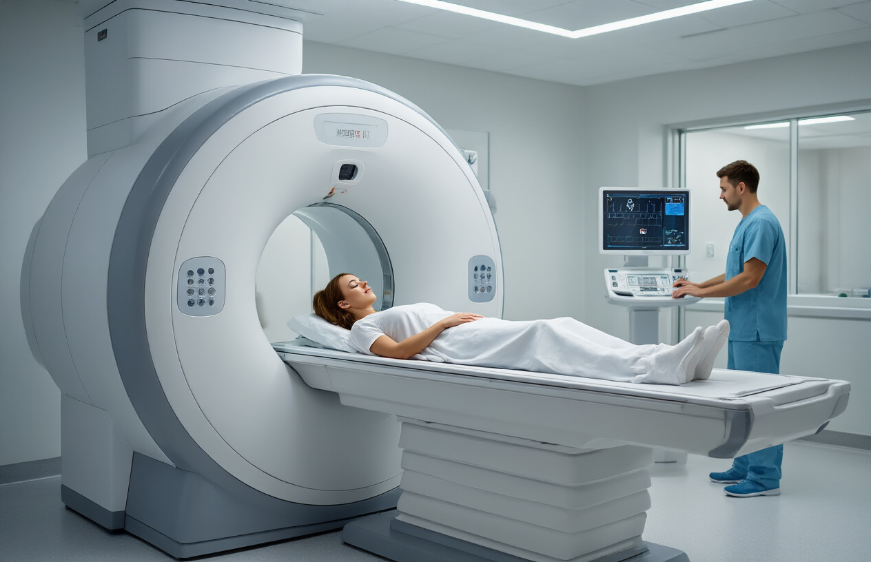
When you arrive for your MRI face and neck scan with contrast, the process starts with check-in and preparation. A technologist will review your medical history, confirm you haven’t eaten for several hours if required, and ensure you’ve removed all metal objects including jewelry, watches, and any removable dental work. You’ll change into a hospital gown and receive an IV line for the contrast injection.
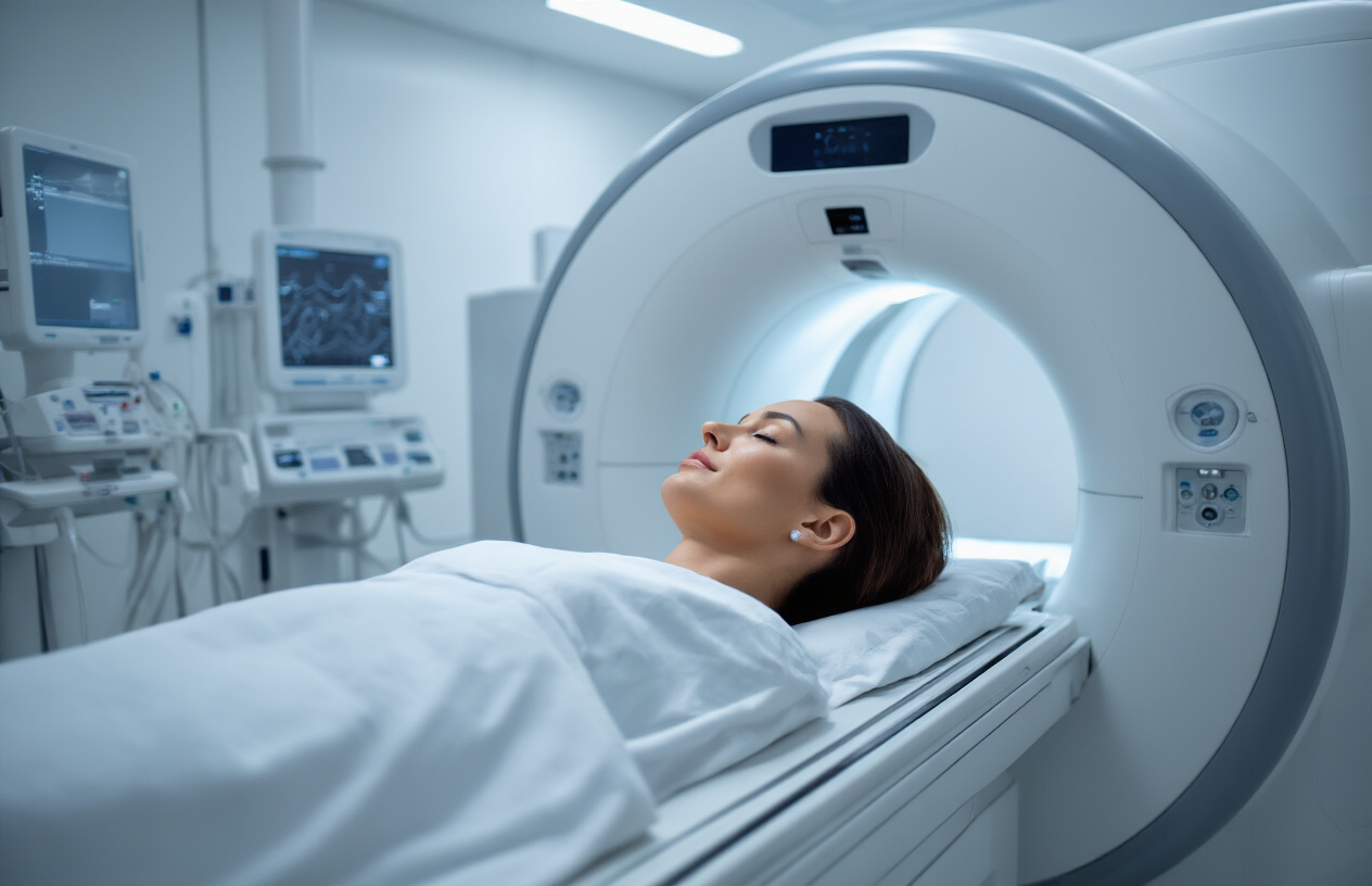
The actual scanning happens in two phases. First, you’ll lie down on the MRI table with your head positioned in a specialized coil designed for face and neck imaging. The technologist will place padding around your head to keep you comfortable and help you stay still. Initial images are taken without contrast, capturing baseline pictures of your facial structures, sinuses, jaw, throat, and neck tissues.
After the first set of images, the contrast agent (typically gadolinium) gets injected through your IV. This happens while you remain on the table – you might feel a cool sensation or slight metallic taste. The contrast helps highlight blood vessels, inflammation, and abnormal tissue that might not show up clearly on regular images.
The second phase captures detailed images as the contrast moves through your system. The entire scan usually takes 45 to 60 minutes, with most of that time spent lying still while the machine takes pictures. You’ll hear various loud knocking and buzzing sounds – this is completely normal and just the machine working.
Managing claustrophobia and staying comfortable
Many people worry about feeling trapped in the MRI machine, but modern scanners are much more open than older models. The face and neck scan requires your head to be inside the machine while your body remains mostly outside, which feels less confining than full-body scans.
If you have concerns about claustrophobia, talk to your doctor beforehand. They might prescribe a mild anti-anxiety medication to take before your appointment. Some facilities offer open MRI machines that provide more space around your body, though these may take longer to complete the scan.
During the procedure, you can communicate with the technologist through an intercom system. They’ll check on you regularly and can stop the scan if you need a break. Many facilities provide music or let you bring your own playlist to help you relax during the longer sequences.
Breathing exercises work well for staying calm. Focus on slow, steady breathing and try to relax your muscles. Some people find it helpful to close their eyes and imagine being somewhere peaceful. The technologist will tell you when each sequence starts and how long it will last, so you know what to expect.
Movement creates blurry images, so staying as still as possible is important for getting clear results. The padding around your head helps with this, but you’ll need to resist the urge to swallow frequently or move your jaw during scanning sequences.
Interpreting Your MRI Results and Next Steps
Understanding Your Radiologist’s Report Findings
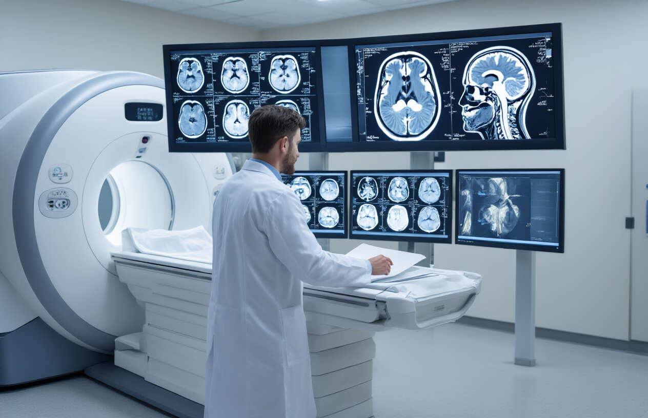
Your MRI report might look like a foreign language at first glance, but breaking it down makes the information much clearer. Radiologists describe what they see using specific medical terminology, and each section of the report covers different anatomical areas and potential findings.
The report typically starts with technical details about the scan itself – the type of contrast used, image sequences captured, and overall image quality. This section confirms that your scan was completed properly and that the images are clear enough for accurate diagnosis.
Next comes the detailed findings section, where the radiologist examines each structure systematically. They’ll describe the appearance of your facial bones, sinuses, soft tissues, blood vessels, and lymph nodes. Normal findings are often described as “unremarkable” or “within normal limits” – these are actually good things to see in your report.
When abnormalities are present, the radiologist will describe their location, size, shape, and how they appear with contrast enhancement. For example, they might note “enhancing mass” or “inflammatory changes,” which tells your doctor about the nature of what was found. Signal characteristics on different MRI sequences provide clues about tissue composition and help distinguish between various conditions.
The impression section at the end summarizes the key findings and often includes the radiologist’s diagnostic opinion. This is where you’ll find the most straightforward interpretation of what the scan revealed.
Follow-up Care Recommendations Based on Results
Your next steps depend entirely on what your MRI revealed, and your doctor will guide you through the appropriate course of action. Normal results typically mean no immediate follow-up imaging is needed, though your doctor might recommend routine monitoring based on your symptoms or medical history.
If inflammatory conditions like sinusitis or soft tissue infections were found, your doctor will likely prescribe antibiotics or anti-inflammatory medications. These conditions often resolve with proper treatment, and follow-up imaging might be scheduled in a few weeks to confirm improvement.
For masses or tumors, the path forward involves more detailed evaluation. Your doctor might recommend a biopsy to determine whether the growth is benign or malignant. This could involve fine needle aspiration or surgical biopsy, depending on the location and characteristics of the finding. Additional imaging studies like CT scans or PET scans might be ordered to better understand the extent of the condition.
Vascular abnormalities such as aneurysms or vascular malformations usually require consultation with specialists like neurosurgeons or interventional radiologists. They’ll discuss treatment options ranging from observation to surgical intervention, depending on the size, location, and risk factors involved.
Some findings require multidisciplinary care involving various specialists. Head and neck cancers, for instance, often need input from oncologists, radiation therapists, and surgeons to develop the most effective treatment plan. Your primary doctor will coordinate these referrals and help you navigate the process.
Regular follow-up appointments are crucial regardless of your results. Even with normal findings, ongoing monitoring ensures any changes are caught early, and with abnormal results, consistent follow-up allows your medical team to track treatment progress and adjust plans as needed.
MRI face and neck scans with contrast give doctors a clear, detailed look at what’s happening in these complex areas of your body. From diagnosing tumors and infections to spotting vascular problems and inflammatory conditions, this imaging technique helps medical professionals make accurate diagnoses and create effective treatment plans. The contrast dye enhances the images, making it easier to spot abnormalities that might otherwise go unnoticed.
Getting ready for your scan is straightforward – just follow your doctor’s prep instructions, arrive early, and don’t worry about the process itself. Most people find the experience much easier than they expected. Once you get your results, work closely with your healthcare team to understand what they mean for your specific situation. If your doctor recommends this type of MRI, trust that it’s an important step in getting the answers you need about your health.