

“MRI Dorsal Spine With Contrast Explained: Detecting Disc, Nerve & Spinal Cord Disorders”
Understanding MRI Dorsal Spine with Contrast: Your Complete Guide
An MRI dorsal spine with contrast is a specialized imaging test that uses contrast dye to create detailed pictures of your thoracic spine – the middle section of your back between your neck and lower back. This advanced scan helps doctors spot problems that might not show up clearly on regular MRI scans.
This guide is for patients scheduled for a contrast-enhanced spine MRI, their family members, and anyone wanting to understand what this important diagnostic test involves. Whether your doctor suspects a herniated disc, spinal infection, or tumor, knowing what to expect can ease your concerns and help you prepare properly.
We’ll walk you through the key medical conditions this scan can detect, from spinal cord compression to inflammatory diseases. You’ll also learn practical spine MRI preparation tips to get the best possible results and understand the thoracic spine MRI contrast procedure from start to finish. Finally, we’ll cover MRI contrast safety considerations so you feel confident about your upcoming scan.
Understanding What MRI Dorsal Spine with Contrast Reveals
Detailed visualization of spinal cord abnormalities
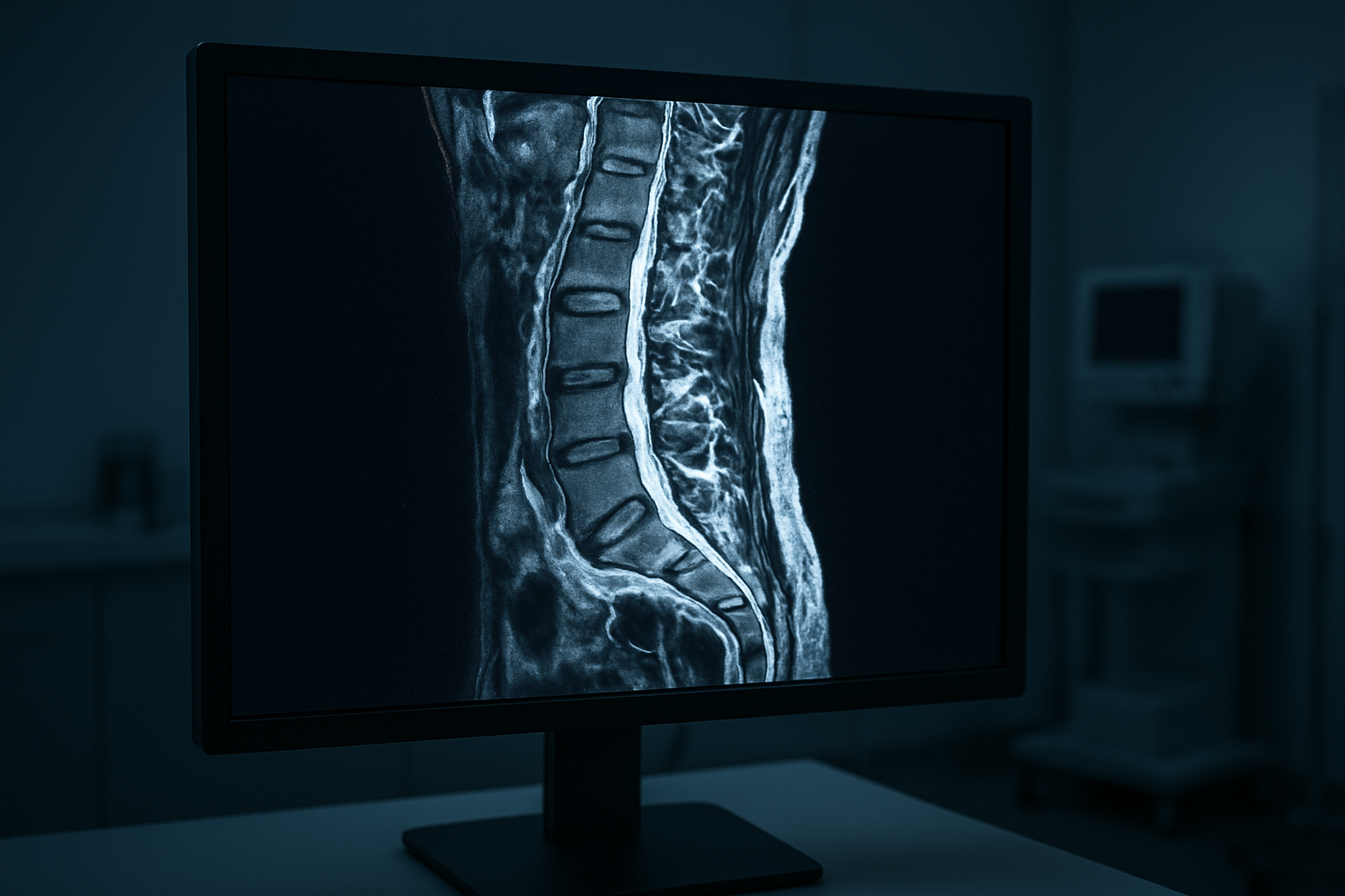
MRI dorsal spine with contrast creates incredibly detailed pictures of your thoracic spine and spinal cord, revealing issues that might stay hidden with regular imaging. The contrast agent, typically gadolinium, acts like a highlighter for your body’s structures, making abnormalities pop out clearly on the scan images.
When doctors examine your contrast-enhanced spine MRI results, they can spot inflammatory conditions like multiple sclerosis with remarkable precision. The contrast helps distinguish between active inflammation and older scar tissue, which looks completely different on the scan. This distinction matters enormously for treatment planning since active lesions require immediate attention while dormant ones might just need monitoring.
Spinal cord compression becomes dramatically visible with contrast enhancement. Whether pressure comes from herniated discs, bone spurs, or swelling, the contrast shows exactly where your spinal cord faces restrictions. The enhanced images reveal how severely the compression affects surrounding tissues and blood flow patterns.
Blood vessel abnormalities also stand out clearly with thoracic spine MRI contrast. Conditions like arteriovenous malformations or spinal cord infarctions become obvious when contrast flows through your vascular system. These blood vessel problems can cause serious neurological symptoms if left undetected.
The contrast agent also highlights areas of blood-brain barrier breakdown, which happens when your spinal cord’s protective barriers become damaged. This breakdown often signals active disease processes that need immediate medical intervention.
Enhanced detection of tumors and lesions
Contrast spine scan technology excels at finding both primary spinal tumors and metastatic cancer that has spread from other body parts. The contrast agent accumulates in areas with increased blood supply, which tumors typically develop to fuel their growth. This makes cancerous tissue light up brightly against the darker background of normal spinal structures.
Primary spine tumors like meningiomas, schwannomas, and ependymomas each have characteristic enhancement patterns that help doctors identify the specific tumor type. Meningiomas show intense, uniform enhancement, while schwannomas often display a more heterogeneous pattern with areas of bright enhancement mixed with darker regions.
Thoracic MRI with gadolinium proves especially valuable for detecting metastatic disease from lung, breast, or prostate cancers. These secondary tumors often appear as multiple bright spots scattered throughout the spine, creating a distinctive pattern that experienced radiologists recognize immediately. Early detection through contrast enhancement can dramatically improve treatment outcomes.
Benign lesions also become more apparent with contrast enhancement. Hemangiomas, which are common benign blood vessel tumors, show characteristic enhancement patterns that help distinguish them from more serious conditions. This prevents unnecessary anxiety and invasive procedures for patients with harmless growths.
The timing of contrast enhancement provides additional diagnostic clues. Some lesions enhance immediately after contrast injection, while others show delayed enhancement patterns. These timing differences help doctors differentiate between various types of tumors and inflammatory conditions, leading to more accurate diagnoses and appropriate treatment strategies.
Key Medical Conditions Diagnosed Through Contrast-Enhanced Dorsal Spine MRI
Spinal Cord Tumors and Metastases
MRI dorsal spine with contrast excels at detecting and characterizing spinal cord tumors and metastatic lesions throughout the thoracic region. The contrast agent, typically gadolinium, enhances blood flow patterns and helps distinguish between different types of abnormal tissue growths.
Primary spinal cord tumors appear as well-defined masses that enhance differently than surrounding healthy tissue. Intramedullary tumors, which develop within the spinal cord itself, show characteristic enhancement patterns that help doctors differentiate between benign and malignant lesions. Ependymomas and astrocytomas, the most common primary spinal cord tumors, each display unique enhancement characteristics that guide treatment decisions.
Metastatic disease presents distinct challenges that contrast-enhanced spine MRI addresses effectively. Cancer cells from breast, lung, prostate, and kidney cancers frequently spread to the spine. The contrast agent highlights these secondary tumors by revealing increased blood supply and disrupted tissue barriers. This enhanced visualization allows radiologists to identify even small metastatic deposits that might be missed on non-contrast studies.
Thoracic spine MRI contrast studies also reveal extradural masses that compress the spinal cord from outside. These lesions, including lymphomas and schwannomas, demonstrate specific enhancement patterns that help distinguish them from inflammatory conditions or infections. The contrast helps map the exact extent of tumor involvement, which proves critical for surgical planning and radiation therapy targeting.
Multiple Sclerosis Lesions
Dorsal spine imaging with contrast plays a vital role in diagnosing and monitoring multiple sclerosis (MS) affecting the thoracic spinal cord. MS lesions in the dorsal spine often appear as focal areas of inflammation that enhance with gadolinium contrast during active disease phases.
Active MS plaques show bright enhancement on contrast studies, indicating ongoing inflammatory activity and blood-brain barrier breakdown. These enhancing lesions typically appear as oval or elongated areas within the spinal cord, often spanning multiple vertebral levels. The enhancement pattern helps distinguish active inflammation from older, inactive scars.
Contrast spine scan technology reveals the characteristic features of MS lesions that help confirm the diagnosis. These lesions typically occupy less than two vertebral segments in length and involve the peripheral portions of the spinal cord. The contrast enhancement helps differentiate MS lesions from other inflammatory conditions like transverse myelitis or neuromyelitis optica.
Chronic MS lesions show different enhancement characteristics compared to acute plaques. Old lesions rarely enhance with contrast, appearing as areas of tissue damage without active inflammation. This distinction proves valuable for tracking disease progression and treatment effectiveness over time.
Spine MRI procedure protocols for MS evaluation include specific sequences that optimize lesion detection. The timing of contrast administration and image acquisition affects lesion visibility, making proper technique essential for accurate diagnosis and disease monitoring.
Preparing Your Body for Optimal Scan Results

Pre-scan dietary restrictions and medications
Your spine MRI preparation starts several hours before the actual scan. Most facilities recommend avoiding food and drinks for 4-6 hours before your contrast-enhanced spine MRI appointment. This fasting period helps reduce potential nausea that can occur when gadolinium contrast enters your system.
If you take regular medications, don’t stop them without talking to your doctor first. Blood pressure medications, heart medications, and diabetes treatments typically continue as normal. However, certain medications like metformin for diabetes may need temporary adjustment. Your healthcare team will provide specific guidance about which medications to take or skip on scan day.
Kidney function plays a crucial role in contrast safety. If you have kidney problems or take medications that affect kidney function, your doctor might order blood tests before your MRI dorsal spine with contrast. These tests ensure your kidneys can safely process the gadolinium contrast material.
Some people worry about allergies to MRI contrast. While rare, allergic reactions can happen. Tell your medical team about any previous reactions to contrast materials, medications, or foods. They might prescribe antihistamines or steroids as precautionary measures.
Hydration matters too. Drinking plenty of water in the days leading up to your scan helps your kidneys flush out the contrast material afterward. Avoid alcohol 24 hours before your procedure, as it can interfere with contrast clearance.
Removing metal objects and jewelry safely
Metal objects create dangerous situations during MRI scans due to powerful magnetic fields. Before entering the MRI suite, you’ll need to remove all metal items from your body. This includes obvious items like jewelry, watches, and glasses, but also less obvious ones like hairpins, underwire bras, and clothing with metal zippers or snaps.
Your thoracic spine MRI contrast procedure requires complete metal removal because even tiny metal fragments can cause serious injuries. The magnetic field can heat metal objects or pull them toward the machine at high speeds. Hair accessories, body piercings, and dental work with removable components all need attention.
Medical devices require special consideration. Pacemakers, insulin pumps, and cochlear implants may prevent you from having an MRI altogether. Some newer devices are MRI-compatible, but your medical team needs to verify this beforehand. Never assume your device is safe without proper verification.
Credit cards, phones, and electronic devices stay outside the scan room. The magnetic field can permanently damage these items and erase stored data. Most facilities provide secure lockers for your belongings.
Some metal implants like hip replacements or surgical screws are usually MRI-safe after healing. Your doctor will review your surgical history and any implanted devices before clearing you for the scan. Bring documentation about any implants or previous surgeries to help your medical team make informed decisions about your spine MRI procedure safety.
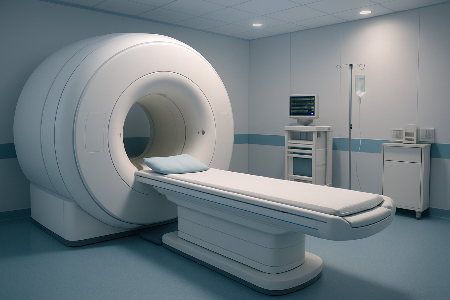
What to Expect During Your Contrast-Enhanced MRI Experience
IV contrast injection procedure and timing
The contrast-enhanced spine MRI procedure begins before you even enter the scanning room. A trained technologist will establish intravenous access, typically in your arm or hand, using a small catheter. This IV line serves as the pathway for delivering gadolinium contrast agent directly into your bloodstream during specific moments of the scan.
The timing of contrast injection is carefully orchestrated to capture optimal images of your dorsal spine structures. Your MRI dorsal spine with contrast session involves multiple imaging sequences – some taken before contrast administration and others captured at precise intervals afterward. The technologist monitors your scan progress from an adjacent control room and injects the contrast agent remotely through your IV line when the protocol calls for it.
You might feel a brief cool sensation or slight metallic taste as the contrast enters your system. This thoracic MRI with gadolinium agent enhances the visibility of blood vessels, inflammation, infections, and abnormal tissue growth around your thoracic vertebrae. The entire injection process takes only seconds, but the enhanced imaging continues for several more minutes to capture how the contrast moves through different tissues.
Most patients experience no discomfort during contrast delivery. The automated injection system ensures consistent flow rates and proper timing coordination with the MRI sequences, making your contrast-enhanced spine MRI both efficient and comfortable.
Positioning techniques for clear dorsal spine imaging
Proper positioning plays a crucial role in obtaining high-quality dorsal spine imaging results. You’ll lie flat on your back on the MRI table, with your arms positioned comfortably at your sides or crossed over your chest, depending on your body size and the specific coil configuration being used.
The technologist places specialized spine coils both underneath and sometimes on top of your thoracic region. These coils act like antennas, capturing the radio frequency signals that create your spine images. Foam padding and positioning aids help maintain proper spinal alignment while keeping you comfortable during the 30-45 minute scan duration.
Your head rests on a cushioned support, and your knees may be elevated with a bolster to reduce lower back strain. The thoracic spine MRI contrast protocol requires you to remain as still as possible, since even small movements can blur the detailed images of your vertebrae, discs, and surrounding soft tissues.
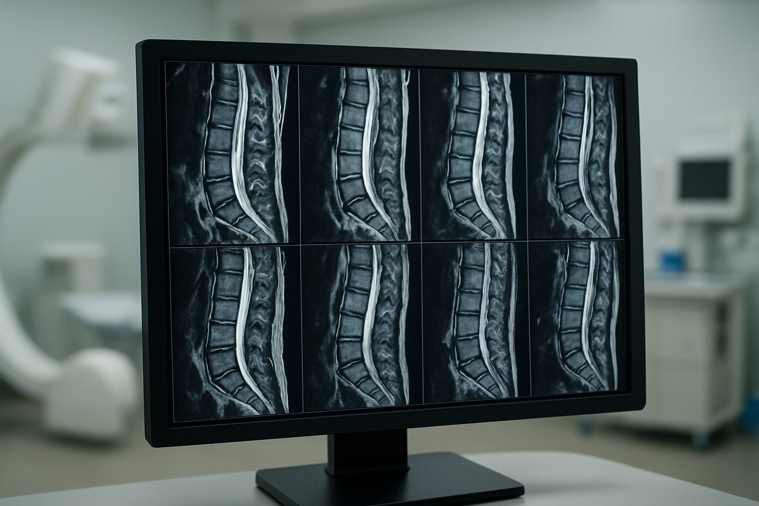
The MRI technologist ensures your dorsal spine sits centered within the magnetic field’s “sweet spot” where image quality is optimal. They mark reference points on your body to guarantee consistent positioning if multiple scan sequences are needed. This precise setup allows the spine MRI procedure to capture clear cross-sectional views from multiple angles, providing your radiologist with comprehensive visualization of your thoracic spine anatomy and any potential abnormalities.
Maximizing Safety and Minimizing Risks

Contrast Agent Allergic Reaction Prevention
Your safety during a contrast-enhanced spine MRI starts with understanding potential allergic reactions to gadolinium-based contrast agents. While serious allergic reactions are rare, occurring in less than 0.1% of patients, taking preventive measures ensures your MRI dorsal spine with contrast proceeds smoothly.
Before your scan, your medical team will review your allergy history thoroughly. If you’ve experienced reactions to contrast agents in previous imaging studies, tell your doctor immediately. Patients with a history of severe allergies, asthma, or multiple drug sensitivities may need premedication with antihistamines or corticosteroids before receiving contrast.
Watch for immediate symptoms during injection, including skin flushing, hives, nausea, or difficulty breathing. Most reactions occur within minutes of injection and are mild, requiring only observation. Your radiologist and technicians are trained to recognize and treat these reactions quickly.
The contrast agent used in thoracic spine MRI contrast procedures is generally well-tolerated, but certain groups face higher risks. Patients with shellfish or iodine allergies don’t automatically face increased risk with gadolinium, as this is a different type of contrast agent than those used in CT scans.
If you’re breastfeeding, discuss timing with your doctor. While gadolinium passes into breast milk in minimal amounts, some women prefer to pump and discard milk for 24 hours after the procedure as a precaution.
Kidney Function Considerations Before Injection
Your kidneys play a crucial role in eliminating contrast agents from your body, making kidney function assessment essential before any contrast spine scan. Patients with reduced kidney function face increased risks, including the rare but serious condition called nephrogenic systemic fibrosis (NSF).
Before scheduling your contrast-enhanced spine MRI, your doctor will likely order blood tests to check your kidney function, particularly your estimated glomerular filtration rate (eGFR). This simple blood test helps determine if your kidneys can safely process the contrast agent. Patients with eGFR below 30 mL/min/1.73m² require special consideration and may need alternative imaging approaches.
Certain medications can affect kidney function and contrast elimination. ACE inhibitors, ARBs, diuretics, and NSAIDs may need temporary adjustment before your scan. Never stop prescribed medications without consulting your doctor, but do inform your medical team about everything you’re taking, including over-the-counter drugs and supplements.
Dehydration significantly impacts kidney function and contrast processing. Drink plenty of water in the days leading up to your MRI contrast safety procedure, unless your doctor advises otherwise. Well-hydrated kidneys work more efficiently to eliminate the contrast agent from your system.
Diabetic patients taking metformin require special attention. While current guidelines are less restrictive than in the past, your doctor may recommend temporarily stopping metformin if your kidney function is borderline. This prevents a rare but potentially serious condition called lactic acidosis.
After your thoracic MRI with gadolinium, maintaining good hydration helps your kidneys eliminate the contrast more effectively. Most healthy patients clear the contrast within 24-48 hours, but those with kidney issues may need longer monitoring periods.
Interpreting Your Results for Better Health Outcomes
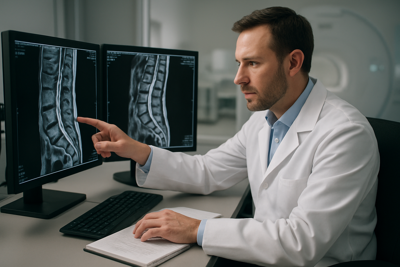
Understanding Radiologist Report Terminology
Deciphering your MRI dorsal spine with contrast report can feel like reading a foreign language, but understanding the key terms helps you make sense of your spine health. Radiologists use specific vocabulary that might seem intimidating at first glance, yet breaking down these terms reveals valuable insights about your thoracic spine condition.
“Enhancement” appears frequently in contrast-enhanced spine MRI reports, referring to areas that absorb the gadolinium contrast agent. This uptake often highlights inflammation, infection, or abnormal tissue growth. When your report mentions “homogeneous enhancement,” it describes uniform contrast absorption, while “heterogeneous enhancement” indicates irregular patterns that may suggest more complex pathology.
Anatomical terms like “vertebral body,” “pedicles,” “laminae,” and “facet joints” pinpoint exact locations within your spine structure. The report might reference specific vertebral levels (T1-T12 for thoracic spine), helping you understand precisely where any issues exist. Signal intensity descriptions such as “hyperintense” (bright) or “hypointense” (dark) on T1 or T2-weighted images provide clues about tissue composition and health.
Technical phrases like “cord compression,” “neural foraminal narrowing,” or “disc protrusion” describe mechanical issues affecting your spinal cord or nerve roots. Mass effect refers to pressure from abnormal structures, while edema indicates swelling that shows up clearly on contrast spine scans.
Distinguishing Normal from Abnormal Findings
Learning to differentiate between normal anatomical variations and concerning abnormalities empowers you to engage meaningfully with your healthcare team about spine MRI results interpretation. Normal thoracic MRI with gadolinium typically shows well-defined vertebral bodies with uniform bone marrow signal, intact disc spaces, and clear neural pathways without compression.
Abnormal findings often stand out due to their contrast enhancement patterns or structural changes. Tumors frequently show intense, irregular enhancement after contrast injection, appearing significantly brighter than surrounding healthy tissue. Infections create characteristic patterns with rim enhancement around abscesses or diffuse enhancement in areas of inflammation.
Degenerative changes represent common age-related findings that may or may not cause symptoms. Disc degeneration appears as loss of normal disc height and signal, while bone spurs (osteophytes) create additional bony growths that can narrow spinal spaces. These changes become more apparent with contrast-enhanced spine MRI, as inflammation around affected areas often enhances.
Vascular abnormalities like arteriovenous malformations show distinctive enhancement patterns with visible blood vessel networks. Herniated discs may enhance if inflammation surrounds the protruding disc material, helping distinguish acute injuries from chronic changes.
Your radiologist considers your symptoms alongside imaging findings when determining clinical significance. Some abnormalities appear dramatic on scans but cause minimal symptoms, while subtle changes might correlate with significant pain or neurological symptoms. Understanding this relationship helps you work with your doctor to develop appropriate treatment strategies based on your specific thoracic spine imaging results.
An MRI dorsal spine with contrast gives doctors a detailed look at your mid-back area, helping them spot everything from herniated discs to spinal tumors that might be missed with regular scans. The contrast dye acts like a highlighter, making inflammation, infections, and blood vessel problems much easier to see. Getting ready for your scan is pretty straightforward – you’ll need to fast for a few hours and let your medical team know about any allergies or kidney issues beforehand.
The actual procedure is safe for most people, though you might feel a warm sensation when the contrast goes in through your IV. Your radiologist will carefully review the images and work with your doctor to explain what they found and what it means for your treatment plan. If your doctor has recommended this type of MRI, don’t put it off – the detailed information it provides can make a real difference in getting the right diagnosis and treatment for your back problems.