

“MRI Cervical Spine in Flexion Explained: Detecting Instability, Disc Issues & Spinal Cord Compression”
MRI Cervical Spine Flexion: Advanced Imaging for Neck Problems
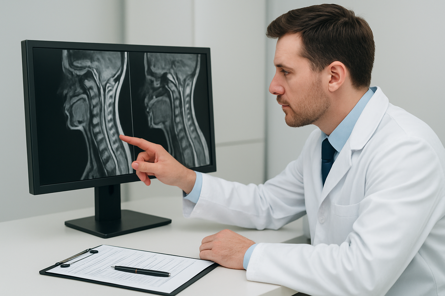
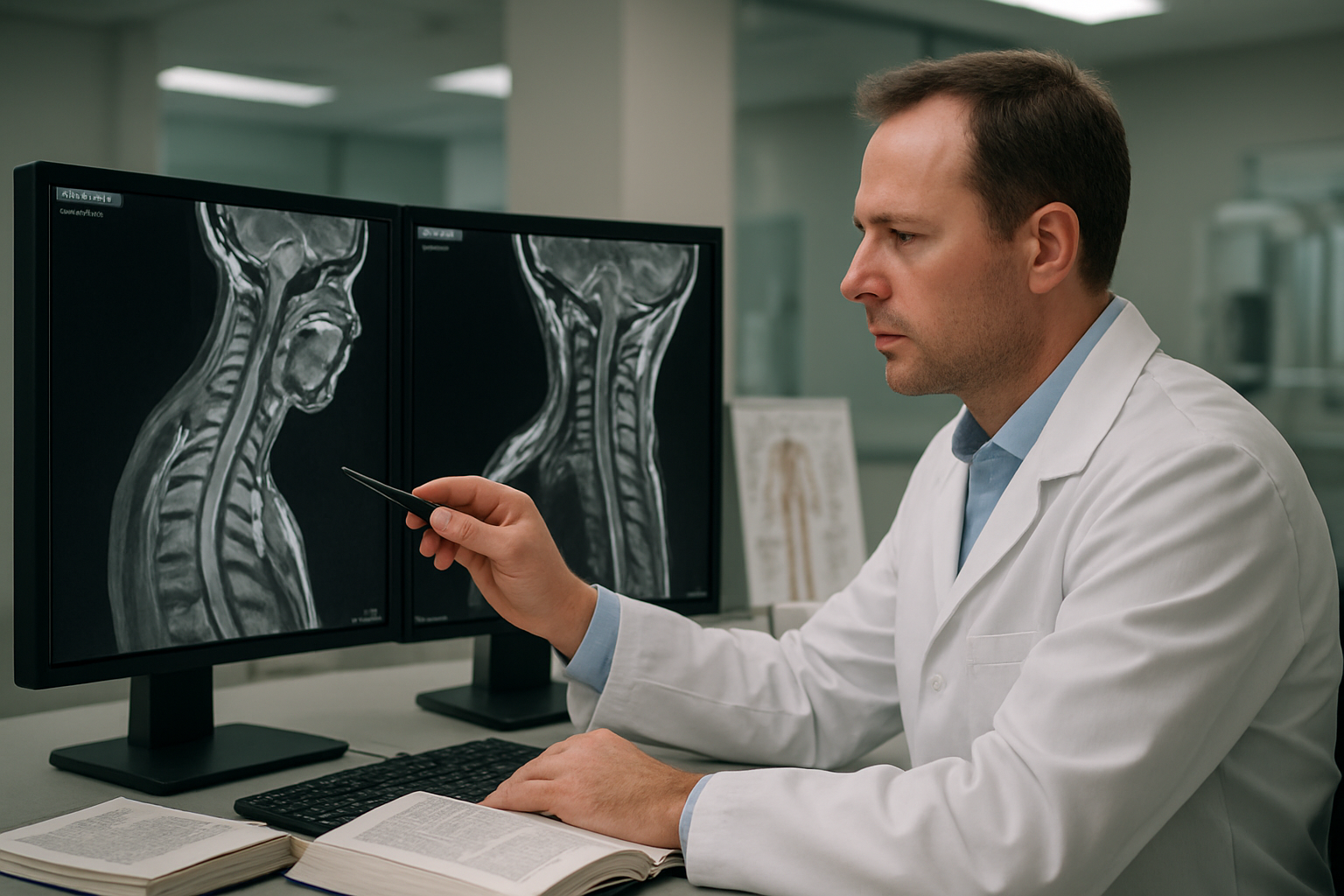
MRI cervical spine flexion is a specialized imaging technique that captures detailed pictures of your neck while it’s bent forward. This dynamic cervical MRI helps doctors spot problems that regular scans might miss, especially issues with cervical spine instability that only show up when your neck moves.
This guide is written for patients who need cervical flexion MRI, their families, and anyone wanting to understand this specific type of cervical spine imaging. Whether your doctor recommended this scan to check for nerve compression or spine instability, you’ll find clear answers about what to expect.
We’ll walk you through how cervical flexion study works and what conditions doctors can diagnose with this advanced imaging method. You’ll also learn how to prepare for your cervical MRI procedure and what happens after your scan is complete. Our facility in Sector 65, Gurugram is a CGHS empanelled centre, making this specialized diagnostic service accessible to government employees and their families.
Digital MRI technology provides clearer images than traditional open MRI systems, giving your doctor the detailed views needed for accurate diagnosis. After your scan, you’ll receive digital reports through WhatsApp, and you can collect your physical films from our centre at your convenience.
Understanding MRI Cervical Spine Flexion Imaging

Key differences between standard and flexion MRI scans
Regular cervical spine MRI scans capture images with your neck in a neutral, straight position. These standard scans work great for spotting many issues like herniated discs, tumors, or general spinal cord problems. But they miss something important – how your spine actually moves and behaves when you bend your neck forward.
MRI cervical spine flexion takes a completely different approach. During this specialized scan, you’ll position your neck in a flexed (chin-to-chest) position while the MRI machine captures detailed images. This forward bending motion puts stress on your cervical spine structures in ways that neutral positioning simply can’t reveal.
The most striking difference lies in what each scan can detect. Standard MRI might show you have a slightly bulging disc that looks manageable. But a cervical flexion MRI could reveal that same disc becomes severely compressed against your spinal cord when you move your head forward – information that changes everything about your diagnosis and treatment plan.
Dynamic cervical imaging also exposes ligament instability that stays hidden during regular scans. Your ligaments might hold everything together perfectly when you’re lying still, but flexion positioning can reveal dangerous looseness or weakness that only appears during movement.
The imaging quality differs too. Flexion scans often provide clearer views of the spinal canal’s posterior elements and can better demonstrate cerebrospinal fluid flow patterns around the spinal cord.
How cervical flexion positioning enhances diagnostic accuracy
Cervical spine instability diagnosis becomes dramatically more accurate when doctors can see how your spine behaves under mechanical stress. Think about it – most neck problems happen during movement, not while you’re perfectly still. Flexion positioning mimics real-life scenarios where symptoms actually occur.
When you flex your cervical spine during the cervical flexion study, several important changes happen inside your spinal canal. The ligamentum flavum (a key stabilizing ligament) thickens and can buckle inward, potentially compressing neural structures. Disc material that seemed stable in neutral position might shift dramatically, creating new pressure points on nerve roots or the spinal cord itself.
Flexion extension MRI cervical spine studies excel at catching what doctors call “dynamic stenosis” – spinal canal narrowing that only shows up during specific movements. You might have perfectly normal canal dimensions in neutral position, but dangerous compression when your neck flexes forward. This explains why some patients feel fine at rest but develop severe symptoms during daily activities like looking down at their phone or computer.
The positioning also helps identify subtle instabilities between vertebrae. Small shifts between bones that don’t register on standard imaging become obvious when your spine is under flexion stress. These micro-movements can cause significant nerve compression and explain mysterious pain patterns that stumped doctors using only neutral-position scans.
Flexion imaging proves especially valuable for evaluating surgical candidates. Surgeons need to know exactly how spinal structures behave during movement to plan the most effective procedures and avoid unnecessary operations on stable areas.
Medical Conditions Diagnosed Through Flexion MRI
Cervical instability and ligamentous injuries
MRI cervical spine flexion plays a crucial role in diagnosing cervical instability, a condition where the spine’s natural stability is compromised due to damaged ligaments or abnormal joint movement. During standard static imaging, these issues often remain hidden, making dynamic cervical MRI essential for accurate diagnosis.
When you flex your neck during the cervical flexion MRI procedure, damaged ligaments become apparent as they fail to maintain proper spinal alignment. The imaging captures real-time changes in vertebral positioning, revealing excessive movement between adjacent vertebrae that wouldn’t show up in regular scans. This makes flexion extension MRI cervical spine studies particularly valuable for patients experiencing neck pain after trauma or degenerative changes.
Ligamentous injuries affecting the anterior longitudinal ligament, posterior longitudinal ligament, and ligamentum flavum create instability patterns that become visible only when the spine moves through its range of motion. These soft tissue structures normally act as the spine’s natural restraint system, and when damaged, they allow abnormal movement that can compress neural structures.
Patients who have suffered whiplash injuries, sports-related trauma, or degenerative disc disease often benefit from this specialized imaging approach. The cervical spine imaging technique helps doctors distinguish between stable and unstable injuries, directly influencing treatment decisions. Without this dynamic assessment, many cases of cervical instability would go undiagnosed, potentially leading to chronic pain and neurological complications.
Dynamic spinal stenosis detection
Dynamic cervical imaging revolutionizes how medical professionals detect spinal stenosis that only becomes symptomatic during movement. Unlike static stenosis visible on standard MRI, dynamic stenosis occurs when the spinal canal narrows during specific neck positions, particularly during flexion.
During a cervical flexion study, the ligamentum flavum buckles inward, and facet joints can shift position, creating temporary but significant narrowing of the spinal canal. This phenomenon explains why some patients experience symptoms only during certain activities like looking down or bending their neck forward, while appearing normal on static imaging.
The cervical spine MRI preparation for dynamic studies requires patients to maintain specific positions during scanning, allowing visualization of these movement-related changes. This approach captures how the spinal cord and nerve roots become compressed during everyday activities, providing a more complete picture of the patient’s condition.
Healthcare facilities offering advanced cervical MRI procedure options, including those empanelled with CGHS, can provide this specialized imaging service. Many CGHS empanelled centres now offer digital MRI technology that delivers superior image quality compared to traditional open MRI systems, ensuring accurate detection of these subtle dynamic changes.
Patients receive their diagnostic reports digitally through WhatsApp, while physical films can be collected from the imaging centre at their convenience. This streamlined approach ensures quick access to results while maintaining the quality needed for precise diagnosis of dynamic spinal conditions.
Preparing for Your Cervical Flexion MRI Procedure
Pre-scan instructions and safety requirements
Before your cervical flexion MRI, you’ll need to follow specific guidelines to ensure accurate imaging results and your safety. Remove all metal objects including jewelry, watches, belts with metal buckles, and clothing with metal fasteners. Even small items like hairpins, dental work with metal components, or body piercings can interfere with the magnetic field. Your healthcare team will provide you with a metal-free gown if needed.
Inform your medical team about any implanted medical devices such as pacemakers, cochlear implants, or surgical clips. While most modern devices are MRI-compatible, your doctor needs this information to determine if your cervical spine MRI preparation requires special protocols. Also mention any history of working with metal fragments, as these could pose safety risks during the scan.
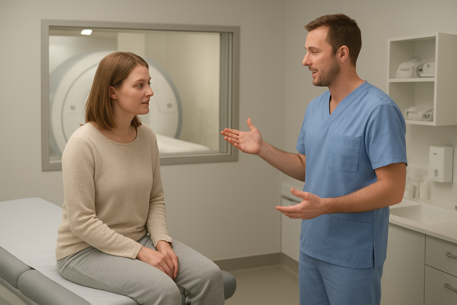
Arrive at least 30 minutes early to complete paperwork and change into appropriate clothing. You can eat normally before the procedure unless your doctor provides different instructions. Bring a list of current medications and any previous cervical spine imaging studies for comparison. If you experience claustrophobia, discuss sedation options with your physician beforehand, as the cervical flexion study requires you to remain still in specific positions for extended periods.
What to expect during the positioning process
The positioning phase for your cervical flexion MRI requires careful attention to achieve optimal imaging of cervical spine instability diagnosis. You’ll lie on a comfortable padded table that slides into the MRI machine. The technologist will position your head and neck using specialized cushions and supports to maintain the flexed position throughout the scan.
During the dynamic cervical imaging process, your neck will be gently flexed forward, bringing your chin closer to your chest. This positioning allows the MRI to capture detailed images of how your cervical vertebrae move and align during flexion. The technologist will ensure you’re comfortable while maintaining the precise angle needed for effective cervical flexion study results.
Expect to hold this position for 15-30 minutes while the machine takes multiple sequences of images. The MRI machine produces loud knocking and tapping sounds, so you’ll receive earplugs or headphones to reduce noise. Communication with the technologist remains possible through an intercom system throughout the procedure.
Some patients experience mild neck stiffness from holding the flexed position, but this typically resolves quickly after the scan. The technologist monitors your comfort level and can make minor adjustments if needed while maintaining the diagnostic quality of your dynamic cervical MRI. Clear instructions about breathing and staying still will help produce the sharpest images for accurate diagnosis.
Interpreting Flexion MRI Results Effectively
Normal versus abnormal cervical alignment patterns
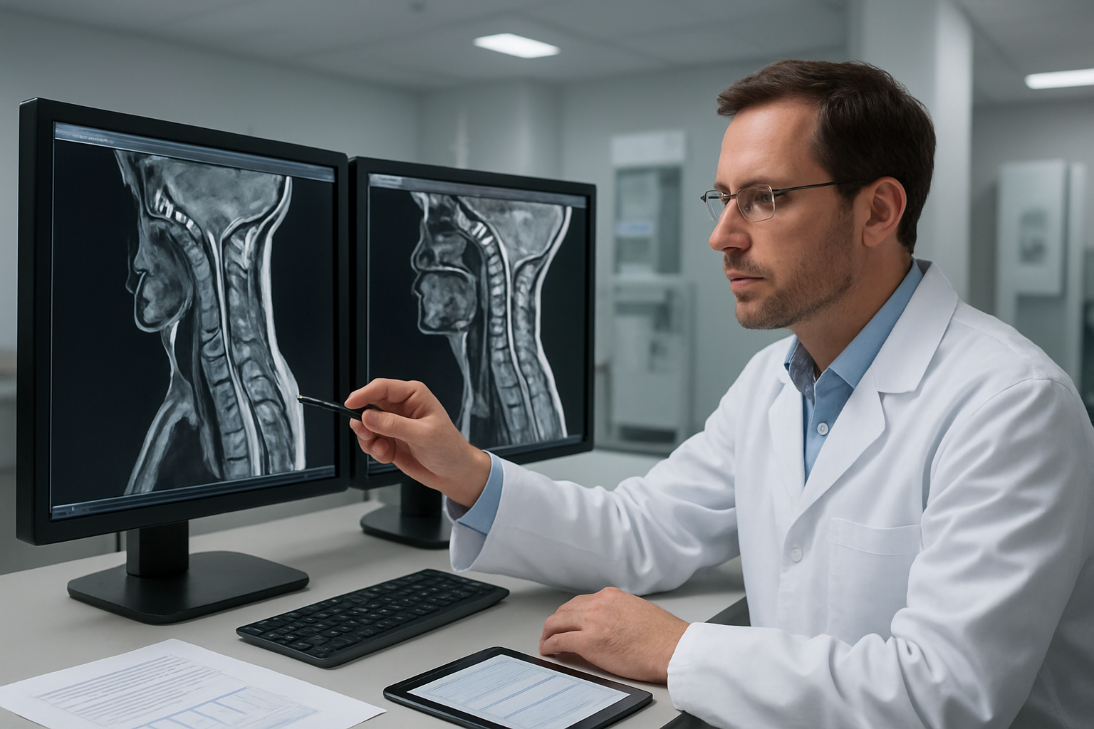
Understanding what normal cervical spine alignment looks like during flexion helps radiologists spot problems quickly. In a healthy cervical spine, the vertebrae maintain their natural curve and move smoothly together when bending forward. The cervical flexion MRI shows each vertebra gliding in a coordinated pattern, maintaining proper spacing between bones.
Normal flexion patterns reveal vertebrae that tilt forward uniformly, creating a gentle forward curve. The spinal cord remains centered within the canal, and ligaments stretch evenly without excessive bulging. Joint spaces stay consistent, and there’s no sudden kinking or sharp angles between vertebrae.
Abnormal patterns tell a different story. Cervical spine instability diagnosis becomes clear when vertebrae slip out of proper alignment during flexion. You might see one vertebra sliding too far forward over another, creating what doctors call spondylolisthesis. Sharp angular changes between bones signal ligament damage or disc problems.
Radiologists also look for areas where movement stops abruptly. This suggests fusion from arthritis or injury. Sometimes vertebrae tilt in opposite directions, creating a zigzag pattern that indicates multiple levels of instability. Dynamic cervical MRI captures these movement problems that static scans might miss completely.
CGHS empanelled facilities often see patients with whiplash injuries where normal alignment patterns break down. The flexion extension MRI cervical spine reveals hidden instabilities that explain persistent neck pain even when regular MRI scans look normal.
Measuring spinal canal dimensions and changes
Precise measurements during cervical flexion study help determine how much space your spinal cord has to work with. Radiologists measure the canal diameter at multiple levels, checking how dimensions change when your neck bends forward. Normal canals maintain adequate space even during maximum flexion.
The anteroposterior diameter – the distance from front to back of the canal – becomes critical during flexion. When this measurement drops below 10mm, spinal cord compression becomes likely. Dynamic cervical imaging shows these changes happening in real-time as vertebrae move.
Cross-sectional area measurements give the complete picture. Advanced software calculates the total canal space available at each level during different phases of flexion. Some patients show dramatic narrowing that only appears during specific positions, explaining why symptoms come and go with neck movement.
Our digital MRI technology captures these measurements with exceptional accuracy, providing clearer data than older open MRI systems. The detailed images allow radiologists to track even small changes in canal dimensions that might indicate early problems.
Cervical spine imaging at our Sector 65 facility includes comprehensive measurement protocols. Patients receive digital reports via WhatsApp showing exact measurements and how they compare to normal ranges. The physical films can be collected from the centre at your convenience, giving you complete documentation of your spinal canal dimensions and any changes detected during the flexion sequence.
Treatment Planning Based on Flexion MRI Findings
Conservative Management Strategies for Mild Instability
When your cervical flexion MRI reveals mild instability, doctors often start with non-surgical approaches that can effectively manage symptoms and prevent progression. Physical therapy forms the cornerstone of conservative treatment, focusing on strengthening the deep cervical flexor muscles and improving neck stability through targeted exercises.
Cervical collars may provide temporary support during acute episodes, but long-term use should be limited to prevent muscle weakness. Anti-inflammatory medications help reduce swelling around affected vertebrae, while muscle relaxants can address spasms that often accompany cervical spine instability.
Activity modification plays a crucial role in managing mild instability detected through cervical flexion MRI. Patients typically need to avoid sudden neck movements, heavy lifting, and contact sports that could worsen the condition. Ergonomic adjustments at work and home, including proper monitor height and pillow support, help maintain neutral spine alignment.
Many facilities offering cervical spine imaging, including CGHS empanelled centres, can provide detailed reports through digital channels like WhatsApp, making it easier for patients to share findings with their treatment team quickly. This streamlined communication helps coordinate care between imaging centres and healthcare providers managing conservative treatment plans.
Heat therapy, gentle stretching, and postural training complement other conservative measures. Regular monitoring through follow-up imaging helps track progress and determine if conservative management remains appropriate for each patient’s specific instability pattern.
Surgical Intervention Criteria and Timing
Surgical intervention for cervical spine instability becomes necessary when conservative management fails to provide adequate symptom relief or when imaging shows progressive deterioration. Dynamic cervical MRI findings help surgeons determine the most appropriate surgical approach and timing.
Key surgical indicators include severe cord compression visible on flexion extension MRI cervical spine studies, progressive neurological symptoms like weakness or numbness, and significant instability measurements exceeding established thresholds. Myelopathy symptoms, including difficulty with fine motor tasks and balance problems, often prompt earlier surgical consideration.
Fusion procedures remain the gold standard for treating significant cervical spine instability. Anterior cervical discectomy and fusion (ACDF) addresses disc-related instability, while posterior fusion techniques stabilize facet joint problems identified through cervical flexion study results.
Timing becomes critical when neurological symptoms progress rapidly. Emergency surgical intervention may be required if patients develop severe myelopathy or if flexion MRI shows dangerous cord compression that threatens permanent neurological damage.
Modern surgical techniques offer excellent outcomes when properly timed. Facilities with advanced imaging capabilities can provide detailed pre-surgical planning using high-resolution MRI data. Digital MRI technology offers superior image quality compared to older open MRI systems, providing surgeons with clearer visualization of anatomical structures and instability patterns essential for surgical planning.
Post-surgical monitoring often includes follow-up cervical spine imaging to ensure proper fusion and stability restoration, with digital reports facilitating ongoing communication between surgical teams and patients throughout recovery.
Cervical spine flexion MRI plays a crucial role in diagnosing conditions that regular spine imaging might miss. This specialized scan helps doctors see exactly what happens when your neck moves forward, revealing problems like disc herniation, spinal stenosis, or ligament damage that only show up during movement. Getting the right diagnosis early can make all the difference in your treatment plan and recovery time.
If you’re dealing with neck pain or mobility issues, consider getting your MRI done at a CGHS empanelled facility for cost-effective care. Digital MRI technology offers clearer images than traditional open MRI systems, giving your doctor better insight into your condition. Once your scan is complete, you’ll receive digital reports directly on WhatsApp for convenience, while physical films can be collected from the center when it works for your schedule. Don’t let neck problems hold you back – the right imaging can guide you toward the most effective treatment approach.