

“MRI Brain With Venography Explained: Detecting Blood Flow & Vein Disorders”
MRI Brain With Venography (MRV): A Complete Guide to Brain Vein Imaging
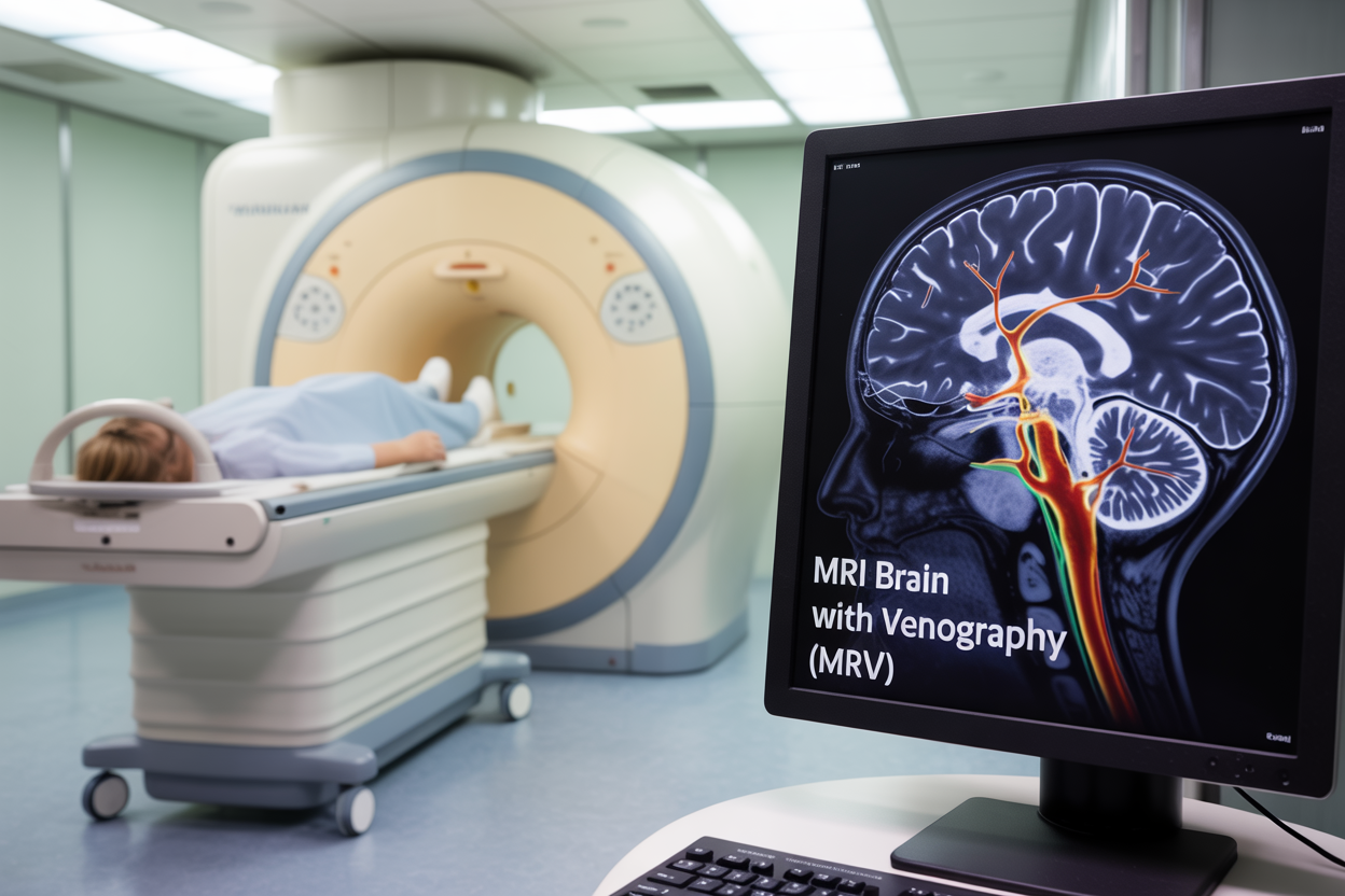
MRI brain venography (MRV) is a specialized magnetic resonance venography scan that creates detailed images of your brain’s blood vessels and cerebral venous system. This non-invasive brain vascular imaging technique helps doctors diagnose problems with veins in your brain, including blood clots, blockages, and structural abnormalities.
This guide is written for patients who need an MRV scan, their families, and anyone wanting to understand this important diagnostic tool. Whether your doctor suspects venous thrombosis or wants to examine your brain’s vascular health, knowing what to expect can reduce anxiety and help you prepare.
We’ll walk you through the MRV procedure from start to finish, including how this brain blood vessel scan works and what medical conditions it can detect. You’ll also learn how to prepare for your appointment and what your results might reveal about your brain’s venous system. Finally, we’ll explore the key benefits and limitations of MRV technology so you can have informed discussions with your healthcare team.
Understanding MRI Brain With Venography Technology
How MRV Combines Traditional MRI With Vascular Imaging
MRI brain venography represents a sophisticated evolution of standard magnetic resonance imaging technology. While traditional MRI scans excel at capturing detailed images of brain tissue, muscles, and organs, MRV takes this capability further by specifically targeting the brain’s venous system. This specialized brain vein imaging technique builds upon the foundational principles of conventional MRI while incorporating advanced pulse sequences designed to highlight blood flow patterns within cerebral veins.
The integration works by using the same powerful magnetic fields and radiofrequency pulses found in standard MRI machines, but with modified imaging protocols that enhance vascular contrast. When blood flows through veins, it creates specific signal patterns that MRV technology can detect and amplify. This allows radiologists to visualize the cerebral venous system with remarkable clarity, showing both the structure and function of these critical blood vessels.
What makes magnetic resonance venography particularly valuable is its ability to capture both anatomical and physiological information simultaneously. The scan reveals not just where veins are located, but also how blood moves through them. This dual capability means doctors can spot both structural abnormalities and functional problems like reduced blood flow or blockages that might indicate serious conditions such as venous thrombosis.
Advanced Magnetic Resonance Technology for Blood Vessel Visualization
The technology behind brain blood vessel scanning relies on sophisticated magnetic field manipulations that differentiate between flowing blood and stationary tissue. Advanced MRV systems use specialized gradient sequences that create contrast between moving blood cells and surrounding brain tissue. As blood flows through veins, it generates unique signal characteristics that the MRI scanner can isolate and enhance.
Time-of-flight techniques represent one cornerstone of modern MRV procedures. This method tracks how blood moves through vessels over time, creating detailed maps of venous flow patterns. The technology can detect even subtle changes in blood velocity, making it possible to identify areas where circulation might be compromised before symptoms become severe.
Phase-contrast imaging adds another dimension to brain vascular imaging capabilities. This technique measures the speed and direction of blood flow by detecting tiny changes in magnetic phase as blood cells move through the scanning field. When combined with contrast-enhanced protocols, these methods can reveal intricate details about venous architecture that would be impossible to see with conventional imaging.
Modern MRV scanners also incorporate real-time processing capabilities that allow technologists to optimize image quality during the scan. These systems can automatically adjust parameters based on patient-specific factors like heart rate, breathing patterns, and blood flow characteristics, ensuring optimal visualization of the venous system for each individual case.
Medical Conditions Diagnosed Through MRV Scanning

Detecting Blood Clots in Brain Veins and Sinuses
Blood clots in the brain’s venous system represent one of the most serious conditions that MRV scanning can detect. Cerebral venous thrombosis occurs when clots form in the brain’s veins or dural sinuses, blocking normal blood flow and potentially causing life-threatening complications. Unlike arterial strokes that cut off blood supply to brain tissue, venous clots prevent blood from draining properly, leading to increased pressure and potential hemorrhage.
MRI brain venography excels at identifying these dangerous clots because it provides detailed images of the entire cerebral venous system without requiring invasive procedures. The scan can detect clots in major dural sinuses like the superior sagittal sinus, transverse sinuses, and sigmoid sinuses, as well as in smaller cortical veins throughout the brain. This comprehensive view allows doctors to see exactly where blockages occur and how extensive they are.
Early detection through MRV scanning makes a huge difference in patient outcomes. When caught quickly, blood clots can often be treated with anticoagulation therapy or other interventions before permanent brain damage occurs. The magnetic resonance venography technique can also monitor treatment progress, showing whether clots are dissolving and blood flow is returning to normal.
Risk factors for cerebral venous thrombosis include pregnancy, birth control pill use, dehydration, certain cancers, and inherited clotting disorders. Patients often present with severe headaches, seizures, or neurological symptoms that might initially seem like other conditions. MRV provides the definitive diagnosis that guides immediate treatment decisions.
Identifying Venous Malformations and Abnormalities
Brain vein imaging through MRV reveals various structural abnormalities in the cerebral venous system that can cause significant health problems. Arteriovenous malformations (AVMs) represent one of the most concerning findings, where abnormal connections form between arteries and veins, bypassing normal capillary networks. These malformations create high-pressure situations that can lead to bleeding in the brain.
Venous angiomas, also called developmental venous anomalies, appear as clusters of enlarged veins that drain into a single collecting vein. While often benign, these formations can sometimes cause headaches or seizures. MRV scanning provides the detailed brain vascular imaging needed to distinguish between harmless variants and potentially problematic malformations.
Cavernous malformations present another category of venous abnormalities that MRV can detect. These lesions consist of enlarged, thin-walled blood vessels that form a berry-like cluster. They’re prone to bleeding and can cause seizures, neurological deficits, or headaches depending on their location in the brain.
The brain blood vessel scan capabilities of MRV also reveal stenosis or narrowing of major venous sinuses, which can occur due to various causes including inflammation, infection, or tumor compression. Idiopathic intracranial hypertension often shows narrowing of the transverse sinuses, and MRV helps determine whether this narrowing is causing increased brain pressure.
Dural arteriovenous fistulas represent another complex vascular abnormality where abnormal connections form between arteries and dural sinuses. These connections can cause dangerous retrograde flow patterns that MRV scanning can clearly demonstrate, helping neurosurgeons plan appropriate treatment approaches.
Preparing for Your MRV Examination

Essential Pre-Scan Medical History Requirements
Your healthcare team needs comprehensive information about your medical background before scheduling your MRV scan. Share your complete history of blood clots, strokes, or any brain-related conditions with your doctor. Previous surgeries, especially those involving your head, neck, or cardiovascular system, play a crucial role in scan interpretation and safety protocols.
Metal implants require special attention during MRI brain venography preparation. Tell your medical team about pacemakers, cochlear implants, aneurysm clips, or any metallic devices in your body. Even small items like dental work with metal components can affect the magnetic resonance venography results. Your doctor will determine if these items are MRI-compatible or if alternative imaging methods are needed.
Allergies deserve particular focus, especially reactions to contrast agents or iodine-based substances. Many MRV procedures use gadolinium-based contrast materials to enhance brain vein imaging quality. Previous allergic reactions to any medical contrast materials should be discussed thoroughly. Your medical history of kidney problems is equally important since contrast agents are processed through your kidneys.
Pregnancy status must be confirmed before any brain blood vessel scan. While MRI technology doesn’t use ionizing radiation, the safety of contrast agents during pregnancy requires careful consideration. Your doctor will weigh the diagnostic benefits against potential risks to make the best decision for your specific situation.
Medication and Dietary Restrictions to Follow
Most medications can continue as prescribed before your MRV procedure, but certain drugs require special attention. Blood thinners like warfarin, heparin, or newer anticoagulants may need temporary adjustments, particularly if your cerebral venous system evaluation is related to suspected venous thrombosis diagnosis. Never stop these medications without explicit instructions from your healthcare provider.
Diabetes medications need careful timing consideration. If your MRV scan includes contrast material, your doctor might adjust insulin or oral diabetes medications to prevent blood sugar complications. Metformin, a common diabetes drug, sometimes requires temporary discontinuation around contrast procedures to protect kidney function.
Dietary restrictions for MRV scanning are generally minimal compared to other medical procedures. You can usually eat and drink normally before the scan unless you’re receiving sedation for anxiety or claustrophobia. If sedation is planned, you’ll need to fast for several hours beforehand, similar to preparation for minor surgical procedures.
Caffeine intake on scan day won’t interfere with the brain vascular imaging process, but some patients find limiting coffee or energy drinks helps reduce anxiety. Stay well-hydrated before your appointment, especially if contrast material will be used. Good hydration helps your kidneys process contrast agents more effectively and can make IV placement easier if needed.
Remove all jewelry, watches, and accessories containing metal before arriving at the imaging center. Nail polish should be removed from fingernails and toenails since some formulations contain metallic particles that could create artifacts on your scan images.
Step-by-Step MRV Procedure Experience
Initial Patient Positioning and Comfort Measures
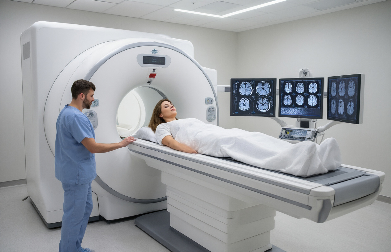
When you arrive for your MRV procedure, the technologist will guide you to the scanning room and help you get positioned on the MRI table. You’ll lie flat on your back with your head placed in a specialized head coil that looks like a helmet or cage-like structure. This coil contains the receivers that capture the detailed images of your brain’s venous system.
The positioning process takes several minutes because precision matters for high-quality brain vascular imaging. Your head will be secured with soft padding and straps to prevent movement during the scan. While this might feel restrictive at first, staying perfectly still is crucial for clear MRV scan results. The technologist will place a small squeeze ball in your hand so you can communicate if you feel uncomfortable or anxious during the procedure.
Comfort measures include providing earplugs or headphones since MRI machines produce loud knocking and buzzing sounds during the magnetic resonance venography process. Some facilities offer music or nature sounds to help you relax. A blanket is typically provided since the scanning room stays cool to keep the equipment at optimal temperature.
The MRI table will slide into the cylindrical scanner, positioning your head and upper body inside the tunnel. Open MRI machines are available for patients with claustrophobia, though they may not provide the same image quality for detailed cerebral venous system evaluation.
Contrast Agent Administration and Timing
Most MRV procedures require intravenous contrast material called gadolinium to enhance visualization of your brain’s blood vessels. Before the scan begins, a nurse or technologist will establish an IV line in your arm or hand. This process involves a small needle stick and takes just a few minutes.
The MRV procedure typically starts with initial images taken without contrast to establish baseline views of your brain anatomy. After these preliminary scans, the contrast agent is administered through your IV line. Gadolinium contrast helps highlight the venous structures and makes abnormalities like venous thrombosis more visible on the images.
Timing is critical during contrast administration. The technologist monitors the injection carefully because different phases of contrast enhancement reveal different aspects of your cerebral venous system. Early images capture the arterial phase, while later images focus on venous drainage patterns. This timing sequence allows radiologists to distinguish between arteries and veins and identify any blockages or abnormal flow patterns.
You might feel a cool sensation or mild metallic taste when the contrast enters your bloodstream. These sensations are normal and temporary. The entire contrast-enhanced portion of the brain blood vessel scan takes approximately 10-15 minutes, with multiple image sequences captured to create a comprehensive view of your venous anatomy.
Throughout the procedure, the technologist communicates with you through an intercom system, letting you know how much time remains and what to expect next during your MRI brain venography experience.
Reading and Understanding Your MRV Results

Normal Versus Abnormal Venous Flow Patterns
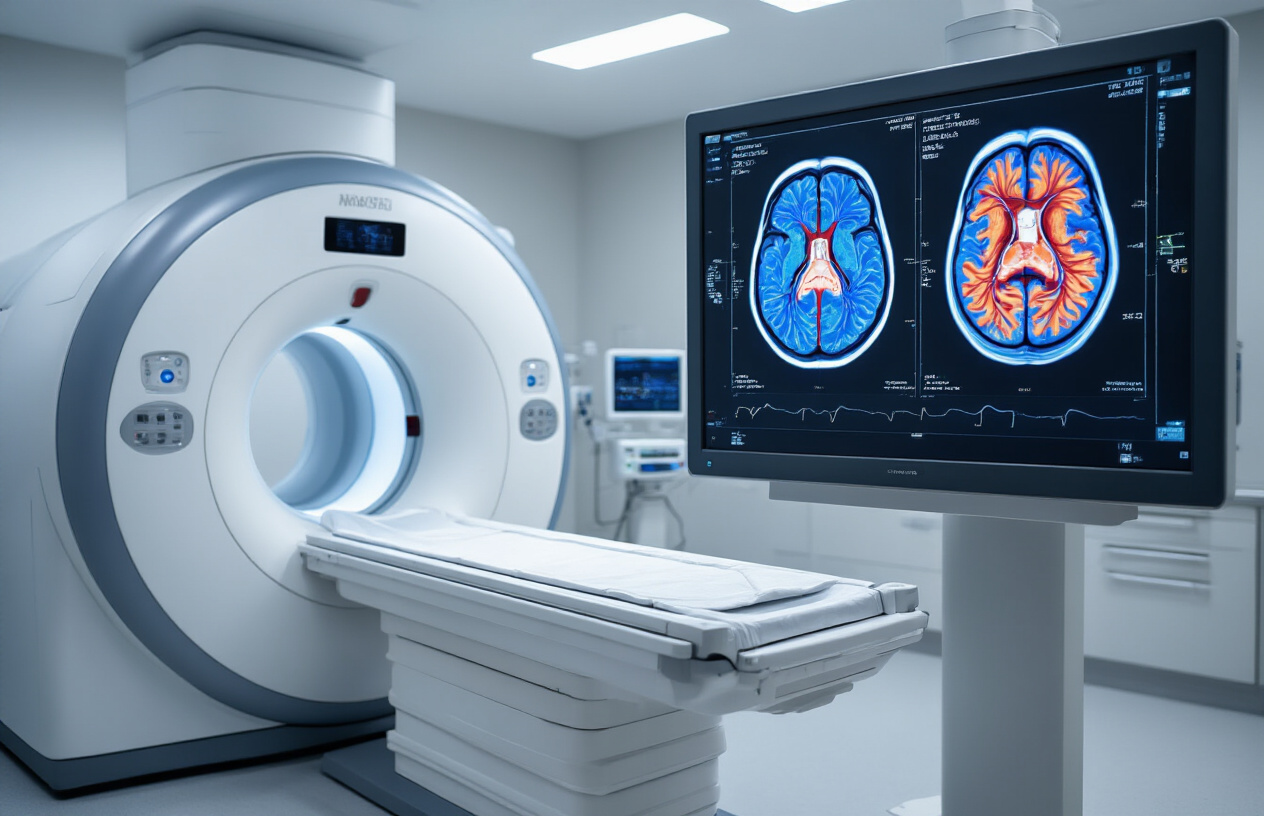
When your radiologist reviews your MRV scan, they’re looking at how blood flows through the intricate network of veins in your brain. Normal venous flow appears smooth and uninterrupted on the magnetic resonance venography images, with all major cerebral venous structures clearly visible and properly connected.
The superior sagittal sinus, transverse sinuses, and jugular veins should show consistent signal intensity, indicating healthy blood flow. Normal brain vein imaging reveals symmetrical patterns on both sides of your head, with no unexpected gaps or narrowing in the vessel pathways.
Abnormal findings typically show up as areas where the expected venous signal is missing, significantly reduced, or appears irregular. These disruptions in the cerebral venous system can indicate blockages, narrowing, or structural abnormalities. Your MRI brain venography might reveal complete occlusion of a vein, where blood flow stops entirely, or partial stenosis, where the vessel becomes narrowed but still allows some blood to pass through.
Collateral circulation often develops when main venous pathways become blocked. Your brain creates alternative routes for blood drainage, which appear as new or enlarged vessels on the MRV procedure images. While this adaptation helps maintain proper drainage, it signals that your primary venous system isn’t functioning normally.
Common Findings and Their Clinical Significance
Cerebral venous thrombosis ranks among the most serious discoveries on brain blood vessel scans. This condition occurs when blood clots form in the brain’s venous system, blocking normal drainage and potentially causing dangerous pressure buildup. The MRV scan shows these clots as areas of missing or altered signal where flowing blood should appear.
Venous malformations represent another significant finding that your MRI with contrast venography can detect. These abnormal connections between arteries and veins bypass the normal capillary network, creating unusual flow patterns visible on the scan. While some malformations cause no symptoms, others can lead to headaches, seizures, or bleeding.
Dural arteriovenous fistulas appear as abnormal connections between arteries and the venous sinuses that drain blood from your brain. These connections create turbulent flow patterns that show up clearly on brain vascular imaging studies. Left untreated, they can cause progressive neurological symptoms or potentially life-threatening complications.
Venous stenosis, or narrowing of brain veins, often relates to conditions like idiopathic intracranial hypertension. Your magnetic resonance venography can pinpoint exactly where these narrowings occur and help determine whether they’re causing your symptoms. Some patients benefit from procedures to open these narrowed vessels.
Developmental variations in venous anatomy also appear on MRV scans. These aren’t necessarily abnormal but represent different ways your venous system formed during development. Understanding these variations helps your doctor distinguish between normal anatomy and pathological conditions requiring treatment.
Benefits and Limitations of MRV Technology

Radiation-Free Imaging for Patient Safety
MRI brain venography stands out as a completely radiation-free imaging technique, making it an exceptionally safe option for patients of all ages. Unlike CT venography or traditional angiography that expose patients to ionizing radiation, MRV scan technology relies purely on magnetic fields and radio waves to create detailed images of the cerebral venous system. This radiation-free approach makes magnetic resonance venography particularly valuable for patients who require multiple follow-up scans, pregnant women where radiation exposure poses risks to the developing fetus, and children whose growing tissues are more sensitive to radiation damage.
The safety profile extends beyond just radiation concerns. MRI with contrast venography uses gadolinium-based contrast agents when needed, which are generally well-tolerated and carry lower risks compared to iodinated contrast agents used in CT scans. Patients with kidney function concerns still need careful evaluation before contrast administration, but the overall safety margin remains significantly higher than radiation-based alternatives.
This radiation-free characteristic allows healthcare providers to perform repeat brain vascular imaging studies without cumulative radiation exposure concerns. Patients with chronic conditions affecting brain veins can undergo regular monitoring through MRV procedure without the long-term health risks associated with repeated radiation exposure, making it an ideal choice for long-term patient care and disease monitoring.
Detailed Visualization of Small Venous Structures
Brain vein imaging through MRV technology excels at visualizing even the smallest venous structures within the brain, providing unprecedented detail of the complex venous anatomy. The technique captures intricate details of cortical veins, deep venous systems, and venous sinuses that measure just millimeters in diameter. This exceptional resolution makes MRV scan technology particularly effective for detecting subtle abnormalities that might be missed by other imaging methods.
The superior soft tissue contrast inherent in magnetic resonance venography allows for clear differentiation between blood vessels, brain tissue, and surrounding structures. Small venous tributaries, anastomotic connections, and even developmental venous anomalies become clearly visible, enabling precise diagnosis of conditions affecting the cerebral venous system. This detailed visualization proves crucial for venous thrombosis diagnosis, where early detection of clots in smaller vessels can prevent serious complications.
Brain blood vessel scan capabilities extend to showing flow patterns and identifying areas of venous congestion or abnormal drainage. The technology can distinguish between normal anatomical variants and pathological changes, providing radiologists with comprehensive information needed for accurate diagnosis. This level of detail supports treatment planning by showing the exact location and extent of venous abnormalities, whether they involve major sinuses or smaller cortical veins.
The multi-planar imaging capabilities allow visualization of venous structures from multiple angles, creating a three-dimensional understanding of the venous architecture that proves invaluable for surgical planning and interventional procedures.

Getting an MRV scan might feel overwhelming at first, but understanding what happens during the procedure and how it works can help ease your concerns. This advanced imaging technique gives doctors a clear view of both your brain tissue and blood vessels, making it easier to spot problems like blood clots, vein abnormalities, or signs of stroke. The scan itself is painless and non-invasive, though you’ll need to lie still for about 30-60 minutes while the machine does its work.
Your doctor will walk you through your results and explain what they found, but remember that having an MRV doesn’t automatically mean something is wrong – it’s often used as a preventive measure or to rule out serious conditions. While MRV technology has some limitations and isn’t right for everyone, it remains one of the best tools available for examining brain blood vessels without surgery or harmful radiation. If your doctor recommends an MRV, ask questions about what they’re looking for and how the results might affect your treatment plan.