

MRI Brain With Diffusion Study: How It Helps Identify Stroke, Tumors & Brain Disorders”
An MRI brain diffusion study combines traditional brain imaging with specialized techniques that track water movement in brain tissue. This advanced neuroimaging diffusion study helps doctors spot strokes, tumors, and other brain conditions that might not show up clearly on regular MRI scans.
This guide is for patients scheduled for a brain MRI with DWI, family members supporting someone through the process, and anyone curious about how diffusion weighted imaging works. You’ll get practical information without the medical jargon that makes your head spin.
We’ll walk you through what medical conditions doctors can catch using diffusion tensor imaging and stroke diagnosis MRI techniques. You’ll also learn how to prepare for your scan and what those DWI brain scan results actually mean when your doctor explains them. Finally, we’ll cover the real benefits of this technology and when it might not give doctors all the answers they need.
Whether you’re nervous about your upcoming appointment or just want to understand what happens during diffusion MRI preparation, you’ll find straightforward answers that make sense.
Understanding MRI Brain Diffusion Studies
What Diffusion MRI Measures in Brain Tissue
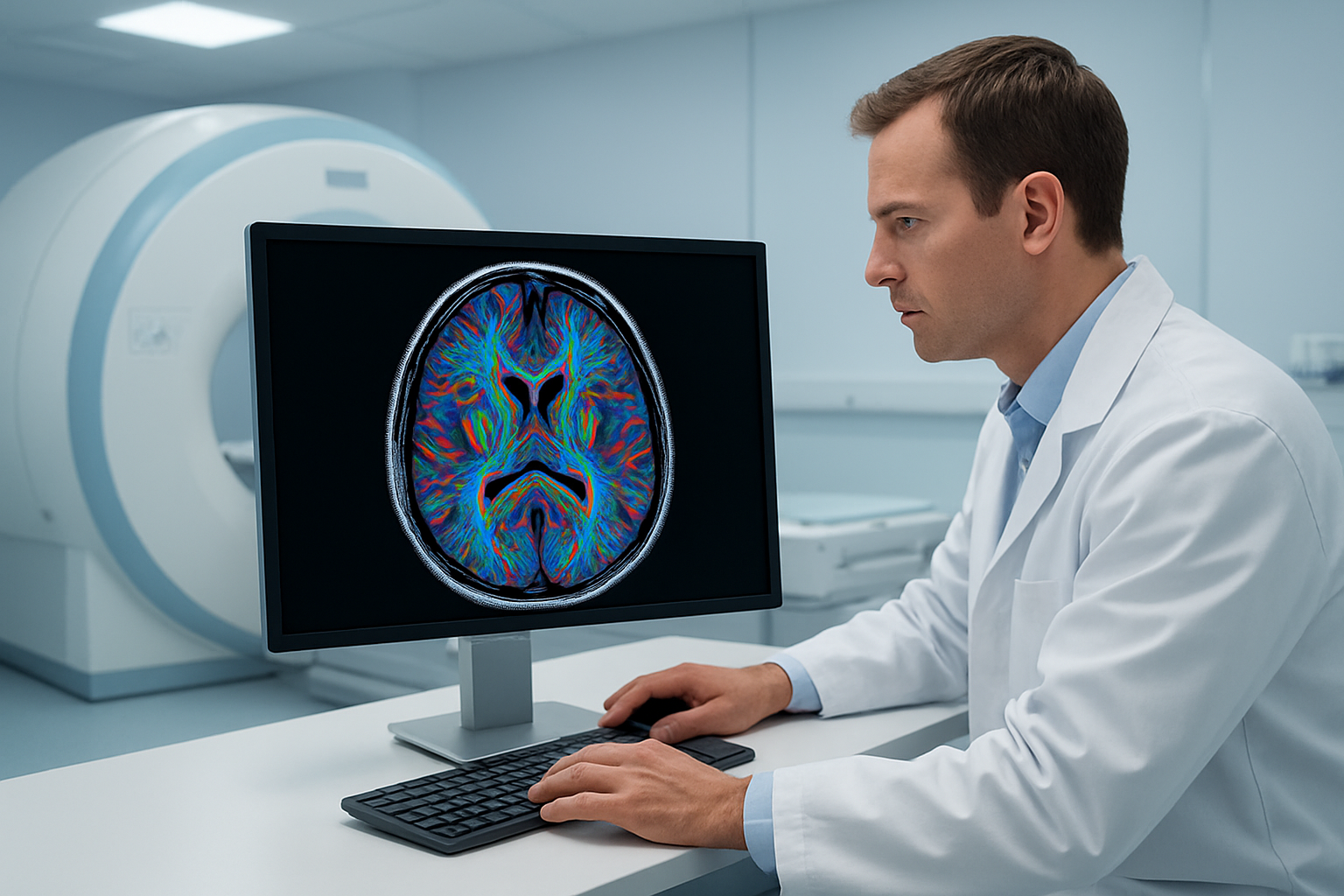
MRI brain diffusion technology captures something remarkable that standard MRI scans miss entirely – the microscopic movement of water molecules within your brain tissue. Every second, billions of water molecules bounce around randomly inside your brain cells and between them, creating what scientists call Brownian motion. Diffusion weighted imaging tracks these tiny movements to create detailed maps of your brain’s internal structure.
When brain tissue is healthy, water molecules move freely in all directions. Think of it like people walking through an open park – they can wander wherever they want. But when something goes wrong, like during a stroke or when scar tissue forms, these pathways become blocked or restricted. The water molecules can’t move as freely, similar to how people would struggle to move through a crowded hallway compared to that open park.
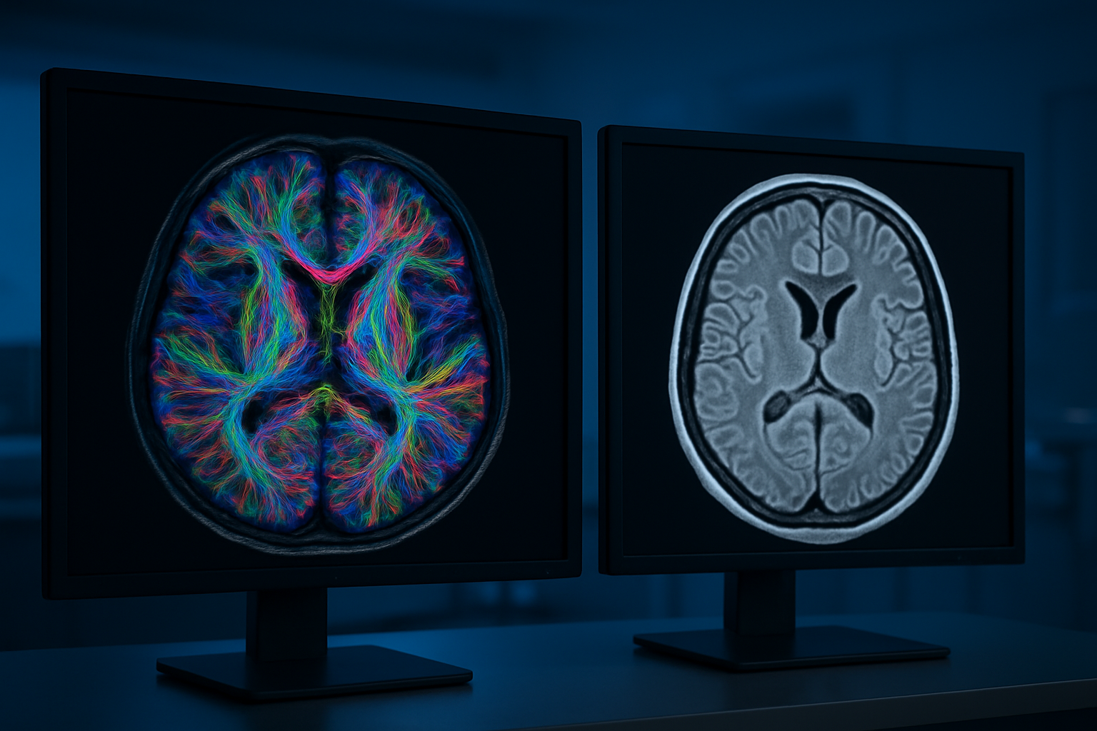
Diffusion tensor imaging, an advanced form of this technology, goes even further by measuring which directions water prefers to move. In healthy white matter tracts – the brain’s information highways – water moves more easily along the nerve fibers rather than across them. This directional preference reveals the integrity and organization of these crucial neural pathways.
The scan creates incredibly detailed images by applying magnetic gradients in multiple directions, typically six or more, allowing radiologists to build comprehensive maps of tissue structure that would be impossible to see with conventional imaging alone.
How Water Movement Reveals Brain Health
Water molecule behavior acts as a sensitive early warning system for brain problems, often detecting issues before they show up on regular MRI scans. When brain cells are damaged or stressed, they swell up and water movement becomes restricted within minutes. This makes brain MRI with DWI incredibly valuable for stroke diagnosis MRI, as it can identify affected tissue within the critical first few hours when treatment options are most effective.
Different types of brain tissue create unique water movement patterns. Gray matter, which contains most of your brain’s nerve cell bodies, allows fairly random water movement in all directions. White matter, packed with long nerve fibers wrapped in fatty insulation, channels water movement along these fiber bundles like water flowing through garden hoses.
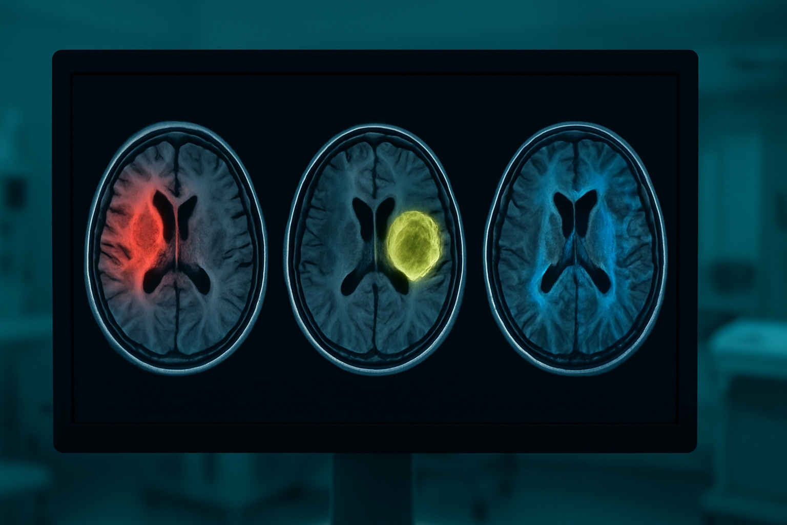
When disease strikes, these patterns change dramatically. Tumors often trap water molecules, creating bright spots on diffusion images. Inflammation from multiple sclerosis disrupts the organized structure of white matter, causing water to leak where it shouldn’t. Even subtle changes from conditions like Alzheimer’s disease alter water movement patterns, potentially allowing doctors to spot problems years before symptoms appear.
The technology also reveals how well different brain regions connect with each other. Damaged connections between areas show up as disrupted water flow patterns, helping doctors understand not just where problems exist, but how they might affect brain function and communication between different regions.
Medical Conditions Diagnosed Through Diffusion MRI
Detecting Acute Stroke Within Critical Time Windows
Diffusion weighted imaging plays a crucial role in emergency stroke diagnosis, often making the difference between effective treatment and permanent brain damage. When someone experiences a stroke, brain tissue begins dying within minutes due to lack of oxygen and nutrients. DWI brain scan results can detect these changes incredibly early—sometimes within 30 minutes of stroke onset—long before conventional MRI sequences show any abnormalities.
The magic happens because diffusion MRI measures how water molecules move through brain tissue. In healthy brain tissue, water moves freely in all directions. During a stroke, brain cells swell and restrict this normal water movement, creating a distinct signal that appears bright on diffusion images. This early detection capability gives doctors precious time to administer clot-busting medications or perform emergency procedures to restore blood flow.
Emergency room physicians rely heavily on stroke diagnosis MRI to distinguish between different types of strokes. Ischemic strokes, caused by blocked blood vessels, show restricted diffusion in affected areas. Hemorrhagic strokes, caused by bleeding, display different patterns that help doctors choose appropriate treatments. The speed and accuracy of diffusion imaging have revolutionized stroke care, allowing treatment decisions within the critical “golden hour” when interventions are most effective.
Identifying Brain Tumors and Their Characteristics
Brain lesion detection through diffusion MRI provides detailed information that goes far beyond simply finding abnormal tissue. Different types of brain tumors have unique diffusion characteristics that help doctors determine their nature, aggressiveness, and treatment approach without invasive procedures.
High-grade malignant tumors typically show restricted diffusion because their rapidly dividing cells create densely packed tissue that limits water movement. Low-grade tumors often demonstrate increased diffusion as their looser cellular structure allows more freedom for water molecules. This distinction helps oncologists plan treatment strategies and predict patient outcomes.
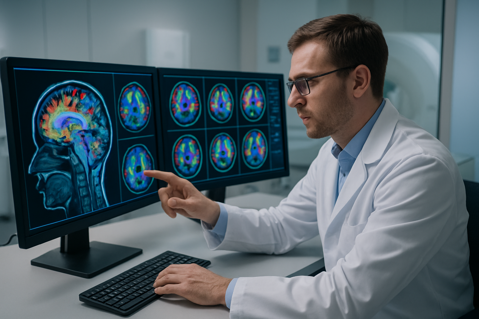
Diffusion tensor imaging takes this analysis even further by mapping the brain’s white matter tracts—the highways that connect different brain regions. When tumors grow near these critical pathways, DTI shows exactly how the tumor affects normal brain connections. Neurosurgeons use this information to plan operations that remove tumors while preserving essential functions like speech, movement, and memory.
Diffusion imaging also helps monitor treatment response. As chemotherapy or radiation therapy works, tumor cells die and tissue structure changes, creating different diffusion patterns. Doctors can track these changes over time to see if treatments are working, often detecting improvements weeks before tumors shrink enough to see on regular MRI scans. This early feedback allows for quick treatment adjustments when needed.
Preparing for Your MRI Brain Diffusion Study
Essential Pre-Scan Safety Screening Requirements
Before your diffusion MRI brain scan, your medical team will conduct a thorough safety screening to ensure the procedure is safe and appropriate for you. This screening process is critical because the powerful magnetic field used in diffusion weighted imaging can interact with certain metals and medical devices.
You’ll need to complete a detailed questionnaire about your medical history, including any previous surgeries, implants, or medical devices. The technologist will ask about pacemakers, cochlear implants, aneurysm clips, or any metal fragments that might be in your body. Even items like permanent makeup, tattoos with metallic ink, or certain types of dental work require disclosure, as these can sometimes interfere with the imaging quality or pose safety risks.
Pregnancy screening is mandatory for women of childbearing age, as the effects of MRI on developing fetuses aren’t fully understood. If you’re pregnant, your doctor will carefully weigh the benefits against potential risks before proceeding with the diffusion tensor imaging.
You’ll also need to remove all metal objects before entering the scanner room. This includes jewelry, watches, hearing aids, dentures with metal components, and clothing with metal fasteners. The facility will provide you with metal-free hospital gowns and lockers for your personal belongings.
Claustrophobia concerns should be discussed during screening. If you have anxiety about enclosed spaces, your doctor might prescribe mild sedation or recommend an open MRI facility if available for brain MRI with DWI.
What to Expect During the 45-60 Minute Procedure
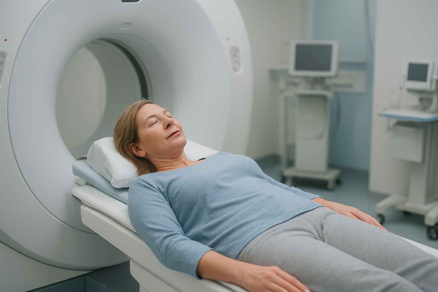
Your diffusion MRI preparation continues once you enter the scanning room. The MRI technologist will help position you on the movable table that slides into the cylindrical scanner. You’ll lie on your back with your head secured in a specialized coil designed to capture detailed brain images. Small foam pads or cushions may be placed around your head to minimize movement, which is crucial for obtaining high-quality diffusion weighted imaging results.
The scanner itself resembles a large, hollow tube with a powerful magnetic field. During the diffusion tensor imaging sequences, you’ll hear various loud knocking, tapping, and buzzing sounds as the machine captures images from different angles and with varying magnetic gradients. These sounds are completely normal and indicate that the scanner is working properly to detect water molecule movement in your brain tissue.
Communication with your technologist remains open throughout the procedure via an intercom system. You’ll receive earplugs or headphones to reduce noise, and some facilities offer music to help you relax. A call button will be placed in your hand so you can alert the staff if you experience any discomfort or need assistance.
The actual neuroimaging diffusion study involves multiple imaging sequences. Standard anatomical images are captured first, followed by the specialized diffusion sequences that measure water molecule movement in different directions. These diffusion measurements are what make this type of brain MRI particularly valuable for stroke diagnosis MRI and brain lesion detection.
Movement during scanning can significantly impact image quality, so staying as still as possible is essential. The technologist will remind you when particularly important sequences are beginning and may ask you to hold your breath briefly for certain images, though most brain diffusion sequences don’t require breath-holding.
Interpreting Your Diffusion Study Results
Understanding Apparent Diffusion Coefficient Maps
When your radiologist examines your MRI brain diffusion study, they’ll spend considerable time analyzing the Apparent Diffusion Coefficient (ADC) maps. These specialized images show how easily water molecules move through different areas of your brain tissue. Think of water molecules like tiny ping-pong balls bouncing around – in healthy brain tissue, they move freely, but when something’s wrong, their movement becomes restricted.
ADC maps appear in grayscale, where brighter areas indicate higher water movement and darker regions show restricted diffusion. Your doctor looks for these dark spots because they often signal acute problems like strokes, where brain cells are swelling and blocking normal water flow. The ADC values are measured in specific units, with normal brain tissue typically showing values between 700-1000 on the measurement scale.
Areas of recent stroke damage appear particularly dark on ADC maps because the affected brain cells swell up, creating barriers that prevent water molecules from moving normally. This restriction happens within minutes of a stroke, making diffusion weighted imaging incredibly valuable for early detection. Your radiologist will compare different brain regions, looking for asymmetry between the left and right sides of your brain, which could indicate developing problems.
Reading Diffusion-Weighted Imaging Findings
Diffusion-weighted imaging findings tell a story about what’s happening inside your brain at the cellular level. When your radiologist reviews your DWI brain scan results, they’re looking for bright white spots that stand out against the darker background of normal brain tissue. These bright areas, called hyperintense signals, can indicate various conditions depending on their location and appearance pattern.
Fresh strokes typically show up as bright white areas on the initial diffusion images, paired with those dark spots we discussed on the ADC maps. This combination – bright on DWI and dark on ADC – creates what doctors call “diffusion restriction,” which is like a red flag pointing to acute brain injury. The timing matters too; these changes can appear within 30 minutes of a stroke, long before they’d show up on standard MRI sequences.
Your brain MRI interpretation will also consider the shape and distribution of any abnormal signals. Stroke patterns often follow specific blood vessel territories, appearing in wedge-shaped areas. Brain lesion detection becomes easier when radiologists can match these patterns to known anatomical regions. Sometimes, small bright spots might represent old, tiny strokes you never noticed, or they could indicate other conditions like infections or tumors.
The radiologist will also examine areas where diffusion appears increased rather than restricted. These regions might indicate older injuries where brain tissue has been replaced by fluid, or areas of inflammation that are resolving. Your complete neuroimaging diffusion study provides a timeline of brain health, showing both recent changes and longer-term patterns.
Benefits and Limitations of Diffusion MRI
Superior Early Detection Capabilities for Stroke
Diffusion weighted imaging stands out as a game-changer when it comes to catching strokes in their earliest stages. While traditional MRI scans might miss acute stroke symptoms in the first few hours, diffusion MRI can detect cellular changes within minutes of a stroke occurring. This early detection capability literally means the difference between life and death, or between full recovery and permanent disability.
The magic happens at the cellular level. When brain cells lose their normal function due to lack of blood flow, water molecules begin moving differently within the tissue. DWI brain scan picks up these microscopic changes long before they show up on standard imaging. Emergency room doctors can spot a stroke within the critical “golden hour” window, allowing them to start life-saving treatments like clot-busting medications or mechanical thrombectomy procedures.
What makes this technology particularly valuable is its ability to distinguish between old and new strokes. Many patients have had previous mini-strokes or silent strokes that don’t show symptoms. Brain MRI with DWI clearly highlights fresh damage, helping doctors focus their treatment on the active problem rather than getting confused by old scar tissue.
The speed factor cannot be overstated. Traditional CT scans often come back normal in the early hours after a stroke, leaving doctors and families in a frustrating waiting game. Diffusion weighted imaging eliminates this uncertainty by providing definitive answers when time matters most.
Enhanced Tumor Characterization Accuracy
Brain lesion detection reaches new levels of precision with diffusion MRI technology. This advanced imaging doesn’t just find tumors – it provides detailed information about their internal structure and behavior. Doctors can tell the difference between aggressive cancers and slower-growing masses, which directly impacts treatment decisions and patient prognosis.
The technique measures how freely water molecules move within different types of tissue. Cancer cells are typically packed more tightly than normal brain tissue, restricting water movement. Diffusion tensor imaging captures these differences, creating detailed maps that show tumor boundaries with remarkable clarity. This information helps surgeons plan operations more precisely, knowing exactly where healthy brain tissue ends and the tumor begins.
Distinguishing between tumor types becomes much more accurate with diffusion imaging. High-grade gliomas, metastatic tumors, and benign masses each have characteristic diffusion patterns. Radiologists can often make educated guesses about tumor grade and type before any biopsy, helping oncology teams prepare appropriate treatment plans from day one.
The technology also excels at monitoring treatment response. As chemotherapy or radiation therapy takes effect, tumor cells begin dying and water movement patterns change. Neuroimaging diffusion study can detect these improvements weeks before changes become visible on standard MRI sequences, giving both doctors and patients earlier feedback about whether treatments are working effectively.
MRI brain diffusion studies give doctors a powerful way to see how water moves through your brain tissue, helping them spot problems that regular MRI scans might miss. These scans are especially good at catching strokes early, finding brain tumors, and checking for conditions that affect the brain’s white matter. The test itself is pretty straightforward – you’ll lie still in the MRI machine for about 30-45 minutes, and the main thing to remember is removing any metal objects beforehand.
When your results come back, your doctor will look at detailed maps showing how healthy your brain tissue is and whether there are any areas of concern. While diffusion MRI isn’t perfect and has some limits – like being sensitive to patient movement – it’s become an essential tool for brain health. If your doctor recommends this type of scan, you can feel confident knowing it’s a safe, non-invasive way to get important information about what’s happening inside your brain.