

“What is an F18 Choline PET CT Scan? Purpose, Procedure & Clinical Benefits”
F18 Choline PET CT Scan: Advanced Imaging for Cancer Detection
An F18 choline PET CT scan combines two powerful imaging technologies to help doctors spot cancer cells, especially in prostate cancer cases. This specialized test is designed for cancer patients, their families, and anyone wanting to understand this advanced diagnostic tool.
Choline PET CT imaging works by tracking a radioactive tracer that cancer cells absorb more readily than healthy tissue. The F18 choline positron emission tomography scan provides detailed pictures that help doctors see exactly where cancer might be hiding in your body.
We’ll walk you through how F18 choline PET CT technology actually works and what medical conditions doctors can diagnose with this scan. You’ll also learn about the key benefits of choosing this imaging method and get practical tips for preparing for your appointment so you feel confident going in.
Understanding F18 Choline PET CT Technology
How F18 Choline Tracer Works in Your Body
F18 choline works like a molecular detective inside your body, targeting areas where cells are rapidly multiplying or metabolically active. When you receive the fluorine-18 choline PET scan injection, this radioactive tracer mimics natural choline, an essential nutrient that cells need for building their membranes and maintaining normal function.
Cancer cells have an insatiable appetite for choline because they’re constantly dividing and growing. The F18 choline tracer gets absorbed by these hyperactive cells at much higher rates than normal tissue. This creates bright spots on the scan images, making it easier for doctors to identify problem areas.
The tracer travels through your bloodstream within minutes of injection, quickly reaching organs and tissues throughout your body. Prostate cancer cells, in particular, show exceptional uptake of choline PET CT imaging compounds, which makes this scan especially valuable for detecting both primary tumors and metastases that might be missed by other imaging methods.
What makes F18 choline unique compared to other PET tracers is its specific affinity for certain types of cancer cells. While FDG (fluorodeoxyglucose) tracers look for sugar consumption, choline tracers focus on membrane synthesis – a different but equally telling sign of malignant activity. The radioactive fluorine-18 component has a relatively short half-life of about 110 minutes, ensuring minimal radiation exposure while providing excellent image quality.
Advanced Imaging Capabilities of PET CT Combination
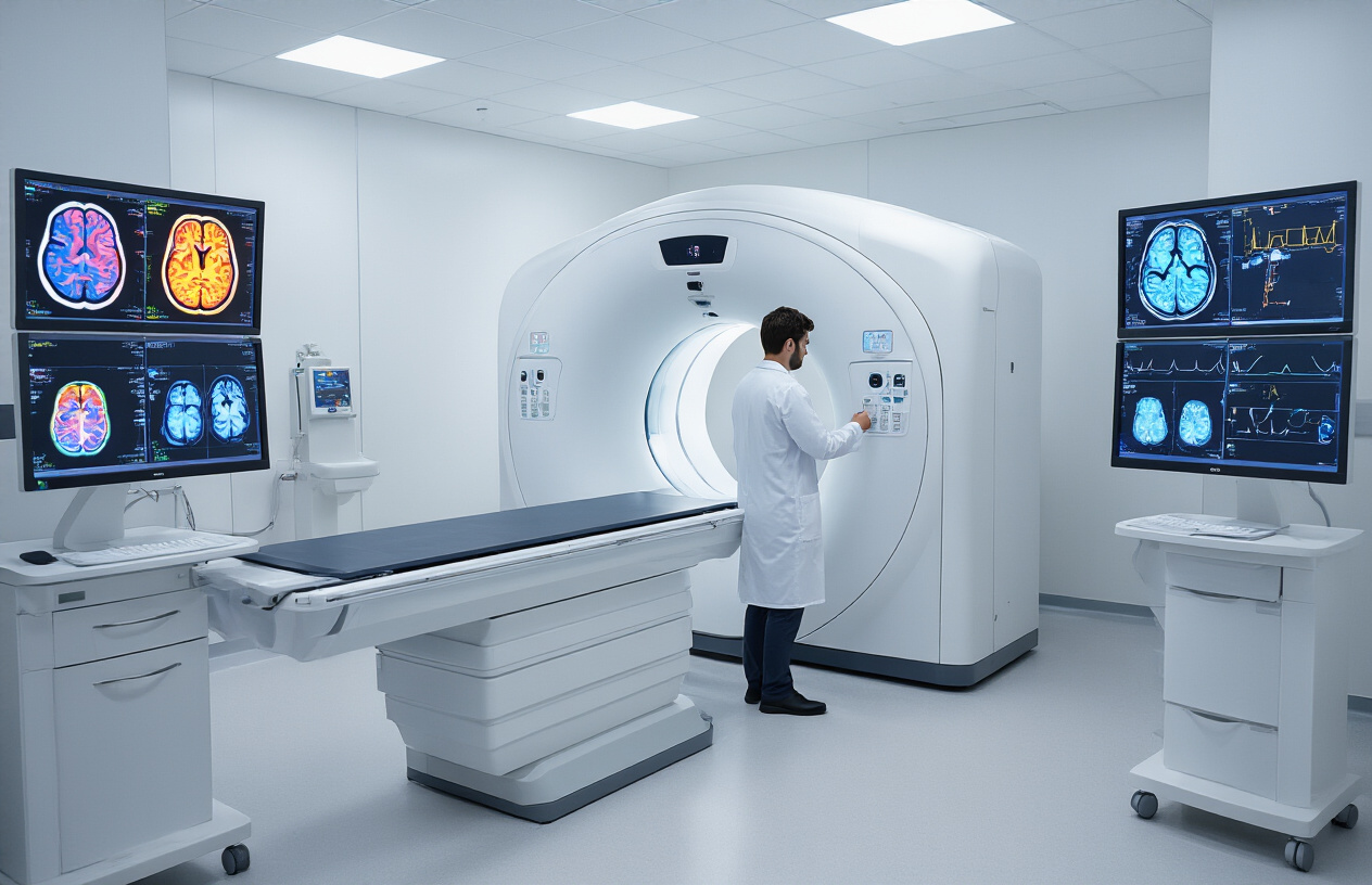
The combination of PET and CT technology in choline PET CT diagnosis creates a powerful dual-perspective view that neither imaging method could achieve alone. The PET component captures the metabolic activity of the F18 choline tracer, showing exactly where abnormal cellular processes are occurring. Meanwhile, the CT portion provides detailed anatomical structure, creating a roadmap of your body’s organs and tissues.
This fusion approach means doctors can see not just that something unusual is happening (from the PET data) but precisely where it’s happening in relation to your anatomy (from the CT data). The fluorine-18 choline PET scan procedure captures images from multiple angles as you lie still on the scanning table, creating cross-sectional views that can be reconstructed into three-dimensional images.
Modern PET CT scanners can detect metabolic changes at the cellular level, often before structural changes become visible on traditional imaging. This early detection capability is particularly valuable for monitoring treatment response and detecting cancer recurrence. The advanced computer processing combines both image sets pixel by pixel, producing fused images where metabolic hotspots are overlaid onto anatomical landmarks.
The spatial resolution of current F18 choline positron emission tomography systems can identify lesions as small as 4-6 millimeters, providing remarkable precision for treatment planning. This level of detail helps surgeons plan procedures more effectively and allows oncologists to target radiation therapy with pinpoint accuracy while sparing healthy surrounding tissue.
Medical Conditions Diagnosed with F18 Choline PET CT
Prostate Cancer Detection and Staging
F18 choline PET CT imaging has become a game-changer for men dealing with prostate cancer concerns. This advanced scanning technique excels at detecting cancer cells that standard imaging methods might miss, especially in cases where prostate-specific antigen (PSA) levels remain elevated after initial treatment.
The choline PET CT procedure works by targeting cancer cells’ increased need for choline, a nutrient essential for cell membrane production. Prostate cancer cells absorb significantly more choline than healthy tissue, making them light up like beacons on the scan. This makes F18 choline positron emission tomography particularly valuable for identifying recurrent disease, even when conventional scans show nothing unusual.
Doctors often recommend fluorine-18 choline PET scan when patients experience biochemical recurrence – a situation where PSA levels rise again after surgery or radiation therapy, but traditional imaging can’t pinpoint where the cancer has returned. The scan can detect small metastases in lymph nodes, bones, and other organs that might be as tiny as a few millimeters.
Staging accuracy improves dramatically with choline PET CT imaging compared to conventional methods. The scan helps determine whether cancer has spread beyond the prostate gland, influencing treatment decisions significantly. Patients with localized recurrence might be candidates for targeted radiation, while those with widespread disease may need systemic therapy.
The precision of F18 choline PET CT benefits both patients and healthcare providers by reducing uncertainty and enabling more personalized treatment approaches.
Liver Tumor Identification and Assessment
Liver tumors present unique diagnostic challenges that choline PET CT diagnosis addresses effectively. The liver’s complex metabolism and dual blood supply can make it difficult to distinguish between benign and malignant lesions using traditional imaging alone. F18 choline PET CT scan technology provides crucial metabolic information that transforms how doctors evaluate liver abnormalities.
Primary liver cancers, including hepatocellular carcinoma, show increased choline uptake patterns that help differentiate them from benign conditions like hemangiomas or focal nodular hyperplasia. The scan’s ability to combine metabolic activity with detailed anatomical images gives doctors a comprehensive view of liver health that neither technique could provide independently.
Metastatic disease detection in the liver becomes more accurate with choline PET CT imaging. Cancer cells that have spread from other organs, particularly colorectal, breast, or neuroendocrine tumors, often demonstrate characteristic choline uptake patterns. This helps doctors determine the extent of disease spread and plan appropriate treatment strategies.
The scan proves especially valuable when evaluating treatment response in liver tumors. Changes in choline uptake can indicate whether chemotherapy, targeted therapy, or other treatments are working effectively, sometimes before conventional imaging shows size changes. This early insight allows for treatment adjustments that can improve patient outcomes.
Pre-surgical planning benefits tremendously from the detailed metabolic mapping that choline PET CT provides, helping surgeons identify the precise location and extent of liver lesions before operating.
Benefits of Choosing F18 Choline PET CT Scanning
Early Disease Detection Before Symptoms Appear
F18 choline PET CT scanning excels at catching diseases in their earliest stages, often years before patients experience any noticeable symptoms. This advanced imaging technology detects metabolic changes at the cellular level, particularly identifying areas where choline uptake is abnormally high. Cancer cells have an increased demand for choline to build their cell membranes, making this radiotracer incredibly sensitive to malignant activity.
When cancer cells begin multiplying, they consume choline at rates far exceeding normal tissue. The F18 choline positron emission tomography picks up these metabolic signatures long before tumors grow large enough to cause physical symptoms or show up on conventional imaging studies. This early detection capability proves especially valuable for prostate cancer patients who may have rising PSA levels but no other signs of disease recurrence.
The ability to spot disease activity before symptoms develop transforms treatment outcomes significantly. Doctors can initiate targeted therapies when cancer burdens are minimal and before the disease spreads to distant organs. Patients benefit from less aggressive treatment protocols and higher success rates when interventions begin during these early stages.
Precise Tumor Location and Size Measurement
Choline PET CT imaging delivers exceptional accuracy in pinpointing exact tumor locations throughout the body. The dual-modality approach combines metabolic information from the PET component with detailed anatomical structures from the CT scan, creating comprehensive three-dimensional maps of disease activity. This precision proves invaluable when planning surgical procedures, radiation therapy, or targeted treatments.
The F18 choline PET CT benefits extend to measuring tumor dimensions with remarkable accuracy. Unlike conventional imaging methods that may miss small lesions or struggle to differentiate between scar tissue and active disease, choline PET scanning highlights metabolically active areas with crystal-clear definition. Radiologists can measure tumors down to millimeter precision and track changes in size over time.
This detailed mapping capability becomes especially important for complex cases involving multiple tumor sites or disease recurrence in previously treated areas. Surgeons use these precise measurements to plan minimally invasive procedures, while radiation oncologists rely on the exact coordinates to deliver focused beam therapy that spares healthy tissue. The fluorine-18 choline PET scan creates treatment roadmaps that maximize therapeutic effectiveness while minimizing side effects.
Preparing for Your F18 Choline PET CT Appointment

Dietary Restrictions and Fasting Requirements
Your F18 choline PET CT preparation involves specific dietary guidelines that directly impact the quality of your scan results. Unlike traditional PET scans that require extended fasting periods, choline PET CT imaging has more moderate dietary restrictions that you can easily follow.
You’ll need to avoid eating for at least 4-6 hours before your F18 choline positron emission tomography appointment. This fasting period helps reduce background noise in your images and allows the choline tracer to concentrate more effectively in areas of interest. Water intake remains unrestricted and actually encouraged throughout your preparation period to stay properly hydrated.
Certain foods can interfere with your choline PET scan procedure results. Avoid high-fat meals for 24 hours before your appointment, as fatty foods can affect how your body processes the radiotracer. Alcohol consumption should also stop 24 hours prior to your scan since it can alter cellular metabolism patterns that the imaging relies on to detect abnormalities.
Diabetic patients need special attention during choline PET CT preparation. Your blood sugar levels should remain stable, so follow your regular medication schedule unless your healthcare provider gives different instructions. The fasting requirements are typically shorter than glucose-based PET scans, making the process more manageable for people with diabetes.
Your medical team will provide specific timing instructions based on your appointment schedule. Most patients find these dietary restrictions straightforward compared to other imaging procedures.
Medication Guidelines Before Your Scan
Managing your medications properly before your F18 choline PET CT scan ensures accurate results and your safety during the procedure. Most of your regular medications can continue as prescribed, but several important exceptions require careful attention.
Blood thinners and anticoagulants typically don’t need adjustment for choline PET CT imaging since the procedure doesn’t involve invasive techniques. However, inform your medical team about all blood-thinning medications including warfarin, aspirin, or newer anticoagulants. They’ll confirm whether any temporary adjustments are needed based on your specific medical situation.
Hormone-related medications, particularly those affecting prostate function, may require temporary modification. Since F18 choline imaging prostate cancer is a common application, certain hormonal therapies could potentially influence tracer uptake patterns. Your oncologist will coordinate with the imaging team to determine if brief medication pauses are beneficial for optimal image quality.
Pain medications and anti-inflammatory drugs can usually continue unchanged. However, high-dose steroids or immunosuppressive medications might affect cellular metabolism in ways that could impact your scan interpretation. Discuss these medications specifically during your pre-scan consultation.
Supplements and over-the-counter medications deserve attention too. Stop taking any supplements containing choline or lecithin for 48 hours before your appointment, as these could interfere with the radiotracer distribution. Common multivitamins often contain these compounds, so check ingredient labels carefully or temporarily discontinue all supplements to avoid any potential interference with your fluorine-18 choline PET scan results.
Interpreting Your F18 Choline PET CT Results

Understanding Normal vs Abnormal Uptake Patterns
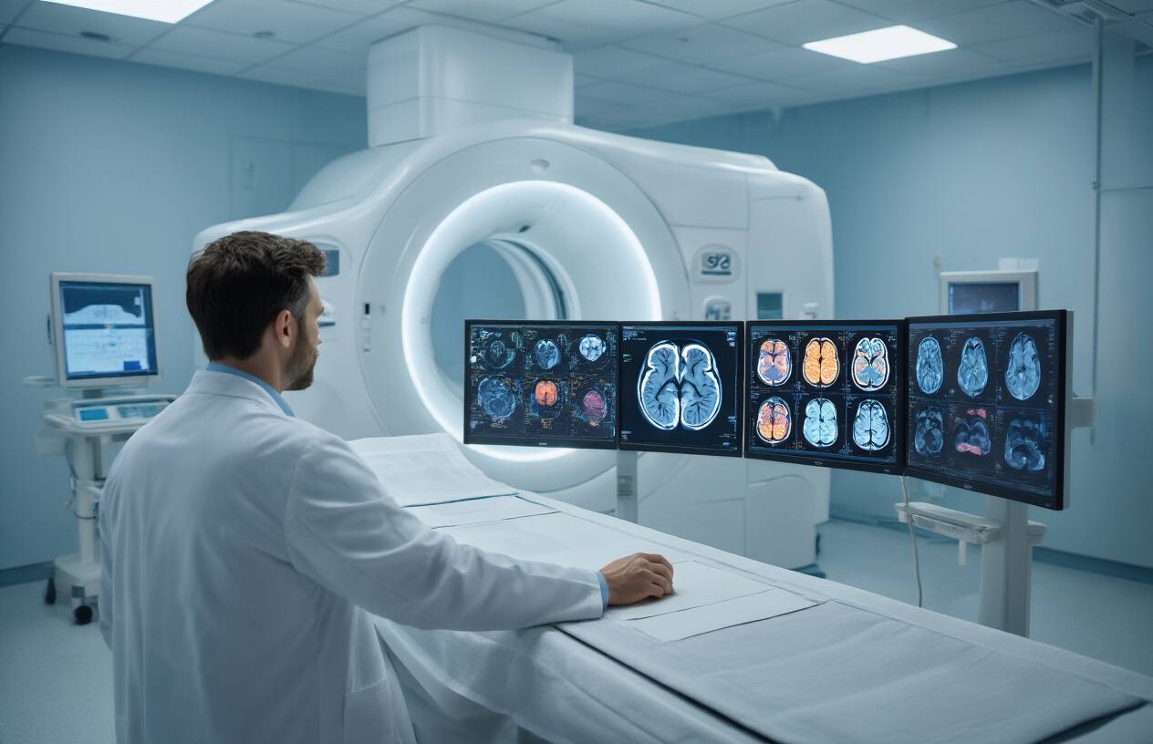
When your doctor reviews your F18 choline PET CT scan, they’re looking for specific patterns that tell a story about what’s happening inside your body. Normal tissue typically shows minimal choline uptake because healthy cells don’t need as much of this building block for rapid growth. Your brain, liver, and kidneys will naturally show some choline activity since these organs are metabolically active, but the uptake remains within expected ranges.
Abnormal uptake patterns appear as bright spots or areas of intense activity on your choline PET CT imaging. Cancer cells are metabolic powerhouses that consume choline at much higher rates than normal tissue. This creates a clear contrast on your scan, making suspicious areas stand out like beacons. Prostate cancer, in particular, shows strong affinity for choline, which makes F18 choline imaging prostate cancer detection highly effective.
Your radiologist will measure something called SUV (standardized uptake value) to quantify how much choline different areas are absorbing. Higher SUV values typically indicate more aggressive cellular activity. Areas with SUV values significantly above background levels warrant closer examination and often represent areas of concern.
The scan also reveals important anatomical details through the CT component, helping doctors pinpoint exactly where abnormal uptake is occurring. This combination of metabolic and structural information provides a comprehensive picture that guides your medical team’s next steps.
How Results Guide Your Treatment Decisions
Your choline PET CT results interpretation directly influences your treatment path. When the scan shows localized uptake confined to the prostate, your oncologist might recommend targeted treatments like surgery or focused radiation. This precise localization helps spare healthy surrounding tissue while effectively treating the cancer.
If your F18 choline positron emission tomography reveals uptake in lymph nodes or distant organs, this information completely changes your treatment approach. Your medical team will shift focus to systemic treatments that can address cancer throughout your body rather than just local therapies.
The intensity of choline uptake also matters for treatment planning. Highly aggressive tumors that show intense uptake might require more aggressive treatment protocols, while areas with modest uptake might respond well to less intensive approaches. Your oncologist uses these uptake patterns to predict how your cancer might behave and respond to different treatments.
Choline PET CT diagnosis results also help monitor treatment effectiveness. Follow-up scans can show whether treatments are working by demonstrating reduced choline uptake in previously active areas. This real-time feedback allows your medical team to adjust treatment plans quickly if needed, switching to more effective therapies or modifying doses based on your tumor’s response.
The detailed anatomical information from your scan helps surgeons plan precise procedures, while radiation oncologists use the metabolic data to target treatment fields more accurately, maximizing cancer destruction while protecting healthy tissue.
F18 Choline PET CT scans offer doctors a powerful way to spot prostate cancer and other conditions that might hide from regular imaging tests. This advanced technology combines two different scanning methods to create detailed pictures of what’s happening inside your body at the cellular level. The scan works especially well for finding cancer that has spread or come back after treatment.
Getting ready for your scan is pretty straightforward – just follow your doctor’s instructions about eating and medications. The results will help your medical team make better decisions about your treatment plan. If your doctor recommends this scan, don’t worry about the process. It’s safe, painless, and could provide the exact information needed to keep you healthy.