
“Wondering what MRI DTI is? It’s an advanced scan that maps the brain’s nerve fibers in 3D.”
MRI Diffusion Tensor Imaging (DTI): Advanced Brain Imaging That Maps White Matter Pathways
MRI DTI represents a breakthrough in brain imaging that goes beyond traditional scans to reveal the brain’s internal wiring. This diffusion MRI technique tracks water movement along nerve fibers, creating detailed maps of white matter pathways that were previously invisible to doctors and researchers.
This guide is designed for healthcare professionals, medical students, researchers, and patients who want to understand how DTI brain scans work and why they matter. You’ll discover how this imaging method is changing the way we diagnose neurological conditions and study the brain.
We’ll explore DTI’s scientific foundation and explain how the technology captures brain fiber tracking data. You’ll learn about DTI clinical applications that are transforming patient diagnosis, from detecting early signs of Alzheimer’s to planning brain surgery. Finally, we’ll cover DTI’s expanding role in neuroscience research and the technical considerations that ensure reliable results for both clinical care and scientific discovery.
Understanding DTI Technology and Its Scientific Foundation

How DTI measures water molecule movement in brain tissue
DTI brain scan technology works by tracking how water molecules naturally move through brain tissue. Water molecules don’t just sit still – they’re constantly bouncing around, moving in different directions. In healthy brain tissue, this movement isn’t random. Water molecules prefer to move along specific pathways, especially within the white matter tracts that connect different brain regions.
Think of it like water flowing through a garden hose versus water spreading out on a flat surface. In the brain’s white matter, water molecules tend to move along the length of nerve fibers rather than perpendicular to them. This directional preference happens because the structure of nerve fibers – with their protective myelin sheaths and organized cellular architecture – creates natural highways for water movement.
The diffusion tensor imaging process captures this directional movement by applying magnetic field gradients in multiple directions. The MRI scanner measures how far water molecules travel in each direction during a specific time period. When water moves freely in all directions (isotropic diffusion), it suggests less organized tissue structure. When water movement shows a strong directional preference (anisotropic diffusion), it indicates well-organized, intact white matter tracts.
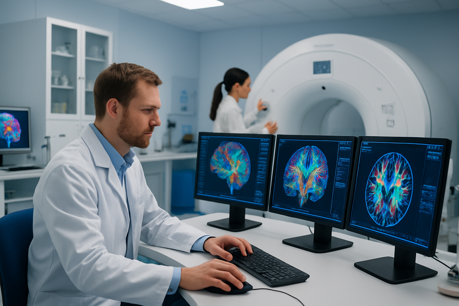
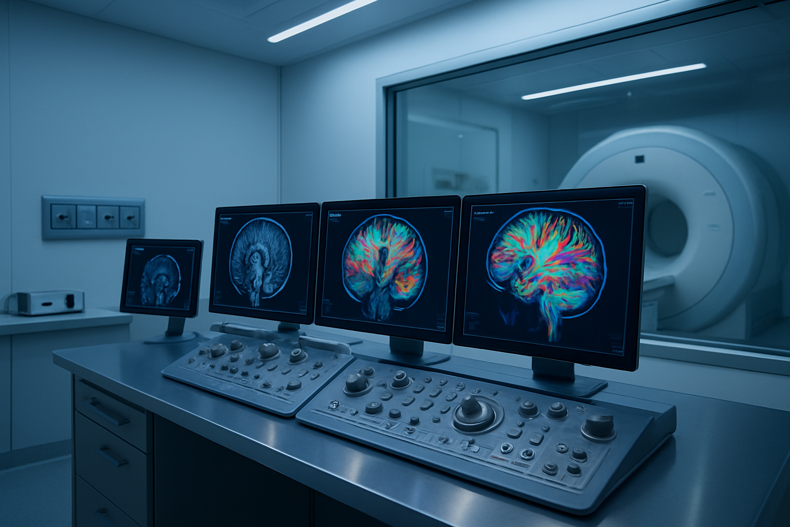
DTI creates detailed maps of these water movement patterns throughout the brain. The data gets processed into colorful brain images where different colors represent the primary direction of water movement – red for left-right, green for front-back, and blue for up-down movement. This allows doctors and researchers to visualize the brain’s internal wiring system in unprecedented detail.
Key differences between DTI and conventional MRI scanning
Standard MRI scans create images based on tissue density and water content, showing anatomical structures in grayscale. These conventional scans excel at identifying tumors, bleeding, or structural abnormalities but can’t reveal the intricate details of white matter organization. Regular MRI is like taking a photograph of a city – you can see the buildings and major landmarks, but you can’t see the traffic patterns or road conditions.
DTI technique goes several steps beyond conventional imaging by measuring the microscopic movement of water molecules. While traditional MRI might show that brain tissue looks normal, DTI can detect subtle changes in white matter integrity that aren’t visible on standard scans. This makes DTI particularly valuable for detecting early-stage neurological conditions or tracking disease progression.
The scanning process itself differs significantly between the two approaches. Conventional MRI typically takes 15-30 minutes and uses standard pulse sequences. DTI requires longer scan times, usually 45-60 minutes, because the scanner must apply magnetic gradients in at least six different directions to calculate water movement patterns. Some advanced DTI protocols use 30 or more gradient directions for higher precision.
Image interpretation also varies dramatically. Radiologists can quickly read conventional MRI scans using established patterns they’ve learned through years of training. DTI images require specialized software and expertise in diffusion MRI technique analysis. The resulting images show brain fiber tracking and white matter integrity maps that reveal information completely invisible to traditional imaging methods.
DTI’s sensitivity to microscopic changes makes it both more informative and more challenging to interpret than conventional MRI scanning.
Clinical Applications That Transform Patient Diagnosis

Detecting White Matter Damage in Stroke and Traumatic Brain Injury
DTI brain scan technology has revolutionized how doctors diagnose and monitor white matter damage following stroke and traumatic brain injury (TBI). Unlike traditional MRI scans that might miss subtle fiber damage, diffusion tensor imaging reveals disruptions in the brain’s white matter tracts with remarkable precision.
When stroke occurs, blood flow interruption damages neural pathways that appear normal on conventional imaging. DTI detects these microscopic changes by measuring how water molecules move along nerve fibers. Damaged areas show altered diffusion patterns, allowing clinicians to identify affected regions and predict recovery potential. This capability proves especially valuable for patients with mild cognitive symptoms following stroke, where standard imaging may appear normal despite significant functional impairment.
DTI clinical applications in traumatic brain injury offer similar diagnostic advantages. Following head trauma, shearing forces often tear white matter fibers throughout the brain. These diffuse axonal injuries frequently escape detection on standard CT or MRI scans, leaving patients with unexplained cognitive deficits. DTI mapping reveals these hidden injuries by highlighting areas where normal fiber architecture becomes disrupted.
The technology enables precise localization of damage, helping medical teams understand why specific cognitive functions remain impaired. For instance, damage to association fibers might explain memory problems, while injury to projection fibers could account for motor difficulties. This detailed mapping guides treatment planning and helps set realistic expectations for recovery, transforming patient care from guesswork to evidence-based medicine.
Monitoring Brain Tumor Growth and Treatment Response
White matter imaging through DTI provides critical insights into how brain tumors affect surrounding neural tissue and respond to treatment interventions. Unlike traditional imaging that focuses primarily on tumor size, MRI DTI reveals how malignancies infiltrate and disrupt normal white matter architecture.
Brain tumors rarely respect anatomical boundaries, often sending microscopic extensions along white matter pathways far beyond what conventional imaging can detect. DTI tractography maps these invasion patterns, showing surgeons exactly which neural pathways face compromise and helping plan surgical approaches that preserve critical functions. This information proves invaluable when tumors grow near eloquent brain regions responsible for speech, movement, or memory.
Diffusion MRI technique also excels at monitoring treatment response in ways that standard imaging cannot match. During chemotherapy or radiation therapy, tumor tissue often shows changes in water diffusion patterns before size reduction becomes visible. These early indicators help oncologists adjust treatment protocols quickly, potentially improving outcomes while reducing unnecessary exposure to ineffective therapies.
The technology distinguishes between treatment effects and tumor progression with greater accuracy than conventional methods. Radiation necrosis, for example, can appear identical to tumor recurrence on standard MRI but shows distinctly different diffusion characteristics on DTI. This differentiation capability prevents unnecessary surgical interventions and guides appropriate treatment decisions.
Brain fiber tracking capabilities allow physicians to monitor how treatments affect surrounding healthy tissue. Some therapies that successfully shrink tumors may simultaneously damage important neural connections. DTI tracking reveals these effects early, enabling treatment modifications that balance tumor control with preservation of neurological function, ultimately improving quality of life for cancer patients.
DTI’s Role in Advancing Neuroscience Research
Mapping neural connections to understand brain networks
DTI neuroscience research has opened up an entirely new way to see how our brains are wired together. Think of your brain as having billions of tiny highways connecting different regions – DTI brain scans can actually trace these pathways and show scientists exactly where they go and how strong they are.
White matter imaging through diffusion tensor imaging reveals the intricate network of nerve fibers that carry information between brain areas. Scientists can now create detailed maps showing how the frontal cortex connects to memory centers, or how visual areas link up with language regions. These brain fiber tracking capabilities have completely changed how researchers understand conditions like autism, where certain connections might be stronger or weaker than typical.
The technology has been particularly eye-opening in studying mental health disorders. Researchers have discovered that depression often involves disrupted connections between emotional processing areas and decision-making regions. Schizophrenia research has shown altered pathways in networks responsible for reality testing and social cognition. Even subtle changes in connectivity patterns can now be detected and studied.
Large-scale brain mapping projects use DTI to create comprehensive atlases of human brain connectivity. These maps help researchers identify which pathways are most critical for specific functions and how individual differences in brain wiring might affect behavior, learning abilities, and disease susceptibility. The data collected from thousands of DTI scans has created a foundation for understanding normal brain variation across populations.
Studying brain plasticity and recovery mechanisms
Brain plasticity research has been revolutionized by diffusion MRI techniques. Scientists can now track how neural pathways change and adapt over time, whether someone is learning a new skill, recovering from injury, or dealing with neurological conditions.
Stroke recovery studies using DTI have shown exactly how the brain reorganizes itself after damage. When traditional pathways are blocked, the brain creates new routes around the damaged areas. Researchers can watch these alternate pathways strengthen over weeks and months of rehabilitation. This knowledge has led to more targeted therapy approaches that specifically encourage the formation of beneficial new connections.
Learning and skill acquisition studies reveal fascinating changes in white matter structure. Musicians show enhanced connectivity in areas linking motor control with auditory processing. Medical students develop stronger pathways between memory regions during intense studying periods. Even simple motor learning tasks create measurable changes in brain fiber organization.
Childhood brain development research benefits enormously from DTI’s ability to track connectivity changes without invasive procedures. Scientists can follow how critical pathways mature during different developmental stages and identify when disruptions might have lasting effects. This research has practical implications for understanding developmental disorders and optimizing educational approaches.
Recovery from traumatic brain injury becomes much more predictable when doctors can see which pathways remain intact and which ones show potential for rebuilding. The diffusion tensor imaging data helps predict recovery outcomes and guide rehabilitation strategies toward the most promising therapeutic targets.
Practical Benefits for Patients and Healthcare Providers

Non-invasive assessment of brain structure and function
DTI brain scan technology offers patients a completely painless and radiation-free way to examine their brain’s internal wiring. Unlike traditional methods that might require invasive procedures or expose patients to harmful radiation, MRI DTI uses magnetic fields and radio waves to create detailed maps of white matter pathways. Patients simply lie still in the scanner for about 30-60 minutes while the machine captures thousands of images that reveal how water molecules move through brain tissue.
This non-invasive approach means patients can receive comprehensive brain assessments without surgical risks, recovery time, or the anxiety that comes with more invasive diagnostic procedures. Healthcare providers can visualize brain fiber tracking patterns that were previously invisible, allowing them to detect conditions like multiple sclerosis, stroke damage, traumatic brain injury, and various neurological disorders much earlier than before.
The comfort factor cannot be overstated. Patients appreciate that diffusion tensor imaging requires no contrast agents or special preparations in most cases. They can return to their normal activities immediately after the scan. For healthcare providers, this means they can schedule more patients per day and provide faster diagnoses, leading to quicker treatment decisions and better patient outcomes.
Improved surgical planning for brain and spine procedures
Neurosurgeons now rely heavily on DTI clinical applications to map critical brain pathways before operating. The technology shows exactly where important nerve bundles run through the brain, helping surgeons navigate around vital areas that control speech, movement, and memory. This precise mapping dramatically reduces the risk of accidentally damaging healthy brain tissue during tumor removal or other procedures.
Spine surgery has been revolutionized through white matter imaging capabilities. Surgeons can see how spinal cord fibers are organized and identify which pathways might be compressed or damaged. This information helps them plan the most effective surgical approach while minimizing potential complications. Patients benefit from shorter recovery times and better functional outcomes because surgeons can work with unprecedented precision.
The diffusion MRI technique also helps surgical teams predict how patients might recover after surgery. By analyzing the integrity of brain pathways before the operation, doctors can set realistic expectations and develop targeted rehabilitation plans. This proactive approach leads to better communication between medical teams and patients, reducing anxiety and improving overall satisfaction with surgical outcomes.
Healthcare providers report that DTI imaging has become essential for complex cases involving brain tumors near critical areas, arteriovenous malformations, and spinal cord injuries. The technology helps them distinguish between damaged and healthy tissue, leading to more conservative surgical approaches that preserve function while effectively treating the underlying condition.
Technical Considerations That Ensure Accurate Results
Optimal scanning parameters for different clinical scenarios
Getting the best DTI brain scan results requires careful attention to scanning parameters that match each specific clinical situation. When examining stroke patients, radiologists typically use higher spatial resolution settings with b-values around 1000 s/mm² to capture subtle white matter damage. The number of diffusion directions plays a crucial role – while 6 directions might work for basic screening, complex cases involving brain fiber tracking need at least 30 directions for reliable results.
Pediatric DTI imaging demands shorter acquisition times to prevent motion artifacts. Echo times should be minimized to around 80-90 milliseconds, while repetition times can be adjusted between 6000-8000 milliseconds depending on the child’s cooperation level. For elderly patients with potential cognitive impairment, increasing the number of signal averages compensates for reduced image quality caused by involuntary movement.
Clinical scenarios involving tumor assessment require different approaches entirely. Higher b-values up to 3000 s/mm² help distinguish between tumor tissue and surrounding edema. Slice thickness typically ranges from 2-3mm for routine clinical work, but research protocols may use thinner slices for enhanced detail.
Temperature fluctuations in the MRI scanner room can affect DTI measurements significantly. Maintaining consistent room temperature within 1-2 degrees ensures reproducible fractional anisotropy values across different scanning sessions.
Image processing techniques that enhance data quality
Raw DTI data undergoes several critical processing steps before clinical interpretation becomes possible. Eddy current correction represents the first essential step, addressing distortions caused by rapidly switching magnetic field gradients. Modern diffusion MRI technique protocols incorporate advanced algorithms that simultaneously correct for subject motion and eddy current effects.
Motion artifact removal requires sophisticated algorithms that can detect and compensate for both sudden head movements and gradual drift during scanning. Quality assessment tools automatically flag volumes with excessive motion, allowing technicians to repeat specific acquisitions rather than the entire scan. This selective approach saves significant time while maintaining data integrity.
Noise reduction techniques have evolved dramatically in recent years. Advanced denoising algorithms can improve signal-to-noise ratios by 40-50% without compromising spatial resolution. These methods prove particularly valuable when working with challenging populations like young children or patients with movement disorders.
DTI technical considerations must account for susceptibility artifacts near air-tissue boundaries. Specialized processing pipelines can recover valuable information in regions traditionally considered problematic, such as areas near the frontal sinuses or ear canals. Distortion correction using field mapping data helps restore anatomically accurate white matter imaging results.
Tensor fitting algorithms determine how water molecules move in different directions within brain tissue. Robust fitting methods can handle outlier data points caused by noise or artifacts, producing more reliable DTI clinical applications. Quality control metrics automatically identify regions where fitting failed, allowing radiologists to focus their attention on areas with reliable measurements.
DTI technology represents a groundbreaking shift in how we understand and diagnose brain conditions. From mapping white matter pathways to tracking disease progression, this advanced imaging technique gives doctors unprecedented insight into the brain’s intricate structure. Patients benefit from more precise diagnoses, while researchers can explore neurological conditions in ways that were previously impossible.
The real power of DTI lies in its ability to see what traditional MRI scans miss. By measuring water movement along nerve fibers, doctors can detect early signs of stroke, multiple sclerosis, and traumatic brain injuries before they become severe. For anyone facing neurological concerns or working in healthcare, DTI offers hope for better outcomes and more targeted treatments. As this technology continues to evolve, it’s reshaping our entire approach to brain health and opening doors to discoveries that could transform lives.