

“Curious About Digital X-Rays? Here’s What You Need to Know”
Remember the last time you were at the doctor’s office waiting for X-ray results? That anxious hour spent flipping through outdated magazines? Those days are disappearing fast.
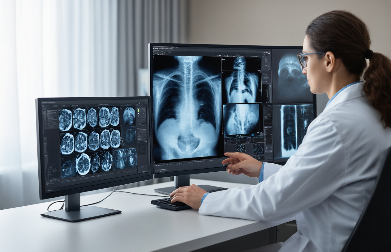
Digital X-ray technology has revolutionized medical imaging, and if you haven’t experienced it yet, you’re in for a pleasant surprise. Imagine walking into your appointment and seeing your images appear on screen within seconds—no chemical processing, no waiting rooms.
For both patients and healthcare providers, this advancement means quicker diagnoses, lower radiation exposure, and images that can be enhanced, shared, and stored electronically in seconds.
But here’s what most people don’t realize about digital X-rays: the technology isn’t just faster—it’s fundamentally changing how doctors detect conditions that once required much more invasive procedures.
Understanding Digital X-Ray Technology
A. Definition and Basic Principles
Digital X-ray technology captures images of your body’s internal structures without using traditional film. Instead, it uses electronic sensors to receive X-ray radiation that passes through your body, instantly converting these signals into digital images on a computer screen.
The magic happens when X-ray radiation travels through your body parts with different densities. Bones absorb more radiation than soft tissues, creating those characteristic white areas on a dark background. But unlike old-school X-rays, digital systems capture these differences with incredible sensitivity.
Think of it like the jump from film cameras to digital photography. The same basic physics apply, but the execution is dramatically improved.
What makes digital X-rays revolutionary is their ability to:
- Produce images in seconds rather than minutes
- Adjust contrast and brightness after the image is taken
- Require up to 80% less radiation than traditional methods
- Allow image sharing between healthcare providers instantly
B. Evolution from Traditional to Digital X-Rays
Remember those huge film X-rays your doctor would clip onto light boards? That technology barely changed for nearly a century after Röntgen’s discovery in 1895.
Traditional X-rays required chemical processing similar to darkroom photography. Technicians had to perfectly expose films, process them in chemical baths, and pray nothing went wrong. If the image was too dark or light? Start over and expose the patient to more radiation.
Digital systems burst onto the scene in the 1980s but didn’t become mainstream until the early 2000s. The transformation was dramatic – no more chemical processing, no more lost films, no more repeat exposures for technical errors.
Today’s digital systems offer advantages that film never could:
| Traditional X-Ray | Digital X-Ray |
|---|---|
| Chemical processing required | Instant electronic processing |
| Fixed image quality | Adjustable contrast/brightness |
| Physical storage needed | Electronic storage with backups |
| Manual film handling | Seamless electronic sharing |
| Higher radiation doses | Significantly reduced exposure |
This shift didn’t just improve image quality – it transformed workflow efficiency and patient safety in radiology departments worldwide.
Benefits of Digital X-Ray Imaging
Superior Image Quality and Detail
Gone are the days of squinting at fuzzy X-ray films. Digital X-rays blow traditional radiography out of the water when it comes to image quality.
The resolution is simply amazing – we’re talking about seeing bone structures with crystal clarity that would have been impossible to detect on conventional films. Doctors can zoom in, enhance contrast, and adjust brightness with a few clicks.
What does this mean for you? More accurate diagnoses. That tiny hairline fracture that might have been missed before? Now it’s clear as day. The subtle signs of early osteoporosis? They’re right there on the screen.
Reduced Radiation Exposure for Patients
Here’s something worth celebrating – digital X-rays typically cut radiation exposure by 70-80% compared to traditional film X-rays.
That’s a massive difference, especially for kids, pregnant women, and patients who need multiple images. The digital sensors are more sensitive, capturing the same diagnostic information with significantly less radiation.
Your body will thank you for this technological upgrade.
Immediate Image Availability and Review
Remember waiting around for X-ray films to be developed? Those days are history.
With digital systems, your images appear on screen within seconds. No chemical processing, no waiting rooms, no delays in diagnosis. Your doctor can review the results immediately, often while you’re still in the exam room.
This speed doesn’t just save time – it can be life-saving in emergency situations where every minute counts. Plus, your doctor can instantly share these images with specialists for second opinions without you needing to physically transport films anywhere.
Applications Across Medical Fields
A. Diagnostic Uses in General Medicine
Digital X-rays have completely changed how doctors diagnose everyday health issues. Gone are the days of waiting hours for film to develop! Now, physicians can spot pneumonia in minutes by seeing clear images of fluid-filled lungs. They can identify gastrointestinal blockages without the guesswork of traditional films.
The beauty of digital X-rays? Doctors can zoom in on suspicious areas, adjust contrast on the fly, and share images instantly with specialists across town or around the world. A patient with unexplained chest pain doesn’t have to wait days for a consultation – their images can be reviewed by a cardiologist within hours.
For primary care doctors, digital X-rays help rule out serious conditions quickly. That persistent cough? A quick chest X-ray shows whether it’s just bronchitis or something more concerning. The technology also means less radiation exposure for patients who need multiple images – a win for everyone involved.
B. Specialized Applications in Orthopedics
Orthopedic specialists absolutely love digital X-rays. They can see bone fractures with incredible detail, down to the tiniest hairline crack that might be missed on traditional film.
For surgeons planning joint replacements, these images are game-changers. They can measure bone dimensions precisely, ensuring perfect fit for artificial joints. During follow-up visits, comparing sequential digital images side-by-side shows healing progress with remarkable clarity.
Sports medicine has particularly benefited. Athletes with suspected stress fractures get faster diagnoses and treatment plans. Doctors can spot subtle alignment issues in runners’ ankles or cyclists’ knees that traditional X-rays might miss.
Digital technology also shines in pediatric orthopedics, where growth plate injuries require especially careful assessment. The enhanced visualization helps doctors distinguish between normal growth patterns and potential problems that need intervention.
Advanced Features of Modern Digital X-Ray Systems
3D Imaging Capabilities
Gone are the days when X-rays only gave us flat, 2D images that doctors had to mentally reconstruct into three dimensions. Modern digital X-ray systems now offer incredible 3D imaging that’s changing the game completely.
These systems use techniques like tomosynthesis, which captures multiple images from different angles and stitches them together into a detailed 3D model. Think about how much more a doctor can see when they can literally rotate your shoulder joint on screen instead of looking at a static image!
The benefits? Huge. Doctors spot tiny fractures that would’ve been invisible on traditional X-rays. They see exactly how bones align in space. And they detect small tumors that might have been hidden behind other structures in a flat image.
For patients, this means more accurate diagnoses the first time around. No more “come back in a week if it still hurts” when the initial X-ray looks inconclusive.
AI-Assisted Diagnosis Tools
AI is revolutionizing X-ray interpretation, and it’s about time. Radiologists are brilliant, but they’re human. They get tired. They might miss subtle patterns after reviewing hundreds of images.
Today’s digital X-ray systems come equipped with AI algorithms that act like a second set of eyes. These tools can:
- Flag potential abnormalities for closer review
- Compare your images against thousands of similar cases
- Calculate precise measurements automatically
- Suggest possible diagnoses based on image patterns
The AI doesn’t replace the radiologist—it enhances their capabilities. In studies, radiologists working with AI catch significantly more issues than either could alone.
What this means for you: faster results, fewer return visits, and peace of mind knowing your X-rays have been thoroughly analyzed by both human expertise and machine precision.
Choosing the Right Digital X-Ray Solution
Key Factors to Consider for Healthcare Facilities
Switching to digital X-ray isn’t a decision to take lightly. Your facility’s specific needs should drive your choice.
First, think about your patient volume. A busy emergency department needs a system with rapid image acquisition and processing. Meanwhile, a small orthopedic practice might prioritize different features.
Image quality is non-negotiable. Look for systems offering high spatial resolution (2.5+ lp/mm) and greater bit depth (14-16 bit) for better contrast visualization.
Workflow integration matters tremendously. The system should play nice with your existing PACS and EMR. Nothing kills efficiency faster than software that doesn’t communicate.
Don’t overlook physical space requirements. Some DR systems need less room than CR, which could be crucial in tight quarters.
Staff training requirements vary widely between systems. Some interfaces are intuitive, while others demand extensive training. Consider your team’s technical comfort level.
Cost-Benefit Analysis of Digital vs. Traditional Systems
The upfront sticker shock of digital systems can make anyone wince. But looking at the complete financial picture tells a different story.
| Factor | Digital X-Ray | Traditional Film |
|---|---|---|
| Initial Cost | $100,000-$250,000 | $30,000-$60,000 |
| Annual Film/Chemistry | $0 | $15,000-$50,000 |
| Storage Space | Minimal | Significant |
| Exam Time | 3-5 minutes | 15-20 minutes |
| Patient Throughput | 3-4x higher | Baseline |
| Repeat Rate | 3-5% | 10-15% |
Most facilities recover their investment within 2-3 years through:
- Eliminated film and chemistry costs
- Increased patient throughput
- Reduced staff time per exam
- Lower radiation doses (meaning fewer liability concerns)
- No darkroom maintenance
Digital also opens revenue streams through teleradiology and improved diagnostic capabilities that film simply can’t match.
Digital X-ray technology has revolutionized medical imaging, offering clearer images, lower radiation exposure, and faster results than traditional film-based methods. The benefits extend across numerous medical specialties, from orthopedics to dental care, allowing healthcare providers to make more accurate diagnoses while improving patient comfort and safety. Modern systems feature advanced capabilities like image enhancement, 3D reconstruction, and integration with electronic health records.
When selecting a digital X-ray solution, healthcare facilities should consider their specific needs, patient volume, and budget constraints. Investing in the right digital X-ray technology not only improves diagnostic capabilities but also enhances workflow efficiency and patient care. As this technology continues to evolve, it remains an essential cornerstone of modern medical imaging.