
CT Pulmonary Angiography (CTPA): Gold Standard for Detecting Pulmonary Embolism
When a blood clot blocks arteries in your lungs, every minute counts. CT Pulmonary Angiography (CTPA) has become the go-to imaging test, saving lives by quickly and accurately detecting pulmonary embolism. This advanced diagnostic tool gives doctors the straightforward answers they need to start treatment fast.
This guide is for healthcare professionals, medical students, and patients who want to understand why CTPA is the gold standard for diagnosing pulmonary embolism. We’ll explore how CTPA technology works to spot even small blood clots that other tests might miss. You’ll also learn about the clinical advantages that make doctors choose CTPA over older diagnostic methods, as well as what patients need to know about preparation and safety during the procedure.
Understanding Pulmonary Embolism and Its Life-Threatening Impact

Recognizing the critical nature of blood clots in the lung arteries
Pulmonary embolism occurs when blood clots travel through the circulatory system and block arteries in the lungs, creating a medical emergency that can kill within hours. These clots typically form in deep leg veins before breaking loose and moving to the lungs, where they obstruct blood flow and prevent oxygen exchange.
Identifying high-risk patient populations most vulnerable to PE
Several patient groups face elevated risks for developing pulmonary embolism. Post-surgical patients, especially those undergoing orthopedic procedures, cancer patients receiving chemotherapy, and individuals with prolonged immobility from long flights or bed rest, show significantly higher incidence rates. Pregnant women and those taking hormone replacement therapy also face increased vulnerability due to changes in blood clotting mechanisms.
Understanding how delayed diagnosis leads to increased mortality rates
| Diagnosis Timing | Mortality Rate |
| Within 1 hour | 5-8% |
| 1-6 hours | 15-20% |
| 6-24 hours | 25-30% |
| Beyond 24 hours | 35-50% |
When doctors miss early signs such as sudden shortness of breath, chest pain, or a rapid heart rate, patients face dramatically worse outcomes. Each hour of delay allows clots to grow larger and block more lung tissue, pushing the heart into dangerous strain patterns that can lead to cardiac arrest.
Why CTPA Revolutionized Pulmonary Embolism Diagnosis
Comparing CTPA accuracy to traditional diagnostic methods
CTPA delivers exceptional diagnostic precision, with sensitivity exceeding 95% for detecting pulmonary embolism, dramatically outperforming older methods such as pulmonary angiography and chest X-rays. Traditional chest imaging often misses smaller clots entirely, while CTPA captures detailed visualization of subsegmental arteries where many life-threatening emboli lodge.
Eliminating the limitations of ventilation-perfusion scans
Ventilation-perfusion scans frequently produce inconclusive results, particularly in patients with existing lung disease or abnormal chest X-rays. CTPA provides definitive answers regardless of underlying respiratory conditions, eliminating the frustrating uncertainty that has plagued emergency physicians who rely on V/Q scanning for critical diagnostic decisions.
Providing rapid results for emergency medical decisions
Modern CTPA scans are complete within minutes, delivering crucial diagnostic information when every second counts in emergency departments. This speed allows physicians to initiate anticoagulation therapy immediately for confirmed cases or safely discharge patients with negative results, preventing dangerous delays in treatment protocols.
Delivering superior image quality for confident diagnosis
High-resolution CTPA images reveal intricate vascular anatomy with remarkable clarity, enabling radiologists to distinguish between actual emboli and artifacts that confuse other imaging modalities. The technology’s ability to visualize contrast enhancement patterns provides unmatched confidence in diagnostic interpretation, reducing false positives and missed diagnoses.
How CTPA Technology Works to Detect Blood Clots
Understanding the contrast-enhanced imaging process
CTPA works by injecting iodinated contrast material directly into a patient’s bloodstream through an intravenous line. This contrast agent flows through the circulatory system and highlights blood vessels on CT images, creating a detailed roadmap of the pulmonary circulation. The timing of the scan is crucial – images are captured during peak contrast enhancement when the pulmonary arteries are optimally opacified.
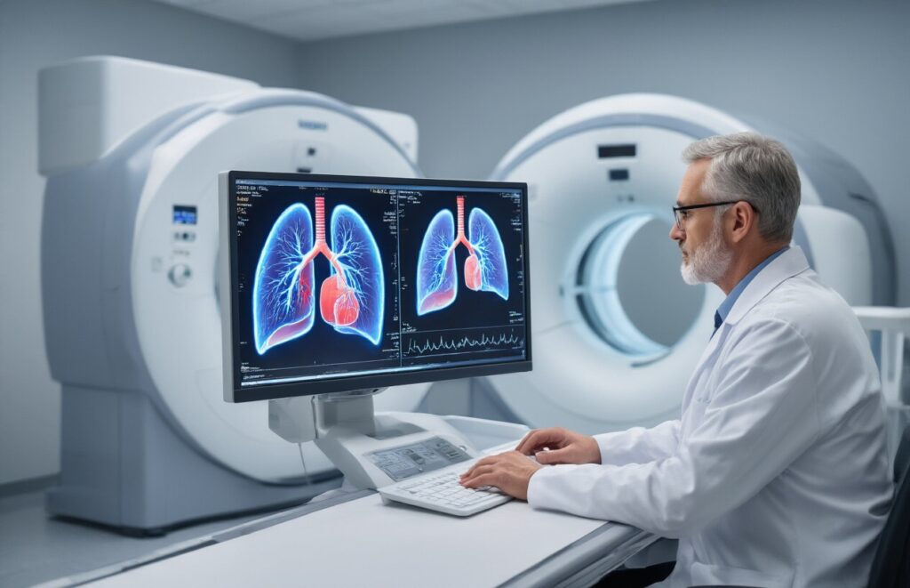
Visualizing pulmonary arteries with unprecedented clarity
Modern multi-detector CT scanners acquire thin-slice images in seconds, generating high-resolution cross-sectional views of the chest. The contrast-enhanced blood appears bright white against darker lung tissue, making even small pulmonary vessels clearly visible. Advanced reconstruction techniques allow radiologists to examine arteries from multiple angles and create three-dimensional models for comprehensive evaluation.
Identifying both central and peripheral embolisms effectively
CTPA excels at detecting blood clots throughout the entire pulmonary arterial tree, from large central vessels to smaller peripheral branches. Central emboli appear as dark filling defects surrounded by bright contrast within major pulmonary arteries. Peripheral clots are visible as abrupt vessel cutoffs or focal areas where contrast fails to flow, indicating complete obstruction of smaller arterial segments.
Clinical Advantages That Make CTPA the Preferred Choice

Achieving diagnostic confidence in emergencies
Emergency departments rely on CTPA’s rapid scan times to make critical decisions within minutes rather than hours. The examination delivers definitive results in under 30 seconds, allowing physicians to immediately rule in or rule out pulmonary embolism with over 95% accuracy. This speed proves essential when patients present with chest pain, shortness of breath, or hemodynamic instability.
Reducing false positive and false negative results
CTPA demonstrates superior diagnostic performance compared to ventilation-perfusion scanning, with sensitivity rates exceeding 96% and specificity approaching 95%. The technology’s ability to directly visualize clots in pulmonary arteries eliminates the interpretive challenges of indirect imaging methods. Modern multi-detector scanners can detect emboli as small as 2-3mm in subsegmental arteries.
Enabling simultaneous detection of alternative diagnoses
Beyond pulmonary embolism detection, CTPA reveals alternative pathologies that explain patient symptoms. The scan identifies pneumonia, pneumothorax, aortic dissection, pericardial effusion, and lung masses during the same examination. This comprehensive approach prevents missed diagnoses and guides appropriate treatment pathways when pulmonary embolism is absent.
Minimizing patient discomfort during the examination process
CTPA requires minimal patient positioning and no special breathing maneuvers beyond a single breath hold lasting 15-20 seconds. Patients remain supine throughout the procedure, avoiding the lengthy positioning required for nuclear medicine studies. The examination doesn’t require fasting, and most patients can resume normal activities immediately afterward.
Providing detailed anatomical information for treatment planning
High-resolution images reveal clot burden, location, and extent throughout the pulmonary vasculature, informing treatment decisions between anticoagulation and thrombolysis. CTPA identifies right-heart strain patterns and quantifies pulmonary arterial pressures, helping clinicians assess disease severity. Detailed anatomical mapping is valuable for interventional procedures such as catheter-directed thrombolysis or embolectomy planning.
Patient Preparation and Safety Considerations for CTPA

Ensuring Optimal Contrast Enhancement Through Proper Protocols
Achieving excellent image quality requires precise timing and proper contrast injection protocols. Patients should fast for 4-6 hours before the scan to reduce the risk of nausea and improve contrast circulation. The injection rate typically ranges from 4 to 5 mL/second through an 18-gauge IV catheter, preferably in the antecubital vein.
Managing Potential Allergic Reactions to Contrast Media
Healthcare teams must screen patients for previous contrast reactions and shellfish allergies before administration. Pre-medication with corticosteroids and antihistamines may be necessary for high-risk patients. Emergency medications, including epinephrine, corticosteroids, and bronchodilators, should remain immediately accessible during all procedures.
Evaluating Kidney Function Before Contrast Administration
| Risk Factor | Creatinine Level | Action Required |
| Normal function | <1.5 mg/dL | Proceed with standard protocol |
| Mild impairment | 1.5-2.0 mg/dL | Hydration recommended |
| Moderate-severe | >2.0 mg/dL | Consider alternative imaging |
Serum creatinine levels must be checked within 48 hours of the procedure, especially in elderly patients or those with diabetes and hypertension.
Addressing Radiation Exposure Concerns in Specific Populations
Pregnant women require alternative imaging methods, like ventilation-perfusion scans or ultrasound, to avoid fetal radiation exposure. Young patients and those requiring repeat scans need dose-optimization techniques, including reduced tube voltage and iterative reconstruction algorithms, to minimize lifetime radiation exposure while maintaining diagnostic accuracy.
Interpreting CTPA Results for Optimal Patient Outcomes

Recognizing Direct Signs of Pulmonary Embolism on Imaging
The most telling sign on CTPA scans is the presence of intravascular filling defects that appear as dark areas within bright contrast-filled pulmonary arteries. These defects represent blood clots that partially or entirely block arterial flow. Radiologists also look for the “railway track sign,” in which clots create parallel lines within vessels, and acute vessel cutoffs, where arteries suddenly terminate.
Understanding the Significance of Filling Defects in Arteries
Complete arterial occlusion appears as an abrupt vessel cutoff with no contrast beyond the clot site, indicating total blockage. Partial filling defects show contrast flowing around the clot edges, creating characteristic central low-density areas surrounded by contrast material. The location and size of these defects help determine treatment urgency and guide anticoagulation decisions.
Evaluating Right Heart Strain Indicators for Severity Assessment
Right heart strain manifests as ventricular dilation, septal flattening, and increased pulmonary artery diameter on CTPA images. These findings signal hemodynamically significant pulmonary embolism requiring immediate intervention. Additional markers include reflux of contrast into the inferior vena cava and hepatic veins, suggesting elevated right-sided pressures that can lead to cardiovascular collapse without prompt treatment.
CTPA has completely changed how doctors diagnose pulmonary embolism, turning what used to be a challenging, sometimes dangerous diagnostic process into a fast, accurate, and reliable one. This imaging technology gives healthcare teams the clear, detailed pictures they need to spot blood clots in the lungs quickly, which can literally be the difference between life and death for patients. The speed and precision of CTPA mean that people get the proper treatment faster, leading to better outcomes and fewer complications.
If you’re experiencing symptoms like sudden shortness of breath, chest pain, or unexplained leg swelling, don’t wait to seek medical attention. Early detection through CTPA can catch pulmonary embolism before it becomes more serious, and knowing what to expect from the procedure can help you feel more prepared and confident about getting the care you need. Your health is worth taking seriously, and CTPA gives both you and your medical team the best possible chance of catching and treating this condition effectively.