
“Clariscan for MRI: How This Contrast Agent Enhances Diagnostic Imaging”
What is Clariscan (MRI)?
Clariscan is an advanced MRI contrast agent that helps doctors get clearer, more detailed images during magnetic resonance imaging scans. This gadolinium-based contrast medium is designed for patients, healthcare providers, and medical facilities looking to improve diagnostic accuracy in various medical conditions.
If you’re scheduled for an MRI with Clariscan or want to understand this contrast agent better, you’ll find answers here. We’ll explore how Clariscan enhances MRI imaging quality and its specific medical applications across different body systems. You’ll also learn about the safety considerations and how this contrast agent compares to other options available in medical imaging.
Our CGHS empanelled centre in Sector 65, Gurugram, uses digital MRI technology that delivers superior image quality compared to open MRI systems. After your scan, you’ll receive digital reports on WhatsApp for quick access, while physical films can be collected from our facility at your convenience.
Understanding Clariscan as an Advanced MRI Contrast Agent
Key components and chemical composition of gadoterate meglumine
Clariscan’s active ingredient is gadoterate meglumine, a sophisticated contrast agent built around a gadolinium ion at its core. This gadolinium atom sits locked within a macrocyclic chelate structure called DOTA (1,4,7,10-tetraazacyclododecane-1,4,7,10-tetraacetic acid), creating an incredibly stable molecular framework. The meglumine salt component acts as a counterion, making the entire compound water-soluble and safe for intravenous administration.
The macrocyclic structure represents a significant advancement in contrast agent design. Unlike linear chelates, this ring-like formation wraps completely around the gadolinium ion, creating multiple binding points that hold it securely in place. This design prevents the toxic gadolinium from separating and accumulating in body tissues, a concern with some older contrast agents.
Each molecule contains one gadolinium ion surrounded by eight coordinating atoms from the DOTA chelate, plus one water molecule that can exchange rapidly with surrounding tissue water. This water exchange is what creates the enhanced contrast visible on MRI scans. The molecular weight of gadoterate meglumine is approximately 753 daltons, making it small enough for efficient kidney filtration while remaining stable in the bloodstream.
How Clariscan differs from traditional MRI contrast agents
Clariscan stands apart from earlier generation contrast agents through its superior stability profile and reduced risk of gadolinium retention. Traditional linear gadolinium agents like gadodiamide or gadoversetamide have a more open molecular structure that can potentially release free gadolinium ions under certain physiological conditions. Clariscan’s macrocyclic design eliminates this risk almost entirely.
The stability difference becomes particularly important for patients with kidney problems or those requiring multiple MRI scans over time. While linear agents showed concerning gadolinium deposits in brain tissue and other organs in some patients, macrocyclic agents like Clariscan demonstrate minimal tissue retention even after repeated administrations.
Clariscan also offers enhanced imaging capabilities compared to older agents. Its optimized relaxivity properties provide clearer tissue differentiation and better lesion detection, particularly in neurological and cardiovascular imaging. The agent’s distribution characteristics allow for more precise timing of image acquisition, giving radiologists better control over scan protocols.
The dosing requirements often differ as well. Clariscan’s high stability and imaging efficiency frequently allow for lower doses while maintaining diagnostic quality, reducing the overall gadolinium burden on patients. This becomes especially valuable in pediatric imaging or when multiple follow-up scans are necessary for monitoring treatment response.
Medical Applications and Diagnostic Benefits

Enhanced brain and spinal cord imaging capabilities
Clariscan transforms how medical professionals examine the brain and spinal cord, offering unprecedented clarity in neurological imaging. When injected during an MRI scan, this advanced contrast agent crosses the blood-brain barrier selectively, highlighting areas where normal brain tissue has been compromised. This selective enhancement makes it particularly valuable for detecting subtle changes in brain structure that might otherwise remain invisible.
The contrast agent excels at revealing detailed anatomical structures within the central nervous system. Radiologists can now distinguish between healthy brain tissue and areas affected by disease with remarkable precision. The enhanced contrast helps identify inflammation, vascular abnormalities, and tissue damage that standard MRI scans might miss. This level of detail proves essential when evaluating conditions like multiple sclerosis, stroke, brain infections, or traumatic brain injuries.
Spinal cord imaging receives similar benefits from Clariscan enhancement. The contrast agent illuminates spinal lesions, herniated discs, and inflammatory conditions affecting the spine. Medical professionals can assess nerve root compression, spinal cord tumors, and degenerative changes with greater accuracy. This improved visualization helps create more targeted treatment plans and allows for better monitoring of disease progression over time.
Digital MRI technology paired with Clariscan delivers superior image quality compared to open MRI systems, providing the detailed visualization needed for complex neurological diagnoses. Patients receiving scans at CGHS empanelled facilities can access these advanced imaging capabilities while benefiting from standardized quality protocols.
Improved detection of tumors and lesions
Brain tumor detection reaches new levels of accuracy with Clariscan-enhanced MRI imaging. The contrast agent accumulates in areas where the blood-brain barrier has been disrupted by tumor growth, creating bright signals that clearly outline malignant and benign masses. This enhanced visualization allows radiologists to determine tumor size, location, and relationship to surrounding healthy brain tissue with exceptional precision.
The contrast agent proves particularly effective at identifying small metastatic lesions that might escape detection on standard MRI scans. These tiny secondary tumors often measure just a few millimeters but can significantly impact treatment decisions. Clariscan’s ability to highlight these minute lesions ensures comprehensive tumor staging and helps oncologists develop appropriate treatment strategies.
Beyond tumor detection, Clariscan enhances the visibility of various brain lesions associated with different neurological conditions. Demyelinating plaques characteristic of multiple sclerosis become clearly visible, allowing neurologists to track disease activity and treatment response. Infectious lesions, inflammatory conditions, and vascular malformations also show enhanced contrast, enabling more accurate diagnoses.
The improved lesion detection capabilities extend to follow-up imaging as well. Medical professionals can monitor treatment effectiveness by comparing enhanced images over time, identifying changes in lesion size, number, or enhancement patterns. This monitoring capability proves invaluable for adjusting treatment protocols and assessing patient response to therapy.
Centres offering Clariscan-enhanced MRI provide patients with digital reports delivered via WhatsApp for convenient access, while physical films remain available for collection at the facility when needed for specialist consultations or second opinions.
How Clariscan Works During MRI Procedures
Injection Process and Timing Considerations
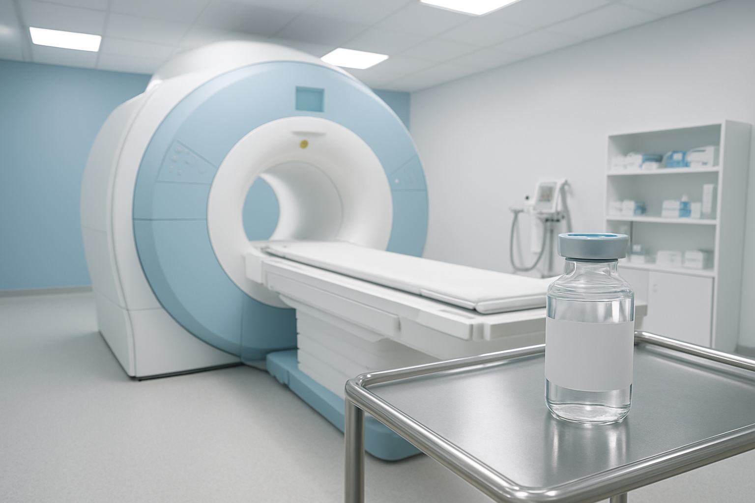
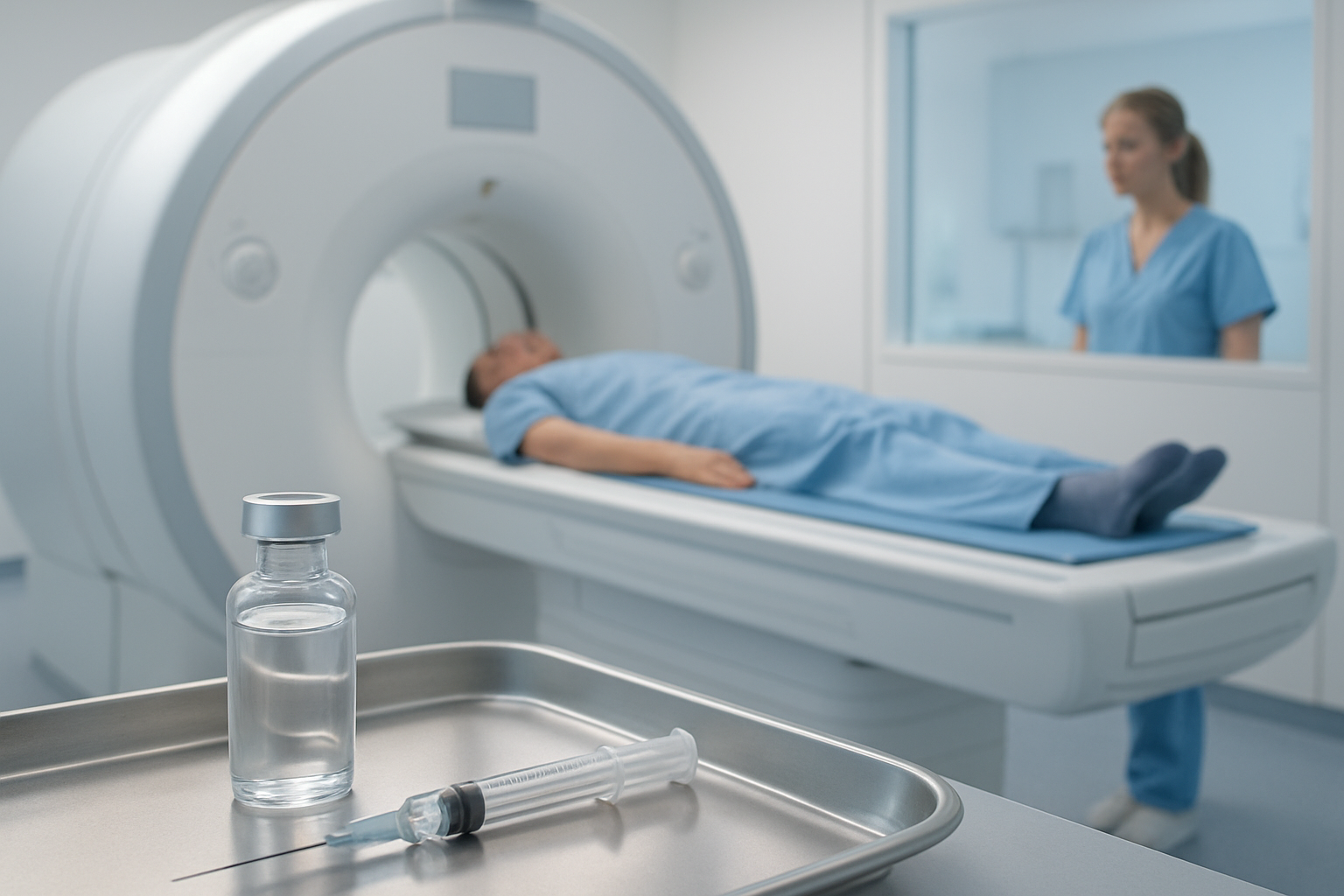
Clariscan administration begins with careful preparation at the diagnostic centre. The medical team establishes intravenous access through a small catheter, typically in your arm or hand. This process takes just a few minutes and feels similar to a routine blood draw.
The timing of Clariscan injection depends on the specific body area being examined. For most brain and spinal cord scans, the contrast agent is administered midway through the procedure. The radiologist monitors your scan in real-time and decides the optimal moment for injection based on the initial images captured.
The injection itself is remarkably quick, usually completed within 10-15 seconds. You might experience a brief warm sensation or metallic taste in your mouth – both completely normal reactions that fade quickly. The automated injector delivers the precise dose calculated for your body weight, ensuring optimal image enhancement while maintaining safety.
At CGHS empanelled facilities, the entire process follows strict protocols. The medical team coordinates timing perfectly with the MRI sequence, ensuring the contrast reaches target tissues exactly when needed for image capture. This precision makes digital MRI scans superior to open MRI alternatives, as the enclosed environment provides better contrast distribution control.
Emergency cases requiring immediate Clariscan-enhanced scans can be accommodated, though centres maintain regular operational hours rather than 24/7 service. The streamlined injection process means minimal delays in urgent diagnostic situations.
Distribution Through Blood Vessels and Tissues
Once injected, Clariscan travels rapidly through your bloodstream, reaching most body tissues within 30-60 seconds. The gadolinium-based compound moves through your circulatory system like a specialized messenger, highlighting areas where blood vessels might be damaged or where abnormal tissue growth exists.
The contrast agent doesn’t distribute evenly throughout your body. It concentrates heavily in areas with increased blood flow or where the blood-brain barrier has been compromised. This selective accumulation makes Clariscan particularly valuable for detecting brain tumors, inflammation, and vascular abnormalities that might otherwise remain invisible on standard MRI images.
Healthy tissue shows different enhancement patterns compared to diseased areas. Tumors often display intense contrast uptake due to their abnormal blood supply, while scar tissue exhibits distinct enhancement characteristics. This differential uptake creates the diagnostic contrast that radiologists need for accurate interpretation.
The distribution process continues throughout your scan, with peak enhancement typically occurring 2-5 minutes after injection. Digital MRI technology captures these enhancement patterns with exceptional clarity, providing superior image quality compared to open MRI systems. The controlled environment of enclosed MRI scanners optimizes contrast distribution visualization.
After completing diagnostic imaging, patients receive digital reports via WhatsApp for immediate access. Physical films and detailed reports can be collected from the centre at your convenience, ensuring you have complete documentation of your enhanced MRI results.
Safety Profile and Patient Considerations

Common side effects and adverse reactions
Clariscan is generally well-tolerated by most patients, but like any contrast agent, it can cause some side effects. The most common reactions are mild and typically resolve on their own without treatment. These include a temporary metallic taste in the mouth, which many patients experience during or shortly after the injection. Some people also report mild nausea or a brief feeling of warmth spreading through their body as the contrast agent circulates.
Headaches can occur in about 5-10% of patients, usually appearing within a few hours of the procedure and lasting no more than 24 hours. A small percentage of patients might experience minor skin reactions like mild redness or itching at the injection site. These local reactions are usually minimal and disappear within a few hours.
More serious adverse reactions are rare but can include allergic responses. Mild allergic reactions might present as skin rash, hives, or mild breathing difficulties. Severe allergic reactions, while extremely uncommon, can include significant breathing problems, severe drop in blood pressure, or widespread skin reactions. Medical facilities performing Clariscan-enhanced MRI procedures maintain emergency protocols and medications to handle such situations promptly.
Patients with a history of allergies to contrast agents or those who have experienced reactions to gadolinium-based compounds in the past should inform their healthcare provider before the procedure. Most diagnostic centres, including CGHS empanelled facilities, maintain detailed patient history records to identify potential risks beforehand.
Contraindications for high-risk patients
Certain patient groups require special consideration or may need to avoid Clariscan altogether. Patients with severe kidney disease or kidney failure face the highest risk, as their bodies cannot efficiently eliminate gadolinium-based contrast agents. This can lead to a rare but serious condition called nephrogenic systemic fibrosis (NSF), which causes thickening and hardening of skin and connective tissues.
Pregnant women should avoid Clariscan unless absolutely necessary for critical diagnostic purposes. While studies haven’t definitively proven harm to developing babies, medical professionals typically recommend postponing non-urgent MRI procedures with contrast until after delivery. Breastfeeding mothers can usually resume nursing 24-48 hours after receiving Clariscan, though many doctors consider it safe to continue breastfeeding immediately.
Patients with a history of severe allergic reactions to gadolinium-based contrast agents should not receive Clariscan. Those with multiple severe allergies or asthma may require pre-medication with antihistamines or corticosteroids before the procedure. People taking certain medications, particularly those affecting kidney function, may need dose adjustments or temporary discontinuation of their medications.
Healthcare providers at specialized centres carefully review each patient’s medical history, current medications, and lab results before administering Clariscan. CGHS empanelled facilities maintain protocols for risk assessment and patient screening to ensure safe administration of contrast agents.
Comparing Clariscan to Alternative Contrast Agents
Stability Advantages Over Linear Contrast Agents
Clariscan stands out from linear contrast agents because of its macrocyclic structure, which creates a much more stable chemical environment. Linear agents like OptiMARK and MultiHance have an open-chain design that makes them more prone to releasing gadolinium ions into the body. This structural difference isn’t just academic – it has real clinical implications.
The macrocyclic design of Clariscan creates what’s essentially a cage around the gadolinium ion. This cage-like structure forms stronger bonds that are much harder to break apart, even when exposed to the body’s natural processes. Linear agents, on the other hand, can start losing their grip on gadolinium ions more easily, especially in patients with compromised kidney function or those undergoing multiple MRI scans over time.
Studies show that Clariscan releases significantly less free gadolinium compared to linear alternatives. This matters because free gadolinium can accumulate in tissues throughout the body, including the brain, bones, and skin. While the long-term effects of gadolinium retention are still being studied, the enhanced stability of macrocyclic agents like Clariscan provides an extra layer of safety for patients.
The stability advantage becomes even more pronounced in patients who need repeated imaging. Each MRI scan with a linear agent potentially adds more unstable gadolinium to the body’s burden, while Clariscan’s superior binding characteristics help minimize this accumulation.
Reduced Risk of Nephrogenic Systemic Fibrosis
Nephrogenic systemic fibrosis (NSF) represents one of the most serious complications associated with gadolinium-based contrast agents, and this is where Clariscan’s safety profile really shines. NSF is a devastating condition that causes skin thickening, joint contractures, and can affect internal organs. The good news is that this condition has been almost exclusively linked to linear, high-risk gadolinium agents.
Clariscan falls into the lowest risk category for NSF development. The European Medicines Agency and FDA have classified it as having the best safety profile when it comes to NSF risk. This classification comes from extensive real-world data showing virtually no confirmed cases of NSF associated with Clariscan use, even in high-risk patient populations.
Patients with severe kidney disease face the highest risk of developing NSF, but even in this vulnerable group, Clariscan has demonstrated remarkable safety. The agent’s stable macrocyclic structure means it gets eliminated from the body more efficiently and doesn’t release free gadolinium that could trigger the fibrotic processes seen in NSF.
This safety advantage has practical implications for patient care. Doctors can feel more confident using Clariscan in patients with borderline kidney function, and patients who need multiple follow-up scans can receive them with greater peace of mind. The reduced NSF risk doesn’t eliminate the need for proper screening and monitoring, but it does provide a significant safety margin that wasn’t available with older linear agents.
Clariscan represents a significant advancement in MRI contrast technology, offering clearer diagnostic imaging with an impressive safety profile for patients. This advanced gadolinium-based agent enhances the visibility of tissues and blood vessels during scans, helping doctors make more accurate diagnoses across various medical conditions. The agent’s unique formulation provides excellent image quality while maintaining a lower risk of side effects compared to some traditional contrast agents.
For patients requiring MRI scans with contrast, choosing a facility that uses cutting-edge technology like Clariscan can make a real difference in diagnostic accuracy. CGHS empanelled centers equipped with digital MRI technology ensure patients receive high-quality imaging services with the convenience of receiving reports digitally via WhatsApp. If your doctor recommends an MRI with contrast, discuss whether Clariscan is the right choice for your specific medical needs and always inform your healthcare provider about any allergies or kidney concerns before the procedure.