
“Breast Ultrasound Guide: Why It’s Done, How It Works & What Results Mean”
A breast ultrasound uses sound waves to create detailed images of breast tissue, helping doctors detect lumps, cysts, and other abnormalities that might not show up clearly on mammograms or physical exams.
This guide is for women who need a breast ultrasound, those preparing for their first appointment, and anyone wanting to understand what this common diagnostic test involves.
We’ll walk you through how breast ultrasound technology works and why doctors recommend it, break down what happens during the procedure so you know exactly what to expect, and explain how to read your results and what different findings mean for your health.
Understanding Breast Ultrasound Technology
How sound waves create detailed breast images
Breast ultrasound works by sending high-frequency sound waves through breast tissue using a small handheld device called a transducer. These sound waves, typically ranging from 5 to 15 megahertz, bounce off different structures within the breast and return to the transducer at varying speeds. The machine converts these returning sound waves into electrical signals that create real-time images on a monitor.
The magic happens because different tissues reflect sound waves differently. Dense breast tissue appears bright white on the ultrasound screen because it’s more solid and reflects more sound waves back. Fat tissue shows up as darker gray areas since it’s less dense and allows more sound waves to pass through. Fluid-filled cysts appear completely black because liquid doesn’t reflect sound waves at all – they pass right through.
This technology excels at distinguishing between solid masses and fluid-filled cysts, making it incredibly valuable for breast health evaluations. The sound waves can penetrate deep into breast tissue without any radiation exposure, providing detailed images of structures that might be hidden or unclear on other imaging methods.
Real-time imaging means radiologists can observe how tissues move and change as gentle pressure is applied with the transducer. This dynamic capability helps identify suspicious areas and guides precise needle placement during biopsies when needed.
Key differences from mammograms and other imaging tests
Mammograms use low-dose X-rays to create images of breast tissue, while ultrasounds rely on sound waves. This fundamental difference means mammograms expose patients to small amounts of radiation, whereas ultrasounds are completely radiation-free, making them safe for pregnant women and frequent monitoring.
Mammograms compress the breast between two plates to spread tissue evenly and reduce thickness, which can be uncomfortable or painful for many women. Breast ultrasounds involve gentle contact with a gel-covered transducer that glides smoothly across the skin – no compression required.
The imaging capabilities differ significantly too. Mammograms excel at detecting tiny calcium deposits called microcalcifications, which can be early signs of breast cancer but are invisible on ultrasound. However, ultrasounds shine when examining dense breast tissue, where mammograms often struggle to distinguish between normal tissue and potential abnormalities.
MRI scans provide the most detailed breast images but require contrast dye injections and take much longer to complete. They’re typically reserved for high-risk patients or specific diagnostic situations. Ultrasounds offer immediate results and can be performed quickly in most medical settings.
Ultrasounds also provide unique advantages for guided procedures. Unlike mammograms or MRIs, ultrasound allows real-time visualization during biopsies, cyst aspirations, or other interventions. This makes procedures more precise and comfortable for patients while ensuring accurate targeting of suspicious areas.
Primary Medical Uses and Benefits
Detecting solid masses versus fluid-filled cysts
Breast ultrasound excels at distinguishing between solid lumps and fluid-filled cysts, something that can’t always be determined through physical examination alone. When you feel a lump in your breast, your mind naturally races to worst-case scenarios. The good news is that many breast lumps turn out to be benign cysts filled with fluid, and ultrasound can quickly identify these.
Solid masses appear as dense, echogenic structures on ultrasound images, while cysts show up as dark, echo-free areas with well-defined borders. This distinction is crucial because simple cysts are almost always benign and don’t require treatment. The ultrasound technologist can often tell you right away if what you’re feeling is just a harmless fluid-filled sac.
Complex cysts, which contain both fluid and solid components, require closer evaluation. These might need additional imaging or monitoring over time. Solid masses, on the other hand, may warrant further investigation through biopsy to rule out malignancy.
The real-time nature of ultrasound allows radiologists to examine suspicious areas from multiple angles, compress tissues to see how they respond, and use Doppler technology to evaluate blood flow patterns. Malignant tumors often have irregular shapes, unclear borders, and increased blood supply, while benign masses typically have smoother, more defined edges.
Guiding needle biopsies for accurate tissue sampling
When imaging reveals a suspicious solid mass that needs tissue sampling, ultrasound becomes an invaluable surgical tool. Ultrasound-guided core needle biopsy offers a minimally invasive way to obtain tissue samples without the need for surgical incision.
The process involves real-time visualization of both the suspicious area and the biopsy needle as it advances toward the target. This direct visual guidance ensures the needle reaches exactly the right spot, even for small or deep lesions that might be difficult to locate otherwise. The radiologist can watch the needle tip enter the mass and collect multiple tissue samples from different areas.
This precision is especially important for small lesions or those located near critical structures like the chest wall or nipple. Without ultrasound guidance, there’s a higher risk of missing the target or inadvertently sampling normal tissue instead of the abnormal area.
The ultrasound-guided approach also allows for immediate confirmation that adequate samples have been obtained. If the initial samples don’t capture enough tissue or miss the target area, additional samples can be taken during the same procedure.
Compared to surgical biopsy, this technique results in minimal scarring, reduced discomfort, and faster recovery times. Most patients can return to normal activities within 24 hours, with only a small bandage covering the biopsy site. The accuracy rates for ultrasound-guided biopsies exceed 95%, making them the gold standard for tissue sampling of visible breast masses.
Step-by-Step Procedure Process
Preparing for your ultrasound appointment
Getting ready for a breast ultrasound is pretty straightforward. You won’t need to fast or make major changes to your routine beforehand. However, there are a few things that can make your experience smoother and help get the best possible images.
Skip the lotion, powder, or deodorant on your chest and underarm areas on the day of your exam. These products can interfere with the ultrasound gel and potentially affect image quality. If you forget and apply them out of habit, don’t worry – the technologist can clean the area before starting.
Consider wearing a two-piece outfit or a top that you can easily remove from the waist up. You’ll need to undress from the waist up and put on a hospital gown that opens in the front. This makes it easier for the technologist to access the areas they need to examine.
If you’re still menstruating, try to schedule your ultrasound for the week after your period ends. Your breasts are typically less tender and swollen during this time, which can make the exam more comfortable and provide clearer images.
Bring a list of any medications you’re taking and be ready to discuss your medical history, including any previous breast imaging, family history of breast or ovarian cancer, and current symptoms you might be experiencing.
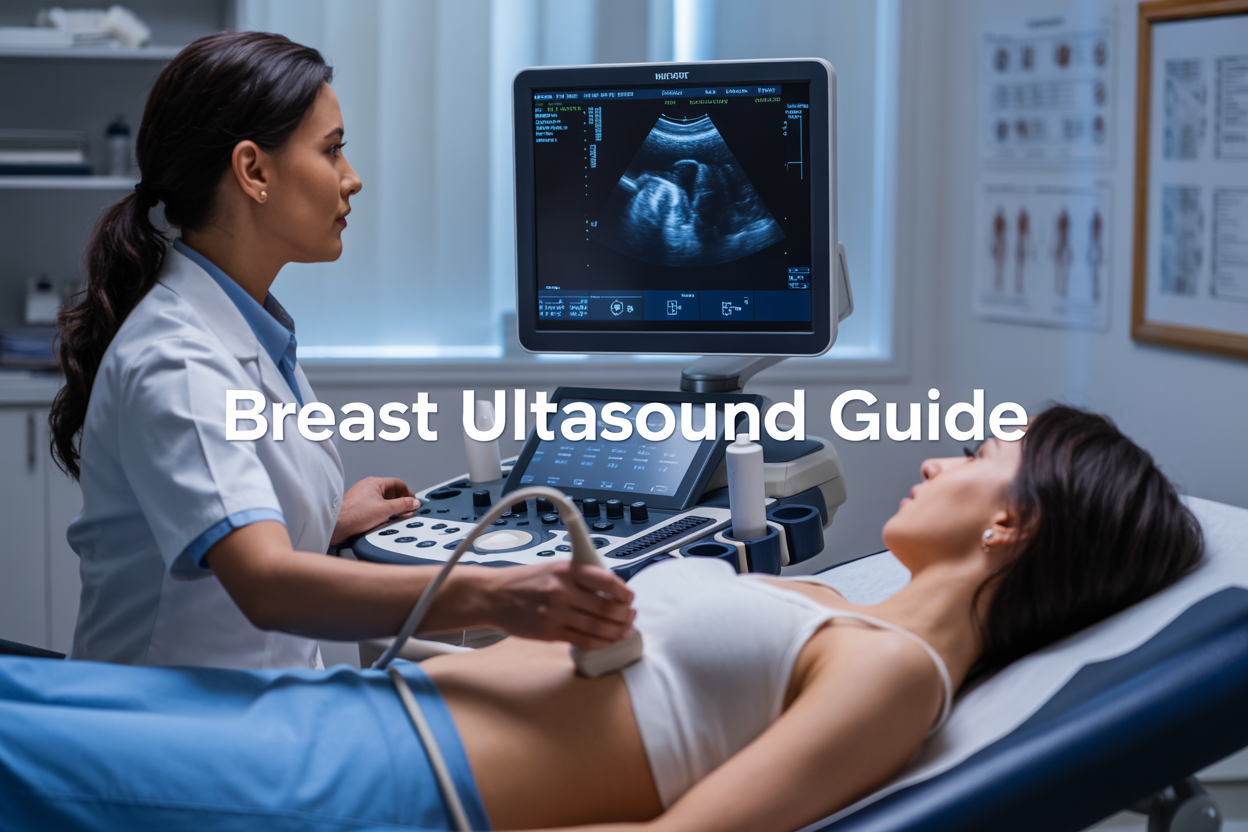
What happens during the imaging session
When you arrive for your appointment, you’ll check in and complete any necessary paperwork. A technologist will escort you to the exam room and explain what to expect during the procedure.
You’ll change into a hospital gown that opens in the front, then lie on your back on the examination table. The technologist will position you comfortably, sometimes with your arm raised above your head on the side being examined. This positioning helps spread out the breast tissue for better visualization.
The technologist applies a clear, water-based gel to your breast. This gel might feel cool at first, but it warms up quickly. The gel serves as a conductor between your skin and the ultrasound transducer, eliminating air pockets that could interfere with the sound waves.
Using a small handheld device called a transducer, the technologist moves it across your breast in various directions. You’ll feel gentle pressure as they press the transducer against your skin to get closer to the tissue being examined. This pressure is necessary to create clear images but shouldn’t be painful.
The transducer sends high-frequency sound waves through your breast tissue. These waves bounce back at different rates depending on the density of the tissue they encounter, creating real-time images on a computer screen. The technologist watches these images closely, taking snapshots of important areas.
The entire scanning process typically takes 15 to 30 minutes, depending on what areas need examination and whether any suspicious areas require additional imaging. You can usually watch the monitor during the exam, and many technologists will explain what they’re seeing if you’re curious.
After completing the scan, the technologist removes the gel with tissues and lets you get dressed. The images are then reviewed by a radiologist who will prepare a detailed report for your doctor.
Understanding Your Ultrasound Results
Normal findings and what they mean for your health
When your breast ultrasound shows normal results, you’ll typically see uniform tissue patterns without any concerning masses or irregularities. Healthy breast tissue appears as a mix of different densities on the ultrasound screen – fatty tissue shows up as darker areas, while glandular tissue appears brighter. Your radiologist looks for smooth, even textures and consistent blood flow patterns throughout the breast.
Normal findings mean the ultrasound didn’t detect any solid masses, cysts with concerning features, or unusual tissue changes. You might still have small, simple cysts, which are completely normal fluid-filled sacs that many women develop. These benign cysts appear as dark, round areas with thin walls and cause no health concerns.
The absence of suspicious findings on your ultrasound is reassuring, but remember that this test works best when combined with other screening methods. Your doctor will consider your ultrasound results alongside your clinical exam, medical history, and potentially your mammogram results to get the complete picture of your breast health.
Normal results don’t guarantee you’ll never develop breast problems in the future, which is why regular screening remains important. Your breast tissue naturally changes throughout your menstrual cycle, with age, and due to hormonal influences, so what’s normal for you might look different at various times.
Abnormal results requiring further investigation
Abnormal ultrasound findings don’t automatically mean cancer, but they do signal that additional testing is needed to determine what’s causing the irregularity. Your radiologist might identify solid masses that appear irregular in shape, have unclear borders, or show unusual internal characteristics. These masses could be benign conditions like fibroadenomas or more complex cysts, but they require closer examination.
Other concerning findings include areas where tissue appears significantly different from the surrounding breast tissue, unusual blood flow patterns detected through Doppler ultrasound, or lymph nodes that appear enlarged or abnormally shaped. Sometimes the ultrasound reveals multiple small masses or areas of architectural distortion where the normal breast tissue pattern seems disrupted.
When abnormal results appear, your doctor will likely recommend additional imaging tests such as diagnostic mammography or MRI. Most importantly, you’ll probably need a biopsy – a procedure where a small tissue sample is removed and examined under a microscope to determine the exact nature of the abnormal area.
The waiting period between discovering abnormal results and getting definitive answers can feel overwhelming, but most breast abnormalities turn out to be benign. Your healthcare team will guide you through each step of the follow-up process, explaining what each test involves and what the results mean for your specific situation. Early detection and prompt evaluation of any abnormal findings give you the best possible outcomes regardless of the final diagnosis.
Safety Considerations and Limitations
Why ultrasound is completely radiation-free
Breast ultrasound stands apart from other imaging methods like mammography or CT scans because it uses sound waves instead of ionizing radiation. The technology works by sending high-frequency sound waves through breast tissue, which bounce back to create detailed images on a monitor. These sound waves are completely harmless to your body and pose no cancer risk whatsoever.
This radiation-free approach makes breast ultrasound incredibly safe for repeated use. Pregnant women can undergo breast ultrasounds without any concern for their developing baby, and there’s no limit to how many ultrasounds you can have throughout your lifetime. Unlike mammograms, which expose you to small amounts of radiation, ultrasounds can be performed as often as medically necessary without accumulating any harmful effects.
The absence of radiation also means there’s no recovery time or special precautions needed after your exam. You can immediately return to your normal activities, drive yourself home, and continue with your day exactly as planned. This safety profile makes ultrasound particularly valuable for monitoring changes in breast tissue over time, especially in younger women or those at higher risk who may need more frequent imaging.
Situations where ultrasound may not provide clear answers
While breast ultrasound excels in many areas, it does have limitations that can affect its diagnostic accuracy. Dense breast tissue creates one of the most significant challenges for ultrasound imaging. When breast tissue is very dense, sound waves may not penetrate as effectively, making it harder to distinguish between normal tissue and potential abnormalities. This same density issue also makes it difficult to detect microcalcifications, which are tiny calcium deposits that can sometimes indicate early breast cancer.
Ultrasound struggles with imaging areas deep within large breasts or tissue near the chest wall. The sound waves may not reach these deeper structures clearly, potentially missing important findings in these regions. Additionally, ultrasound cannot always differentiate between benign and malignant solid masses based on appearance alone, which means suspicious findings often require additional testing like biopsy for definitive diagnosis.
The technology is also highly dependent on the operator’s skill and experience. Unlike mammography, which produces standardized images, ultrasound quality varies significantly based on the technician’s technique and the radiologist’s interpretation skills. Some subtle abnormalities might be missed if the person performing the exam doesn’t have extensive training in breast ultrasound.
Weather conditions and patient positioning can affect image quality too. If you’re unable to lie still or position yourself properly, the images may be less clear, potentially requiring repeat scans or alternative imaging methods.
Breast ultrasound stands out as a safe, painless diagnostic tool that plays a vital role in women’s health care. From detecting lumps and monitoring existing conditions to guiding biopsies, this technology offers doctors and patients valuable insights without radiation exposure. The procedure itself is straightforward and comfortable, typically taking just 15-30 minutes while providing immediate visual feedback about breast tissue health.
Your ultrasound results work hand-in-hand with other screening methods to create a complete picture of your breast health. While the technology has some limitations in detecting certain types of abnormalities, its benefits far outweigh these constraints. If your doctor recommends a breast ultrasound, approach it with confidence knowing you’re taking an important step in maintaining your health. Don’t hesitate to ask questions about your results or discuss any concerns with your healthcare provider – they’re there to help you understand what the findings mean for your specific situation.