

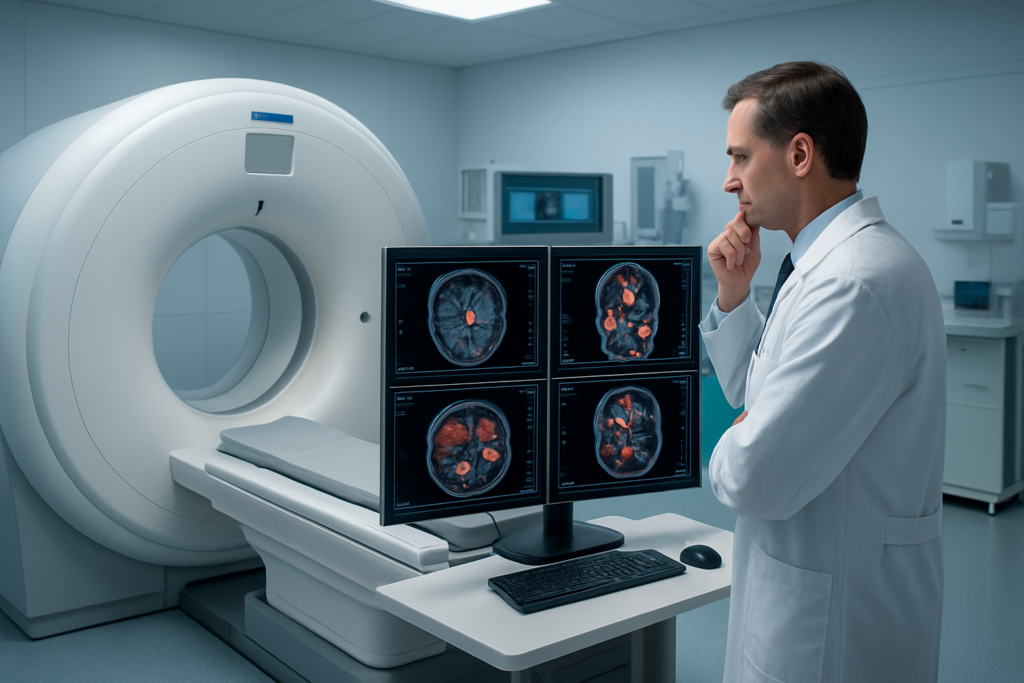
DOPA PET-CT scans have changed how doctors diagnose and treat brain tumors and neuroendocrine cancers. This advanced imaging technique uses a special radiotracer to spot cancer cells that other scans might miss.
This guide is for patients facing a DOPA PET-CT scan, their families, and healthcare professionals who want to understand this diagnostic tool better. We’ll break down complex medical information into easy-to-understand explanations.
You’ll learn how DOPA PET-CT technology works and why it’s become so valuable in cancer care. We’ll explore how this scan helps doctors detect and stage brain tumors more accurately than traditional methods. You’ll also discover how DOPA PET-CT plays a key role in diagnosing neuroendocrine cancers, giving patients and their medical teams clearer answers when facing these challenging conditions.
Understanding DOPA PET-CT Scan Technology

How DOPA Tracers Target Specific Cellular Processes
DOPA tracers work by mimicking the natural amino acid L-DOPA, which gets absorbed by cells that produce dopamine and other catecholamines. When injected into the body, these radioactive tracers travel through the bloodstream and accumulate in tissues with high amino acid uptake, particularly cancer cells and certain brain regions. The tracer’s radioactive properties allow PET scanners to detect and map these areas with remarkable precision.
Advanced Imaging Capabilities Beyond Traditional Scans
Unlike conventional CT or MRI scans that show anatomical structure, DOPA PET-CT combines metabolic activity with detailed anatomical images. This dual approach reveals not just where tumors are located, but how active they are at the cellular level. The technology excels at detecting small lesions that other imaging methods might miss, making it especially valuable for early-stage cancer detection and monitoring treatment response.
Key Advantages Over Conventional Diagnostic Methods
| Feature | DOPA PET-CT | Traditional CT/MRI |
| Detection sensitivity | High for metabolically active tumors | Limited to structural changes |
| Early detection | Identifies cellular changes before structural damage | Requires visible anatomical changes |
| Treatment monitoring | Shows real-time metabolic response | Limited functional information |
| Specificity | Targets specific cellular processes | General anatomical imaging |
The main benefit lies in its ability to catch diseases earlier and track how well treatments work by monitoring cellular activity rather than just size changes.
Detecting and Staging Brain Tumors with DOPA PET-CT
Early identification of malignant brain lesions
DOPA PET-CT excels at spotting malignant brain tumors that other imaging methods might miss. The tracer accumulates in areas where amino acid metabolism is elevated, which happens in growing cancer cells. This allows doctors to catch tumors earlier than conventional MRI or CT scans alone. The technique proves especially valuable for detecting gliomas and other primary brain cancers in their initial stages.
Precise tumor boundary mapping for surgical planning
Brain surgeons rely on DOPA PET-CT to see exactly where tumors begin and healthy brain tissue ends. The scan creates detailed maps showing tumor metabolism patterns, helping surgeons plan the safest removal route. This precision reduces the risk of removing healthy brain tissue while ensuring complete tumor removal. The metabolic information guides surgical decisions that standard imaging cannot provide.
Differentiating between tumor recurrence and treatment effects
| DOPA PET-CT Findings | Interpretation |
| High tracer uptake | Active tumor tissue |
| Low/no uptake | Treatment effects or scar tissue |
| Mixed patterns | Possible partial recurrence |
After radiation or chemotherapy, brain scans often show changes that look suspicious on regular imaging. DOPA PET-CT solves this puzzle by showing whether these changes represent growing cancer cells or just treatment-related scarring. Active tumors consume the DOPA tracer eagerly, while scar tissue shows little to no uptake. This distinction helps doctors decide whether to continue current treatment or switch strategies.
Monitoring treatment response and disease progression
Regular DOPA PET-CT scans track how well treatments are working against brain tumors. Decreasing tracer uptake signals that therapy is shrinking the cancer, while increasing uptake suggests the tumor is growing despite treatment. This real-time feedback allows oncologists to adjust treatment plans quickly rather than waiting for symptoms to worsen. The scans also help predict long-term outcomes and survival rates for patients with different types of brain cancers.
DOPA PET-CT Applications in Neuroendocrine Cancer Diagnosis

Identifying primary neuroendocrine tumor locations
DOPA PET-CT excels at pinpointing the original source of neuroendocrine tumors, particularly when conventional imaging falls short. The scan captures amino acid metabolism patterns unique to these cancer cells, revealing hidden primary tumors in challenging locations like the pancreas, small bowel, or lung. This precision helps doctors develop targeted treatment plans rather than searching blindly for the cancer’s origin point.
Detecting metastatic spread throughout the body
The whole-body imaging capability of DOPA PET-CT maps cancer spread with remarkable accuracy. Unlike traditional scans that might miss small metastatic deposits, this advanced technique detects neuroendocrine cancer cells throughout organs and lymph nodes. Doctors can see the complete disease picture in a single examination, identifying liver metastases, bone involvement, and lymph node spread that could influence treatment decisions and patient prognosis significantly.
Evaluating treatment effectiveness in real-time
DOPA PET-CT transforms treatment monitoring by showing metabolic changes before structural changes become visible on regular scans. Cancer cells that respond to therapy show reduced DOPA uptake, while resistant tumors maintain high activity levels. This early insight allows oncologists to adjust treatment protocols quickly, switch medications when current therapy isn’t working, or continue successful treatments with confidence based on clear biological evidence.
Clinical Benefits for Patients
Improved diagnostic accuracy reduces unnecessary procedures
DOPA PET-CT scans dramatically reduce false positives and negatives compared to traditional imaging methods. This precision helps doctors avoid ordering additional invasive tests, biopsies, or exploratory surgeries that patients don’t actually need. The enhanced accuracy means fewer anxiety-inducing follow-up appointments and reduced exposure to unnecessary radiation or surgical risks.
Personalized treatment planning based on precise imaging data
The detailed metabolic information from DOPA PET-CT allows oncologists to create tailored treatment strategies for each patient’s unique tumor characteristics. Doctors can identify which areas of the brain or body show the highest activity levels, helping them target radiation therapy more effectively or choose the most appropriate chemotherapy protocols. This personalized approach leads to better outcomes with fewer side effects.
Enhanced quality of life through better-targeted therapies
Patients benefit from more focused treatments that spare healthy tissue while attacking cancer cells more aggressively. The precise imaging data helps medical teams avoid over-treatment of slow-growing tumors and ensures aggressive cancers receive appropriate intervention. This balance means patients experience fewer treatment-related complications and maintain better cognitive function and overall well-being during their cancer journey.
Cost-effective long-term patient management strategies
Healthcare systems see significant savings when DOPA PET-CT guides treatment decisions from the start. Early accurate diagnosis prevents costly treatment delays, reduces hospital readmissions, and minimizes the need for multiple imaging studies.
Preparing for Your DOPA PET-CT Examination

Essential pre-scan dietary and medication restrictions
You’ll need to follow specific dietary guidelines before your DOPA PET-CT scan. Stop taking any medications containing L-DOPA, carbidopa, or other Parkinson’s drugs 12-24 hours before the exam, as directed by your doctor. Avoid foods high in protein like meat, dairy, nuts, and beans for 24 hours prior to scanning, since these can interfere with tracer uptake.
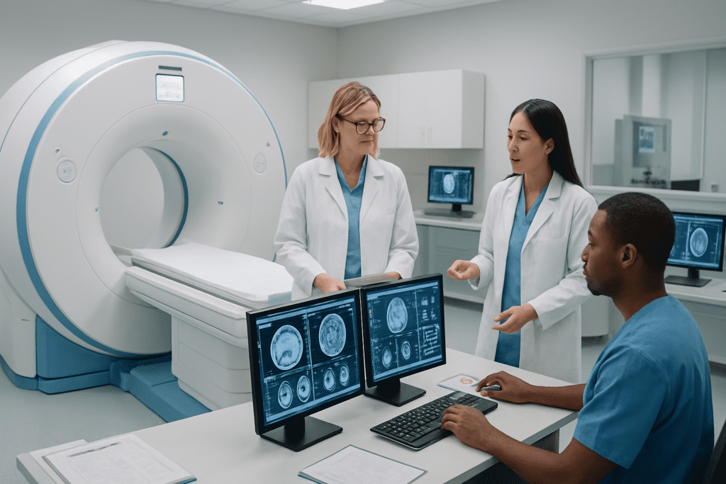
What to expect during the scanning procedure
The entire process takes about 2-3 hours from start to finish. After receiving the radioactive tracer injection, you’ll wait 60-90 minutes in a quiet room while the compound distributes throughout your body. The actual scan lasts 20-30 minutes, during which you’ll lie still on a table that moves through the scanner. The procedure is painless, though some people feel claustrophobic in the machine.
Post-scan care instructions and result timelines
| Timeline | Instructions |
| First 24 hours | Drink plenty of water to flush out the radiotracer |
| 48 hours | Limit close contact with pregnant women and young children |
| 3-5 days | Results typically available for review with your doctor |
Resume normal activities immediately after the scan. The small amount of radiation clears your system quickly, and most people experience no side effects. Your healthcare team will contact you once the radiologist completes the detailed image analysis.
DOPA PET-CT scans represent a major breakthrough in medical imaging, offering doctors unprecedented accuracy when diagnosing brain tumors and neuroendocrine cancers. This advanced technology helps medical teams catch these conditions earlier, plan better treatments, and monitor how well therapies are working. The detailed images it provides can make all the difference between a good treatment plan and a great one.
If your doctor recommends a DOPA PET-CT scan, you’re getting access to one of the most precise diagnostic tools available today. Take time to discuss the procedure with your healthcare team, follow the preparation guidelines carefully, and remember that this scan could provide the crucial information needed for your treatment journey. The investment in this technology means better outcomes and more personalized care for patients facing these challenging diagnoses.