
MRI Cisternography for CSF Rhinorrhea:
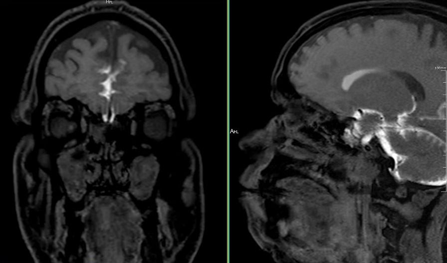
A 48-year-old male presented with right-sided nasal discharge and a clinical suspicion of 𝐂𝐒𝐅 𝐑𝐡𝐢𝐧𝐨𝐫𝐫𝐡𝐞𝐚.
An 𝐌𝐑𝐈 𝐂𝐢𝐬𝐭𝐞𝐫𝐧𝐨𝐠𝐫𝐚𝐩𝐡𝐲 was performed at Scan4Health using high-resolution T2 SPACE sequences, followed by post-intrathecal contrast-enhanced imaging.
On detailed evaluation, no definite contrast-enhanced CSF tract was seen extending across the cribriform plate or skull base into the sinonasal cavity, ruling out active CSF leak or cephalocele.
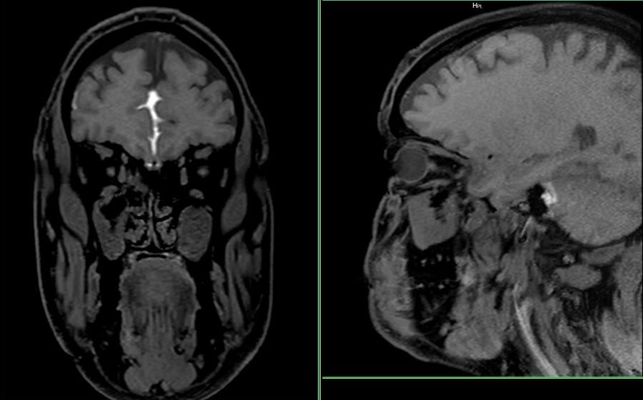
To further validate, a delayed scan done after 36–38 hours also showed no evidence of any contrast leak.
Associated findings included mild to moderate pansinusitis and hypertrophied turbinates.
This case emphasizes how combining advanced MRI protocols with meticulous review can differentiate between sinus pathology and true CSF leak, guiding clinicians toward the right management approach without unnecessary interventions.
At Scan4Health, every case is a step forward in precision and patient-focused diagnostics.
MRI Cisternography for CSF Rhinorrhea