

An MRI spine screening gives doctors a complete view of your entire spinal column in one comprehensive scan. This advanced imaging test helps healthcare providers spot problems from your neck down to your lower back that might be missed with limited scans.
This guide is for patients who’ve been recommended for whole-spine MRI screening, family members seeking to understand the process, and anyone curious about what this comprehensive test entails.
We’ll walk you through the medical conditions that typically lead doctors to order this extensive screening and explain the key anatomical structures the scan reveals. You’ll also learn about the diagnostic benefits this technology offers and get practical tips for preparing for your examination day.
Understanding MRI Spine Whole Screening Technology
How whole spine MRI differs from targeted scans
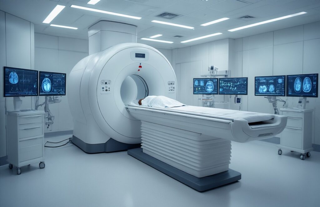
Whole spine MRI captures the complete spinal column from the skull base to the sacrum in a single comprehensive examination. At the same time, targeted scans focus on specific regions of the spine, such as the cervical, thoracic, or lumbar spine. This comprehensive approach allows radiologists to visualize the entire spinal anatomy and identify issues that may span multiple areas or exhibit patterns across different spinal levels.
Advanced imaging capabilities and resolution benefits
Modern whole-spine MRI technology delivers exceptional detail through high-resolution sequences that reveal soft-tissue structures, bone marrow changes, and subtle abnormalities throughout the spine. The comprehensive view enables detection of multi-level pathology, spinal alignment issues, and systemic conditions affecting the spine that could be missed with segmented imaging approaches.
Time efficiency compared to multiple separate scans
| Scan Type | Total Time | Patient Positioning |
| Three separate regional scans | 90-120 minutes | Multiple repositioning required |
| Single whole spine scan | 45-60 minutes | One positioning session |
A complete spine screening eliminates the need for various appointments and reduces overall examination time by nearly half compared to acquiring separate cervical, thoracic, and lumbar studies.
Safety advantages of comprehensive screening
Single-session whole spine imaging reduces radiation exposure concerns and minimizes contrast agent use when needed. Patients avoid repeated visits to medical facilities, thereby reducing infection risk and travel burden. The comprehensive approach also reduces the likelihood of missing interconnected spinal pathology that could require additional follow-up scans, making it both safer and more diagnostically complete.
Medical Conditions That Warrant Whole Spine MRI Screening

Unexplained chronic back pain across multiple regions
Doctors recommend a whole spine MRI when patients experience persistent pain that spans multiple spinal regions – cervical, thoracic, and lumbar areas. This comprehensive imaging helps identify interconnected issues that might be missed with single-region scans, revealing problems like disc degeneration patterns or nerve compression affecting multiple levels.
Suspected spinal tumors or metastatic disease
When cancer spreads to the spine or primary spinal tumors develop, whole spine screening becomes essential for staging and treatment planning. This approach detects multiple lesions simultaneously and helps oncologists understand the full extent of disease progression throughout the vertebral column.
Complex spinal deformities and structural abnormalities
Patients with scoliosis, kyphosis, or congenital spine abnormalities benefit from complete spinal imaging. The scan reveals how deformities affect the entire spinal alignment, helping surgeons plan comprehensive correction strategies.
Inflammatory conditions affecting the entire spine
Autoimmune diseases like ankylosing spondylitis and rheumatoid arthritis often cause inflammation throughout the spine. Whole spine MRI detects early inflammatory changes and monitors disease progression across all spinal segments.
Post-surgical monitoring of extensive spinal procedures
After multi-level spinal surgeries or complex fusion procedures, doctors use whole spine MRI to monitor healing and detect complications. This comprehensive monitoring ensures proper surgical outcomes and identifies any issues requiring additional intervention.
Key Anatomical Structures Revealed by Whole Spine MRI
Detailed visualization of all vertebrae and discs
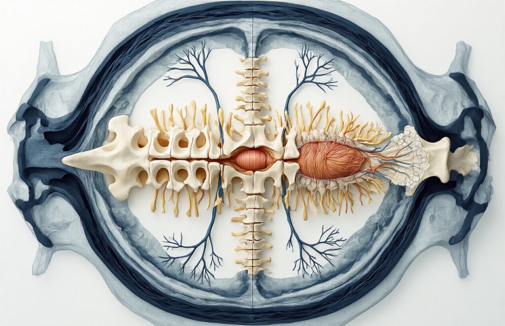
Whole-spine MRI captures every vertebra from the cervical region to the sacrum, providing radiologists with a complete picture of spinal alignment and structure. This comprehensive view reveals disc herniation, bulging, degeneration, and height loss throughout the entire spine. The technology shows bone marrow changes, fractures, and vertebral compression with remarkable clarity.
Spinal cord health and nerve root compression
The scan excels at assessing spinal cord integrity, revealing swelling, lesions, or signal changes that may indicate neurological problems. Nerve root compression becomes visible on MRI, which shows precisely where herniated discs or bone spurs pinch nerve pathways. This detailed nerve visualization helps doctors pinpoint the exact source of pain, numbness, or weakness patients experience.
Surrounding soft tissue and muscle condition
Beyond bones and nerves, a whole-spine MRI reveals the condition of the paraspinal muscles, ligaments, and other soft tissues that support the spine. The scan identifies muscle atrophy, inflammation, or tears that often accompany spinal problems. This soft-tissue assessment provides doctors with a comprehensive understanding of how surrounding structures contribute to a patient’s symptoms.
Diagnostic Benefits for Patients and Physicians

Early detection of serious spinal pathologies
Whole-spine MRI screening detects dangerous conditions before they become life-threatening emergencies. This advanced imaging identifies tumors, infections, and degenerative diseases in their earliest stages when treatment options remain most effective. Doctors can spot spinal cord compression, metastatic cancer, and inflammatory disorders that might otherwise go undetected until symptoms become severe and irreversible.
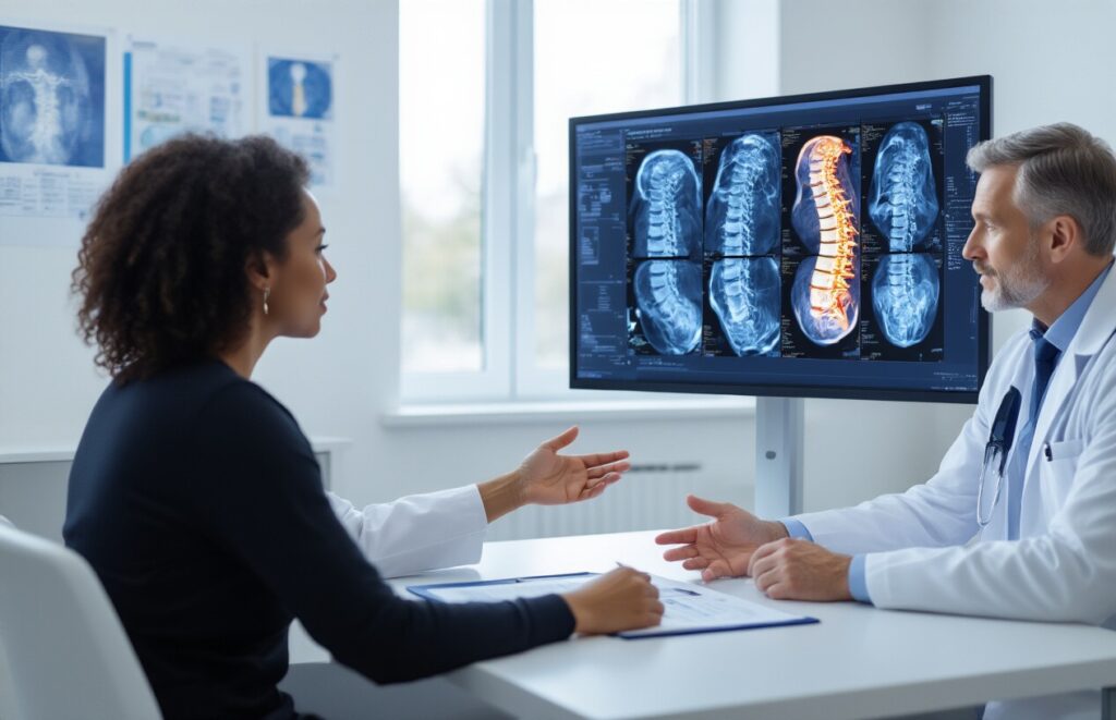
Comprehensive treatment planning advantages
Complete spinal visualization enables medical teams to plan precise surgical approaches and coordinate multidisciplinary care strategies. Surgeons can plan complex procedures with detailed knowledge of anatomical variations, while oncologists use the comprehensive data to stage cancers accurately. This complete picture prevents surgical surprises and enables personalized treatment protocols tailored to each patient’s unique spinal anatomy and pathology distribution.
Preparing for Your Whole Spine MRI Examination
Essential pre-scan preparation steps
Remove all metal objects, including jewelry, watches, hairpins, and clothing with metal zippers or buttons. Wear comfortable, loose-fitting clothes without underwire bras or metallic threads. Inform your doctor about any implanted devices, such as pacemakers, cochlear implants, or metal plates from previous surgeries. Fast for 4-6 hours if contrast dye will be used during your scan.
What to expect during the extended scanning process
Whole spine MRI scans typically take 45-90 minutes, significantly longer than standard single-region scans. You’ll lie flat on a motorized table that moves through the MRI tunnel multiple times to capture images of your cervical, thoracic, and lumbar spine sections. The machine produces loud knocking and buzzing sounds throughout the procedure. Technicians will provide earplugs or headphones and can communicate with you between sequences.
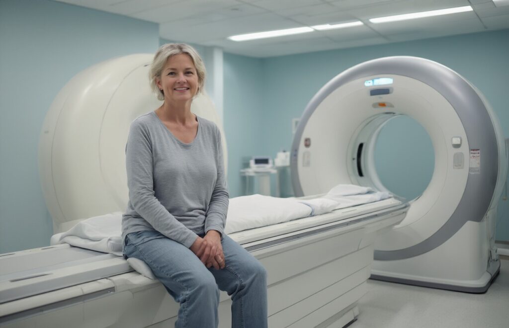
Managing claustrophobia and anxiety during longer scans
| Strategy | Description |
| Breathing exercises | Practice slow, deep breathing to stay calm |
| Visualization | Focus on peaceful scenes or positive thoughts |
| Music/audiobooks | Request headphones for distraction |
| Sedation | Ask about mild anti-anxiety medication if needed |
Many facilities offer open MRI machines or wider bore scanners for claustrophobic patients. Bring a support person who can stay nearby during the procedure. Some centers provide mirrors or prisms that allow you to see outside the machine, reducing the feeling of confinement.
A whole spine MRI gives doctors a fantastic window into your entire spinal column, helping them spot problems they might miss with smaller, targeted scans. This comprehensive screening becomes essential when you’re dealing with unexplained back pain, potential spinal tumors, or complex conditions that could affect multiple areas of your spine. The detailed images show your vertebrae and discs, spinal cord, and nerve roots, making it easier for your medical team to create the right treatment plan.
If your doctor suggests a whole spine MRI, don’t worry about the process – it’s straightforward and completely painless. Take time to prepare properly by removing any metal objects and letting your healthcare team know about any concerns or claustrophobia. This screening could be the key to finally understanding what’s causing your symptoms and getting you on the path to feeling better. Trust your doctor’s recommendation and remember that this detailed look at your spine is one of the best tools available for getting accurate answers about your spinal health.