
If you or a loved one has been diagnosed with lymphoma, your doctor has likely mentioned getting a PET-CT scan for staging. This advanced imaging test combines two powerful technologies to create detailed pictures of your body, helping your medical team determine precisely where the cancer is located and how far it has spread.
This guide is written for lymphoma patients, their families, and caregivers who want to understand what PET-CT staging involves and how to prepare for the best possible results. Getting clear information about this process can help reduce anxiety and ensure you’re ready for your scan.
We’ll walk you through what happens before, during, and after your PET-CT scan, including the preparation steps that can make a real difference in your results. You’ll also learn how doctors use your staging information to create the most effective treatment plan for your specific situation, giving you the knowledge you need to feel confident about the next steps in your care.
Understanding PET-CT Scans and Their Role in Lymphoma Diagnosis

How PET-CT technology combines metabolic and anatomical imaging
PET-CT scans merge two powerful imaging technologies into one comprehensive diagnostic tool. The PET component uses a radioactive tracer that cancer cells absorb more readily than healthy tissue, creating bright spots where lymphoma is present. Meanwhile, the CT portion provides detailed anatomical maps of your body’s structures, showing exactly where organs, bones, and tissues are located.
When these images combine, doctors see both the metabolic activity of cancer cells and their precise anatomical location simultaneously. This dual approach gives your medical team a complete picture that neither scan could provide alone, making it the gold standard for lymphoma staging and monitoring treatment response.
Why PET-CT is superior to traditional CT scans for lymphoma detection
Traditional CT scans rely solely on size and shape to identify suspicious areas, but lymphoma can be tricky. Cancer cells might be present in normal-sized lymph nodes, while enlarged nodes might just be fighting an infection. PET-CT solves this problem by detecting metabolic activity, revealing active lymphoma even in nodes that appear normal on CT alone.
Studies show PET-CT changes staging in about 25% of lymphoma cases compared to CT alone. This accuracy directly impacts your treatment plan, helping doctors avoid under-treating aggressive disease or over-treating less severe cases.
The difference between PET-CT and other imaging methods
| Imaging Method | What It Shows | Best Used For |
| CT Scan | Size, shape, density | Detecting enlarged nodes |
| MRI | Detailed soft tissue contrast | Brain and spinal cord involvement |
| PET-CT | Metabolic activity + anatomy | Staging, treatment monitoring |
| Bone Scan | Bone metabolism | Detecting bone involvement |
PET-CT stands out because it catches lymphoma that other scans miss. While MRI excels at brain imaging and bone scans detect skeletal involvement, PET-CT provides the most comprehensive view for initial staging and tracking how well treatment is working throughout your care journey.
Essential Pre-Scan Preparation Steps for Optimal Results

Dietary restrictions and fasting requirements before your appointment
Your doctor will ask you to fast for at least 6 hours before your PET-CT scan, though some facilities require 12 hours. You can drink plain water during this time, but avoid all food, candy, gum, and beverages containing sugar or calories. This fasting period helps ensure the radioactive glucose tracer distributes properly in your body, giving clearer images of lymphoma activity.
Medication adjustments you may need to discuss with your doctor
Blood sugar medications require special attention before your scan. If you take insulin or diabetes medications, your doctor will provide specific instructions about timing and dosing. Some medications can interfere with scan accuracy, so bring a complete list of everything you take, including supplements and over-the-counter drugs, to discuss with your medical team.
What to wear and bring on scan day
Wear comfortable, loose-fitting clothes without metal components like zippers, buttons, or underwire bras. Leave jewelry, watches, and metal accessories at home. Bring a valid ID, and any previous scan images your doctor requested. Many facilities provide gowns, but comfortable metal-free clothing saves time during preparation.
Managing diabetes and blood sugar levels before the procedure
Diabetic patients need careful blood sugar management before PET-CT scans. Your glucose level should be below 150 mg/dL for optimal imaging results. Check your blood sugar before leaving for your appointment and bring your glucose meter. If levels are too high, your scan may need rescheduling. Work closely with your healthcare team to adjust medication timing while maintaining safe blood sugar levels throughout the fasting period.
Step-by-Step Guide Through Your PET-CT Scanning Experience
The Injection Process and Waiting Period for Tracer Distribution
You’ll receive a small injection of radioactive glucose tracer through an IV line, typically in your arm. After the injection, you’ll need to wait 60-90 minutes in a quiet, dimly lit room while the tracer circulates through your body and accumulates in areas with high metabolic activity, including any lymphoma cells.
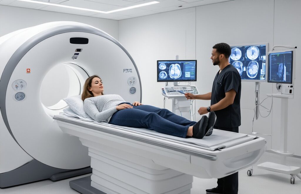
What to Expect During the Actual Scanning Procedure
The PET-CT scanner looks like a large donut with a movable table that slides through the center. You’ll lie still on the padded table as it slowly moves through the scanner, which captures detailed images of your body. The machine makes some clicking and whirring sounds, but it’s not particularly loud or uncomfortable.
How Long Will the Entire Appointment Take
Plan for 2-3 hours total at the imaging center. This includes check-in, tracer injection, the mandatory waiting period, the actual 20-30 minute scan, and a brief post-scan observation. The scanning portion itself is relatively quick, but the preparation and waiting time make up most of your visit.
Comfort Measures and Positioning During the Scan
| Comfort Feature | Description |
| Temperature Control | Blankets available; room kept cool |
| Positioning Aids | Cushions and supports for comfort |
| Communication | An intercom system to talk with the technologist |
| Movement Restrictions | Must remain still; brief position changes allowed |
You’ll be positioned on your back with your arms raised above your head. The technologist will provide pillows, cushions, and blankets to help you stay comfortable during the scan. While you need to remain as still as possible, you can breathe normally, and the staff can hear you if you need anything.
Safety Protocols and Radiation Exposure Considerations
The radiation exposure from a PET-CT scan is similar to what you’d receive from natural background radiation over 2-3 years. The radioactive tracer leaves your body naturally through urine within 24 hours. You’ll be advised to drink plenty of water after the scan and avoid close contact with pregnant women and small children for the rest of the day as a precaution.
Interpreting Your PET-CT Results and Staging Information

How doctors use SUV values to assess lymphoma activity
SUV (Standardized Uptake Value) measures how much glucose your lymphoma cells absorb during the PET scan. Normal tissue typically shows SUV values below 2.5, while lymphoma often shows SUV values above 3-5. Your oncologist looks at both the absolute numbers and changes between scans to track treatment response and disease progression.
Understanding the different lymphoma staging systems
The Ann Arbor staging system classifies lymphoma from Stage I (single lymph node region) to Stage IV (widespread disease in organs). Each stage includes A or B designations – B means you’ve experienced symptoms like night sweats, fever, or weight loss. This classification directly influences your treatment approach and prognosis discussions with your medical team.
What abnormal findings mean for your treatment plan
Abnormal PET-CT results don’t automatically mean poor outcomes. High SUV values in multiple locations might indicate aggressive disease requiring immediate treatment, while isolated hot spots could suggest localized disease with excellent cure rates. Your doctor considers these findings alongside your symptoms, blood work, and overall health to create your personalized treatment strategy.
Timeline for receiving and discussing your results
Most patients receive preliminary results within 24-48 hours, with complete reports available within a week. Your oncologist will schedule a dedicated appointment to review findings rather than delivering results over the phone. This face-to-face discussion allows for detailed explanation of images, staging information, and immediate answers to your questions about next steps.
Making Informed Treatment Decisions Based on Your Staging Results

How staging results influence chemotherapy and radiation choices
Your PET-CT staging results directly shape your treatment plan. Early-stage lymphoma (stages I-II) often responds well to shorter chemotherapy courses or localized radiation, while advanced stages (III-IV) typically require more intensive, systemic treatments. Your oncologist uses the scan’s detailed mapping of disease spread to determine whether you need aggressive combination therapies or if gentler approaches will be effective.
The role of PET-CT in monitoring treatment response
PET-CT scans become your treatment compass, showing how well therapy is working. Most patients receive interim scans after 2-4 cycles of chemotherapy to assess response. These mid-treatment images help your medical team decide whether to continue the current regimen or switch strategies. The scans measure metabolic activity changes in lymph nodes and organs, providing clearer progress indicators than size measurements alone.
When follow-up scans will be scheduled
Your follow-up schedule depends on your specific lymphoma type and treatment response. Most patients undergo scans at treatment completion, then every 3-6 months for the first two years. After achieving remission, scans typically become less frequent – every 6-12 months. High-risk patients or those with aggressive lymphomas may need more frequent monitoring, while slow-growing types require less intensive surveillance schedules.
Questions to ask your oncologist about your specific results
Ask your doctor to explain your staging in simple terms and what it means for your prognosis. Key questions include: “What specific areas show lymphoma activity?” “How does my staging affect treatment duration?” “What response should we expect to see on follow-up scans?” Request copies of your results and ask about any areas of concern or uncertainty in the imaging.

PET-CT scans have become a game-changer for lymphoma patients, offering doctors the detailed roadmap they need to create the most effective treatment plan. From understanding what the scan actually does to preparing properly and knowing what to expect during the procedure, being informed helps reduce anxiety and ensures you get the best possible results. The staging information from your PET-CT isn’t just medical jargon – it’s the foundation for your entire treatment journey.
Armed with knowledge about your PET-CT results, you can have meaningful conversations with your healthcare team and feel confident about the treatment decisions ahead. Don’t hesitate to ask questions about your staging results or request clarification on anything that seems unclear. Your active participation in understanding these results will help you feel more in control of your lymphoma treatment and recovery process.