

“What is MRI Defecography? Purpose, Procedure & Clinical Benefits Explained”
MRI defecography is a specialized imaging test that helps doctors see how your pelvic floor muscles and organs work during bowel movements. This non-invasive procedure gives your healthcare team detailed pictures of what’s happening inside your pelvis when you’re having trouble with bowel control or evacuation.
This guide is for patients who’ve been scheduled for an MRI defecography, their family members, and anyone wanting to understand this important diagnostic tool for pelvic floor dysfunction. If you’re dealing with chronic constipation, fecal incontinence, or other bowel issues that haven’t responded to initial treatments, your doctor might recommend this test to get a clearer picture of what’s going on.
We’ll walk you through everything you need to know about MRI defecography preparation, including the simple steps you can take at home to get ready for your appointment. You’ll also learn about the MRI defecography process itself – what happens during the scan, how long it takes, and what to expect while you’re there. Finally, we’ll cover how doctors read your defecography test results and what treatment options might be available based on what the pelvic floor imaging reveals.
Understanding MRI Defecography and Its Medical Purpose

What MRI defecography reveals about pelvic floor dysfunction
MRI defecography provides doctors with an incredibly detailed view of how your pelvic floor muscles, organs, and tissues work together during bowel movements. This advanced imaging technique captures real-time images as you strain and evacuate contrast material, revealing problems that might be invisible during regular physical exams or other tests.
The procedure excels at diagnosing conditions like pelvic organ prolapse, where organs like the bladder, uterus, or rectum drop from their normal positions. It can spot rectoceles (bulging of the rectal wall), enteroceles (small bowel hernias), and intussusception (when part of the intestine folds into itself). MRI defecography also identifies structural abnormalities in the anal sphincter muscles and evaluates how well different parts of your pelvic floor coordinate during defecation.
What makes this imaging so valuable is its ability to show dynamic function rather than just static anatomy. While you might experience symptoms like incomplete evacuation, straining, or pelvic pressure, the root cause often lies in complex muscle coordination problems that only become apparent when observed in action. The detailed soft tissue contrast that MRI provides helps doctors distinguish between different types of tissue and identify subtle changes that could explain your symptoms.
How this advanced imaging differs from traditional defecography methods
Traditional defecography relies on X-ray imaging with barium contrast, which primarily shows the outline of your rectum and anal canal. While this older method can detect some structural problems, it has significant limitations compared to MRI defecography. X-ray defecography only provides a two-dimensional view and exposes you to ionizing radiation.
MRI defecography offers superior soft tissue contrast without any radiation exposure, making it safer for repeat studies if needed. The multi-planar imaging capability means doctors can view your pelvic floor from multiple angles – front to back, side to side, and top to bottom – creating a comprehensive three-dimensional understanding of the problem.
The enhanced detail of MRI allows visualization of structures that X-ray defecography simply cannot show clearly. This includes the levator ani muscles, different layers of the anal sphincter complex, and surrounding organs like the bladder and uterus. MRI can also detect small amounts of fluid or inflammation that might indicate infection or other complications.
Another major advantage is the ability to perform the study without inserting tubes or catheters into your rectum, which many patients find more comfortable. The contrast material used in MRI defecography is typically a gel that you can insert yourself privately, reducing anxiety and making the experience more tolerable than traditional methods.
Preparing for Your MRI Defecography Procedure
Essential pre-procedure dietary and medication guidelines
Your doctor will provide specific instructions about what to eat and avoid before your MRI defecography procedure. Most patients need to follow a clear liquid diet for 24 hours before the test. This means you can have water, clear broths, plain tea or coffee without milk, clear juices like apple or white grape juice, and gelatin desserts. Avoid anything with red or purple coloring, as these can interfere with the imaging.
Stop eating solid foods at least 12 hours before your appointment. Your medical team might also ask you to avoid certain medications that could affect bowel function, especially anti-diarrheal medications or opioid pain relievers. If you take regular medications for chronic conditions, ask your doctor which ones you should continue taking and which ones to temporarily stop.
Some facilities require patients to take a bowel preparation solution similar to what’s used for colonoscopies, though this varies by location. Always follow your specific center’s guidelines, as MRI defecography preparation can differ between hospitals and imaging centers.
Remember to stay well-hydrated with clear fluids unless your doctor tells you otherwise. Dehydration can make the procedure more uncomfortable and may affect image quality.
What to expect during the bowel preparation process
The bowel preparation for MRI defecography is generally less intensive than what you’d experience before a colonoscopy, but it’s still important for getting clear, accurate images. Your preparation typically begins the day before your scheduled procedure.
You’ll receive detailed instructions about timing your meals and when to start the clear liquid diet. Some patients receive a mild laxative or enema to ensure the rectum is adequately prepared for the contrast material that will be used during the scan. The goal isn’t complete bowel emptying like with other procedures, but rather ensuring optimal conditions for the defecography process.
On the day of your MRI defecography, you might receive additional preparation at the imaging center. This could include a small enema with contrast material that helps visualize the rectal structures during the scan. The contrast material is typically a thick, paste-like substance that mimics the consistency of stool, allowing doctors to see how your pelvic floor muscles and organs function during bowel movements.
Don’t worry if you feel some discomfort or urgency after receiving the contrast material – this is completely normal and expected. The imaging technologist will guide you through the entire process and let you know what to expect at each step. Most patients find the preparation manageable, and any discomfort is temporary.
Step-by-Step Breakdown of the MRI Defecography Process

Initial positioning and contrast material administration
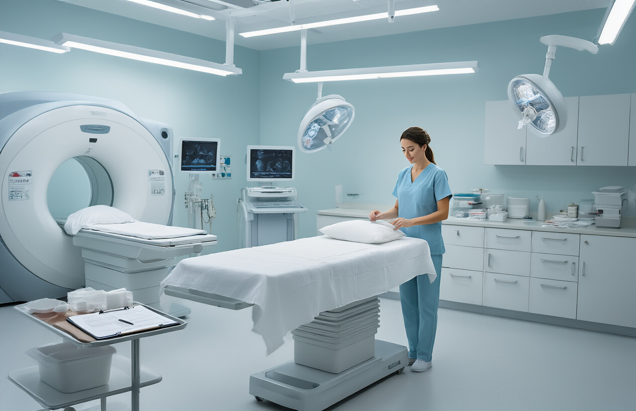
The MRI defecography process begins with careful patient positioning to ensure optimal imaging quality. You’ll lie on your side on the MRI table, typically in a left lateral position, which allows for the best visualization of your pelvic floor structures. The technologist will position you comfortably and may use cushions or supports to help you maintain the proper alignment throughout the procedure.
Before imaging begins, contrast material administration is a crucial step in the defecography procedure. The radiologist or technologist will introduce a thick, gel-like contrast material into your rectum through a small, flexible tube. This contrast agent, often barium-based or gadolinium-enhanced, helps create clear visibility of your rectal walls and surrounding pelvic floor muscles during the MRI pelvic floor dysfunction evaluation.
The contrast material has a consistency similar to thick pudding, designed to mimic the texture of stool while providing excellent image contrast. The amount administered is typically around 120-150 milliliters, enough to adequately distend the rectum without causing excessive discomfort. Some facilities may also use vaginal contrast in female patients to better visualize the relationship between pelvic organs.
During this preparation phase, the medical team ensures you’re comfortable and explains each step. The contrast material may feel cool initially, and you might experience a sensation of fullness, which is completely normal and expected for successful pelvic floor imaging.
Real-time imaging during simulated defecation
The most critical phase of the MRI defecography involves real-time imaging while you perform simulated defecation maneuvers. Unlike traditional static MRI scans, this dynamic imaging captures your pelvic floor muscles and organs in motion, providing invaluable diagnostic information about functional abnormalities.
You’ll be instructed to perform several different maneuvers while the MRI machine captures continuous images. These include squeezing (contracting your anal sphincter muscles), bearing down as if having a bowel movement, and attempting to evacuate the contrast material. The technologist will guide you through each phase, typically asking you to hold each position for 15-30 seconds while images are acquired.
The MRI defecography process captures multiple anatomical changes in real-time. During the evacuation phase, the images reveal how your rectum empties, whether there’s complete evacuation, and if any structural abnormalities impede normal function. The scan shows the movement of your pelvic floor muscles, the descent of pelvic organs, and any prolapse or herniation that occurs during straining.
Advanced MRI sequences can capture images at several frames per second, creating a movie-like representation of your pelvic floor function. This dynamic imaging is what sets MRI defecography apart from other diagnostic tests, as it evaluates both structure and function simultaneously. The entire imaging portion typically takes 20-30 minutes, with multiple sequences capturing different aspects of pelvic floor movement and function.
Interpreting Your MRI Defecography Results

Common conditions detected through dynamic pelvic imaging
MRI defecography excels at revealing a wide range of pelvic floor dysfunctions that might remain hidden with traditional static imaging. Rectoceles rank among the most frequently detected conditions, where the rectal wall bulges into the vaginal canal due to weakened tissue support. These bulges can trap stool and make complete evacuation difficult.
Enteroceles present another common finding, characterized by small intestine dropping down into the pelvic cavity. This condition often develops when the supporting ligaments stretch or tear, creating a pocket where bowel loops can settle. Similarly, sigmoidoceles involve the sigmoid colon descending abnormally low during straining.
Intussusception shows up clearly on defecography MRI as one part of the rectum telescopes into another section. This folding action creates a blockage that prevents normal bowel movements and often causes significant discomfort.
Pelvic organ prolapse encompasses multiple structures dropping from their normal positions. Uterine prolapse, vaginal vault prolapse after hysterectomy, and bladder prolapse (cystocele) all become visible as organs shift during the dynamic imaging sequences.
Anismus, also called dyssynergic defecation, appears when the pelvic floor muscles fail to relax properly during evacuation attempts. The imaging captures this paradoxical muscle contraction that prevents normal stool passage despite adequate pushing efforts.
Perineal descent syndrome shows excessive downward movement of the pelvic floor beyond normal limits. This condition often develops from chronic straining and can lead to nerve damage affecting bowel control.
How radiologists analyze pelvic organ movement and positioning
Radiologists examine defecography MRI results by tracking specific anatomical landmarks throughout the imaging sequence. They measure the position of key structures at rest, during straining, and during evacuation to identify abnormal movement patterns.
The anorectal angle serves as a crucial measurement point. Normally, this angle opens from about 90 degrees at rest to approximately 110-130 degrees during defecation. Angles that remain too acute suggest pelvic floor dysfunction, while excessive opening might indicate muscle weakness.
Pelvic floor descent gets quantified by measuring how far structures drop below the pubococcygeal line – an imaginary reference line drawn from the pubic bone to the tailbone. Descent greater than 3 centimeters typically signals abnormal weakness in the supporting tissues.
The rectum’s ability to empty completely receives careful evaluation. Radiologists look for retained contrast material after the patient completes evacuation attempts. Significant retention often points to mechanical obstructions or coordination problems between different muscle groups.
Organ compartment interactions demand detailed analysis since problems rarely occur in isolation. A rectocele might push against the vaginal wall, affecting bladder function. Multiple compartment prolapses often compound each other’s effects, creating complex symptom patterns.
Real-time sequences allow radiologists to observe the timing and coordination of muscle contractions. They can spot when muscles contract out of sequence or fail to work together properly. This timing analysis proves especially valuable for diagnosing functional disorders that don’t show obvious structural abnormalities.
Radiologists also assess tissue support structures, looking for tears or stretching in the fascia and ligaments that normally hold organs in place. These soft tissue changes often precede more obvious organ displacement.
Treatment Options Based on MRI Defecography Findings

Conservative Management Approaches for Mild Dysfunction
When your MRI defecography results show mild pelvic floor dysfunction, doctors typically recommend non-surgical treatments first. These approaches work well for many patients and can significantly improve quality of life without the risks associated with surgery.
Pelvic floor physical therapy stands as the cornerstone of conservative treatment. Specialized therapists teach targeted exercises to strengthen weakened muscles and improve coordination between different pelvic floor muscle groups. Biofeedback training often accompanies these exercises, helping patients learn proper muscle control through visual or audio cues. Many patients see meaningful improvement within 8-12 weeks of consistent therapy.
Dietary modifications play a crucial role, particularly for those with constipation-related dysfunction. Increasing fiber intake gradually, staying well-hydrated, and identifying trigger foods can make a substantial difference. Some patients benefit from working with nutritionists who specialize in digestive health.
Bowel training programs help establish regular patterns and reduce straining. These programs teach proper positioning, breathing techniques, and timing strategies. Simple changes like using a footstool during bowel movements can improve the anorectal angle and reduce dysfunction.
Medications may support conservative treatment. Stool softeners, fiber supplements, or specific medications for chronic constipation can complement other therapies. For patients with pelvic organ prolapse, pessaries offer non-surgical support and symptom relief.
Lifestyle adjustments round out the conservative approach. Weight management, avoiding heavy lifting, and treating chronic cough can reduce pressure on already compromised pelvic floor structures.
Surgical Intervention Criteria and Procedures
Surgery becomes necessary when MRI defecography reveals severe dysfunction that doesn’t respond to conservative treatment, or when structural abnormalities require surgical correction. The decision involves careful consideration of symptom severity, impact on daily life, and potential surgical risks.
Rectocele repair addresses posterior vaginal wall weakness that allows the rectum to bulge forward. Surgeons typically recommend this when the rectocele measures larger than 2-3 centimeters on MRI pelvic floor dysfunction imaging and causes significant symptoms like incomplete bowel movements or the need for manual assistance.
For rectal prolapse identified through defecography MRI, surgical options include rectopexy procedures that secure the rectum to surrounding structures, or resection surgeries that remove excess bowel tissue. The choice depends on patient age, overall health, and the extent of prolapse shown in MRI defecography results.
Enterocele repair becomes necessary when small bowel herniation into the pelvis causes discomfort or interferes with normal function. These procedures typically involve closing the defect and providing additional support to prevent recurrence.
Complex cases may require combined procedures. Surgeons might address multiple issues during one operation, such as repairing both rectocele and enterocele simultaneously. Recovery varies depending on the specific procedure, but most patients can expect several weeks of limited activity and gradual return to normal function.
Minimally invasive techniques, including laparoscopic and robotic approaches, offer reduced recovery times and smaller incisions compared to traditional open surgery. However, not all patients qualify for these approaches based on their specific anatomy and the extent of repair needed.

MRI defecography offers doctors a clear window into how your pelvic floor muscles and organs work during bowel movements. This specialized imaging test helps identify problems like pelvic organ prolapse, muscle weakness, or structural issues that might be causing your symptoms. The preparation is straightforward, and the actual procedure gives your medical team detailed information they simply can’t get from other tests.
Your results will guide your doctor toward the right treatment plan for your specific situation. Whether that means physical therapy, lifestyle changes, medications, or surgical options, having accurate imaging data makes all the difference in getting you the relief you need. Talk to your healthcare provider about whether MRI defecography could be the key to understanding and treating your pelvic floor concerns.