

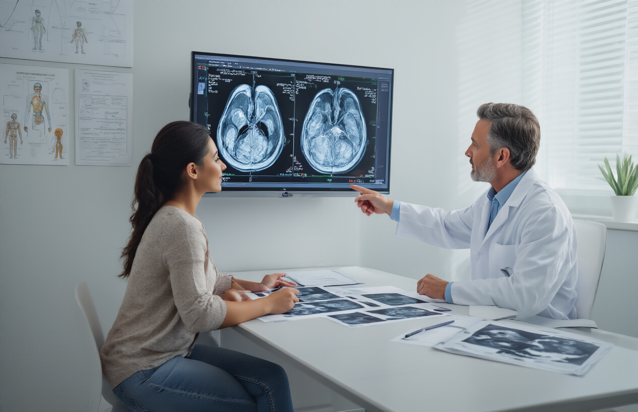
“What is an MRI Brachial Plexus With Contrast? Purpose, Procedure & Benefits”
MRI Brachial Plexus with Contrast: A Complete Guide for Patients and Healthcare Professionals
If you’re dealing with unexplained arm pain, weakness, or numbness, your doctor might recommend an MRI brachial plexus with contrast to get to the bottom of what’s happening. This specialized imaging technique uses gadolinium MRI brachial plexus enhancement to create detailed pictures of the complex network of nerves that control your shoulder, arm, and hand.
This guide is designed for patients preparing for the procedure, healthcare professionals seeking procedural insights, and anyone wanting to understand how contrast-enhanced brachial plexus imaging works. We’ll walk you through the essential brachial plexus anatomy and common nerve disorders that might require this advanced diagnostic tool, explain exactly when doctors turn to contrast enhancement for better visualization of nerve root compression and other issues, and break down what happens during the MRI preparation and contrast injection process.
You’ll also learn how to read and make sense of your brachial plexus MRI contrast enhancement results, giving you the knowledge to have more informed conversations with your healthcare team about your diagnosis and treatment options.
Understanding Brachial Plexus Anatomy and Common Disorders
Essential nerve network structure from neck to arm
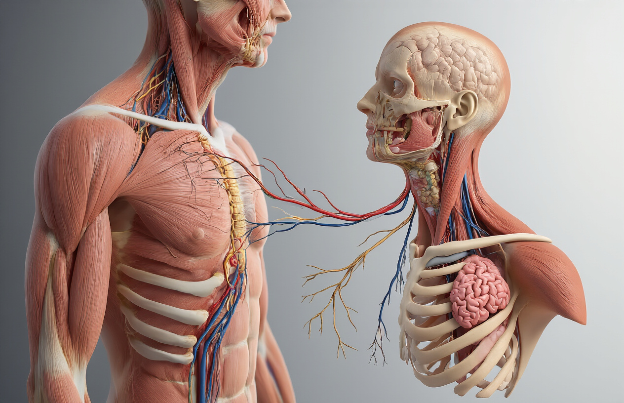
The brachial plexus forms a complex web of nerves that originates from the spinal cord at cervical vertebrae C5 through thoracic vertebra T1. Think of it as the body’s electrical junction box that connects your brain to your arm. These five nerve roots merge and divide multiple times, creating trunks, divisions, cords, and finally the major peripheral nerves that control arm movement and sensation.
MRI brachial plexus anatomy shows this intricate network beginning at the nerve roots as they exit the spinal column. The roots combine to form three main trunks: upper (C5-C6), middle (C7), and lower (C8-T1). Each trunk splits into anterior and posterior divisions, which then reorganize into three cords – lateral, posterior, and medial. These cords give rise to the terminal nerves including the median, ulnar, radial, axillary, and musculocutaneous nerves.
Brachial plexus MRI contrast enhancement helps radiologists distinguish between healthy nerve tissue and surrounding structures like blood vessels, muscles, and scar tissue. The contrast agent improves visualization of nerve inflammation, tumors, or other pathological changes that might not be clearly visible on standard MRI sequences.
The plexus travels through several anatomical regions, making it vulnerable to injury at different locations. It passes between the anterior and middle scalene muscles, under the clavicle, and behind the pectoralis minor muscle before branching into the arm. Each location presents unique challenges for imaging and potential sites for nerve compression or injury.
Most frequent injuries and conditions affecting the plexus
Brachial plexus nerve disorders MRI can detect a wide range of conditions that affect this critical nerve network. Traumatic injuries represent the most dramatic category, often resulting from motorcycle accidents, sports injuries, or difficult childbirth deliveries. These injuries can range from minor stretching (neurapraxia) to complete nerve root avulsions where the nerve tears away from the spinal cord.
Birth-related brachial plexus injuries, including Erb’s palsy and Klumpke’s paralysis, affect different portions of the nerve network. Erb’s palsy involves the upper trunk (C5-C6), causing weakness in shoulder and elbow muscles, while Klumpke’s paralysis affects the lower trunk (C8-T1), impacting hand and finger function. Contrast-enhanced brachial plexus imaging helps determine the extent of injury and guides treatment decisions.
Thoracic outlet syndrome creates another common category of brachial plexus problems. This condition occurs when nerves and blood vessels become compressed in the narrow space between the collarbone and first rib. Patients typically experience numbness, tingling, and weakness in the arm and hand, especially when raising their arms overhead.
Tumors affecting the brachial plexus include both primary nerve tumors like schwannomas and neurofibromas, as well as metastatic disease from breast, lung, or lymphatic cancers. Gadolinium MRI brachial plexus studies excel at identifying these masses and determining their relationship to surrounding structures.
Nerve root compression MRI contrast studies also reveal cervical radiculopathy, where herniated discs or bone spurs compress nerve roots as they exit the spine. This condition can mimic brachial plexus disorders but requires different treatment approaches.
Inflammatory conditions like neuralgic amyotrophy (Parsonage-Turner syndrome) cause sudden, severe shoulder pain followed by weakness and muscle wasting. These episodes can affect single or multiple nerves within the plexus, creating complex patterns of dysfunction that brachial plexus injury diagnosis through MRI helps clarify.
When Contrast-Enhanced MRI Becomes Necessary
Limitations of Non-Contrast MRI for Nerve Visualization
Standard MRI scans without contrast can show the basic structure of the brachial plexus, but they often fall short when doctors need detailed information about nerve inflammation, scar tissue, or subtle abnormalities. Non-contrast imaging struggles to differentiate between healthy nerve tissue and areas affected by inflammation or fibrosis. The natural contrast between nerves and surrounding soft tissues isn’t always sufficient to reveal critical details that could change your treatment plan.
Nerve roots and smaller nerve branches can appear similar to nearby blood vessels or lymph nodes on standard scans, making it challenging for radiologists to pinpoint exactly where problems exist. When you’re dealing with complex conditions affecting the brachial plexus nerve disorders MRI visualization becomes crucial for accurate diagnosis. Without contrast enhancement, subtle changes in nerve tissue that indicate early disease processes might go undetected.
Blood flow patterns around inflamed or compressed nerves also remain invisible on non-contrast scans. This limitation becomes particularly problematic when evaluating whether nerve symptoms stem from active inflammation that might respond to anti-inflammatory treatments, or from permanent structural changes requiring different interventions.
Specific Conditions Requiring Enhanced Imaging Detail
Contrast-enhanced brachial plexus imaging becomes essential when doctors suspect nerve root tumors, including schwannomas or neurofibromas. These growths often enhance dramatically with gadolinium MRI brachial plexus contrast, making them stand out clearly from surrounding tissues. Without contrast, small tumors might blend into the background, potentially delaying critical treatment.
Inflammatory conditions like brachial neuritis or radiation-induced plexopathy require contrast enhancement to assess active inflammation levels. The contrast agent highlights areas where increased blood flow indicates ongoing inflammatory processes, helping doctors determine appropriate treatment timing and monitor response to therapy.
Nerve root compression MRI contrast studies excel at revealing vascular malformations that might compress brachial plexus nerves. These abnormal blood vessel formations become highly visible with contrast injection, allowing surgeons to plan precise interventions while avoiding damage to healthy nerve structures.
Post-surgical evaluation represents another critical application for MRI brachial plexus with contrast. After nerve repair procedures or tumor removal, contrast helps distinguish between expected post-operative changes and concerning complications like infection or tumor recurrence. Scar tissue patterns also become more apparent with contrast enhancement, helping doctors understand whether ongoing symptoms relate to surgical healing or new problems requiring attention.
Pre-Procedure Preparation for Optimal Results

Essential Patient Screening for Contrast Allergies
Before your MRI brachial plexus with contrast, your healthcare team needs to know about any previous reactions to contrast agents. Gadolinium-based contrast materials used in brachial plexus MRI contrast enhancement are generally safer than iodine-based CT contrasts, but allergic reactions can still occur.
Your medical team will ask detailed questions about past MRI or CT scans with contrast. Even mild reactions like nausea, skin rash, or metallic taste from previous procedures matter. These symptoms might seem minor, but they help predict how your body might respond to gadolinium MRI brachial plexus imaging.
People with kidney problems face special risks with contrast agents. Your doctor will review recent blood work, particularly creatinine levels, to ensure your kidneys can safely process the contrast. If you have chronic kidney disease or are on dialysis, alternative imaging approaches might be considered for your brachial plexus nerve disorders MRI.
Asthma patients need extra attention during screening. While not directly related to contrast allergies, severe asthma can complicate any breathing difficulties that might arise during the procedure. Your medical history of drug allergies, especially to shellfish or iodine, will also be carefully reviewed.
Pre-medication with antihistamines or steroids might be recommended if you have a history of mild contrast reactions but still need the enhanced imaging for accurate diagnosis of nerve root compression MRI contrast studies.
Medication Adjustments and Dietary Restrictions
Most medications can continue as normal before your contrast-enhanced brachial plexus imaging, but some adjustments are necessary. If you take metformin for diabetes, your doctor might pause this medication 48 hours before and after the procedure. Metformin combined with contrast can rarely cause a serious condition called lactic acidosis in people with kidney problems.
Blood thinners like warfarin or newer anticoagulants typically don’t need adjustment for MRI procedures since there’s no surgical intervention. However, inform your medical team about all blood-thinning medications you’re taking.
Kidney medications require special attention. ACE inhibitors, ARBs, and diuretics might be temporarily adjusted based on your kidney function tests. Your doctor will provide specific instructions about which medications to continue or pause.
Regarding food and drink, you can eat normally before your MRI preparation contrast injection. Unlike CT scans, MRI procedures don’t require fasting. Staying well-hydrated actually helps your kidneys process the contrast more effectively, so drink plenty of water in the days leading up to your appointment.
Avoid alcohol for 24 hours before the procedure, as it can affect kidney function and potentially interfere with contrast elimination. If you’re breastfeeding, discuss timing with your doctor, though most guidelines suggest you can resume breastfeeding immediately after gadolinium-based contrast administration.
Remove all jewelry and metal objects before arriving, as these can interfere with the magnetic field and affect image quality of your brachial plexus anatomy MRI.
The Contrast-Enhanced MRI Procedure Step-by-Step
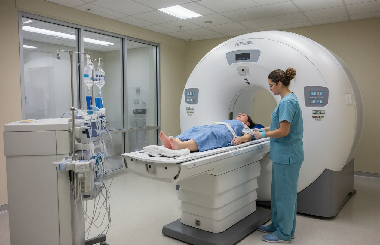
Gadolinium contrast injection process and timing
The gadolinium MRI brachial plexus procedure begins with establishing IV access, typically through a vein in your arm opposite to the affected area. The contrast injection happens in two phases that work together to create detailed images of your nerve structures.
Your technologist will first complete pre-contrast imaging sequences to establish a baseline. This initial phase captures your brachial plexus without enhancement, allowing radiologists to compare before and after images. The actual contrast injection occurs through an automated injector system that delivers gadolinium at a controlled rate, usually around 2-3 mL per second.
Timing plays a critical role in contrast-enhanced brachial plexus imaging. The enhancement peaks at different intervals for various tissues – blood vessels light up almost immediately, while nerve inflammation and scar tissue show maximum enhancement 5-10 minutes post-injection. This delayed enhancement proves especially valuable when diagnosing nerve root compression or distinguishing between active inflammation and chronic changes.
During injection, you might experience a brief metallic taste or slight warmth spreading through your body. These sensations are completely normal and fade quickly. The entire contrast delivery takes less than 30 seconds, but imaging continues for 20-30 minutes afterward to capture the full enhancement pattern.
Multiple imaging sequences and their purposes
MRI brachial plexus with contrast employs several specialized imaging sequences, each designed to highlight different aspects of nerve anatomy and pathology. Understanding these sequences helps explain why your scan takes 45-60 minutes to complete.
T1-weighted sequences with gadolinium provide excellent anatomical detail and show enhanced blood flow to inflamed areas. These images appear bright where contrast accumulates, making inflamed nerve roots and vascular malformations stand out clearly against surrounding tissues. Fat-suppressed T1 sequences eliminate the bright signal from fat, making contrast enhancement even more obvious.
T2-weighted sequences remain the backbone of brachial plexus nerve disorders MRI evaluation. These images show fluid and inflammation as bright signals, helping identify nerve swelling, cysts, or fluid collections around nerve roots. Short tau inversion recovery (STIR) sequences combine T2 weighting with fat suppression, creating exceptional contrast between normal and abnormal tissues.
Diffusion tensor imaging (DTI) sequences track water movement along nerve fibers, providing insights into nerve fiber integrity that conventional imaging cannot reveal. This advanced technique proves particularly valuable for evaluating brachial plexus injury diagnosis when standard sequences appear normal but symptoms persist.
Three-dimensional sequences create detailed reconstructions of the entire brachial plexus network, allowing radiologists to trace individual nerve roots from the spinal cord through the shoulder region. These reconstructions prove invaluable for surgical planning and understanding complex nerve disorders that affect multiple levels simultaneously.
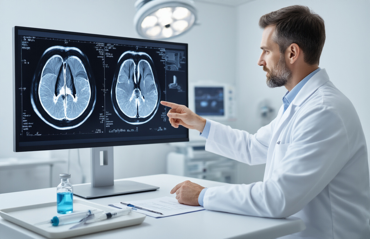
Reading and Interpreting Your MRI Results
Normal versus abnormal contrast enhancement patterns
Normal contrast enhancement patterns in brachial plexus MRI with contrast show minimal to no uptake of gadolinium within healthy nerve tissues. The contrast agent typically highlights blood vessels surrounding the nerve roots and plexus branches, creating a clear outline that helps distinguish neural structures from surrounding soft tissues. Healthy nerve roots appear as dark, well-defined structures against the enhanced vascular background.
Abnormal contrast enhancement patterns present differently depending on the underlying pathology. Inflammatory conditions like brachial neuritis or plexitis show diffuse enhancement along affected nerve segments, appearing brighter on post-contrast images compared to normal tissue. This enhanced signal indicates increased vascular permeability and blood-nerve barrier breakdown, which occurs during active inflammation.
Nerve root compression typically shows focal enhancement at the site of impingement, often accompanied by nerve swelling and signal changes on T2-weighted images. The contrast helps identify the exact location and extent of compression, whether from herniated discs, bone spurs, or other space-occupying lesions.
Tumors demonstrate characteristic enhancement patterns that help differentiate between benign and malignant processes. Schwannomas typically show heterogeneous enhancement with areas of non-enhancing cystic or necrotic components, while neurofibromas often display more uniform contrast uptake.
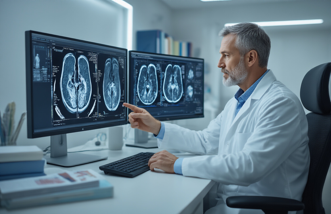
Common findings and their clinical significance
Brachial plexus injury diagnosis through contrast-enhanced imaging reveals several key findings that correlate with specific clinical conditions. Post-traumatic changes following motor vehicle accidents or birth injuries show as nerve discontinuity, pseudomeningoceles, or scarring with abnormal enhancement patterns. These findings help surgeons plan reconstruction procedures and predict functional outcomes.
Thoracic outlet syndrome presents with characteristic enhancement patterns around the scalene muscles and first rib area, where nerve compression occurs. The contrast helps identify vascular compromise alongside neural compression, which influences treatment decisions between conservative management and surgical intervention.
Radiation-induced brachial plexopathy shows delayed enhancement patterns that develop months to years after treatment. Early detection through contrast-enhanced MRI allows for timely intervention before permanent nerve damage occurs. The enhancement patterns help distinguish radiation effects from tumor recurrence, a critical diagnostic challenge in cancer survivors.
Infectious processes like epidural abscesses or osteomyelitis show rim enhancement around fluid collections and intense enhancement of inflamed tissues. These findings guide antibiotic therapy and surgical drainage decisions, particularly important given the proximity to vital neural structures.
Interpreting brachial plexus MRI results requires correlation between enhancement patterns, signal characteristics, and clinical symptoms. The combination provides a comprehensive picture that guides treatment planning and helps predict patient outcomes across various brachial plexus disorders.
Getting an MRI of your brachial plexus with contrast might seem overwhelming at first, but knowing what to expect makes the whole process much smoother. The contrast helps doctors see detailed images of your nerve network, making it easier to spot injuries, tumors, or other issues that might be causing your symptoms. From understanding the basic anatomy to preparing properly and knowing how the procedure works, each step plays an important role in getting accurate results.
Your healthcare team will guide you through every part of the process, from the initial preparation to explaining your results afterward. If your doctor has recommended this scan, it’s because they need the clearest possible view of what’s happening with your nerves. Don’t hesitate to ask questions about anything you’re unsure about – your medical team wants you to feel comfortable and informed throughout your MRI experience.