

“MRI Brain With Diffusion Imaging Explained: Detecting Stroke and Brain Disorders”
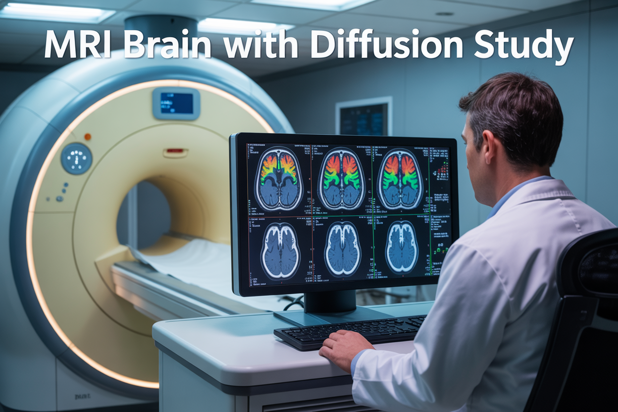
An MRI brain with diffusion study combines traditional brain imaging with advanced diffusion weighted imaging technology to detect medical conditions that regular scans might miss. This specialized brain imaging technology tracks water movement in brain tissue, making it especially powerful for stroke detection MRI and identifying early signs of neurological problems.
This guide is designed for patients scheduled for a DWI brain scan, their families, and anyone wanting to understand how diffusion MRI results can reveal critical health information. We’ll walk you through the essential brain MRI preparation steps you need to know before your appointment, explain what medical conditions doctors can diagnose using this advanced scanning method, and show you how diffusion tensor imaging compares to standard MRI vs CT scan brain options.
You’ll discover why diffusion MRI has become a game-changer in detecting strokes, tumors, and other brain conditions faster and more accurately than ever before.
Understanding MRI Brain Scans with Diffusion Technology
How diffusion MRI reveals brain tissue microstructure
Diffusion weighted imaging (DWI brain scan) works by tracking how water molecules move through brain tissue at a microscopic level. When you think about it, water molecules naturally bounce around in random directions – a process called Brownian motion. In healthy brain tissue, these molecules move freely in all directions, but when tissue is damaged or diseased, their movement becomes restricted.
The MRI brain with diffusion technology measures these subtle changes in water movement by applying special magnetic gradients during the scanning process. Brain tissue acts like a complex maze for water molecules. In areas with healthy white matter, molecules can move easily along nerve fibers but face barriers when trying to cross them. Gray matter has a different structure, so water molecules move more freely in multiple directions.
Diffusion tensor imaging takes this concept even further by mapping the preferred directions of water movement throughout the brain. This creates detailed pictures of white matter tracts – the brain’s internal highways that connect different regions. Radiologists can literally see the brain’s wiring diagram, identifying areas where connections might be damaged or disrupted.
The beauty of this brain imaging technology lies in its sensitivity. Diffusion MRI can detect tissue changes within minutes of a stroke, often before standard MRI shows any abnormalities. It reveals microstructural damage in conditions like traumatic brain injury, where conventional scans might appear normal despite significant neurological symptoms.
Key differences between standard MRI and diffusion studies
Standard MRI creates images based on how different tissues respond to magnetic fields and radio waves. These scans show excellent anatomical detail – you can clearly see the brain’s structure, identify tumors, and spot obvious damage. However, they primarily capture what tissues look like, not necessarily how well they’re functioning.
Diffusion weighted imaging adds a functional dimension to brain scanning. While traditional MRI might show you a road, diffusion MRI tells you whether traffic is flowing smoothly or if there are roadblocks. This functional information proves invaluable for stroke detection MRI protocols, where time is critical.
The scanning process itself differs significantly. Standard brain MRI sequences typically take 20-30 minutes and focus on creating sharp, detailed anatomical images. Diffusion studies require additional time – usually 10-15 minutes extra – because the scanner needs to apply motion-sensitive gradients in multiple directions.
MRI vs CT scan brain comparisons often highlight speed differences, but diffusion MRI occupies a unique middle ground. While CT scans are fastest for emergency situations, and standard MRI provides superior soft tissue contrast, diffusion MRI offers real-time insights into tissue health that neither traditional method can match.
The data processing also sets diffusion studies apart. Standard MRI images can be interpreted immediately after scanning, but diffusion data requires specialized software to create maps showing water movement patterns. These maps reveal information about tissue integrity that remains invisible on conventional scans, making diffusion MRI an essential tool for detecting early disease changes and monitoring treatment responses.
Medical Conditions Diagnosed Through Diffusion MRI

Early stroke detection and treatment planning
MRI brain with diffusion technology has revolutionized how doctors detect strokes, particularly in the critical first hours when every minute counts. Diffusion weighted imaging picks up brain tissue damage within minutes of a stroke, often before traditional MRI scans show any changes. This early detection capability makes the difference between successful treatment and permanent brain damage.
When someone experiences stroke symptoms, DWI brain scan results help emergency teams determine whether the stroke is caused by a blood clot (ischemic) or bleeding (hemorrhagic). The scan reveals areas where water movement becomes restricted in brain cells, indicating tissue that’s dying or already damaged. This information guides doctors in choosing the right treatment approach immediately.
Stroke detection MRI using diffusion imaging also shows the “penumbra” – brain tissue that’s struggling but still salvageable. This zone around the stroke core represents the area doctors can potentially save with quick intervention. Without diffusion MRI, identifying this treatable tissue would be nearly impossible in the acute phase.
Treatment planning becomes more precise when doctors can see exactly which brain regions are affected and how severely. The scan helps determine if clot-busting medications will be effective or if mechanical removal procedures are needed. For patients arriving at the hospital hours after symptom onset, diffusion MRI can still detect recent strokes that might benefit from extended treatment windows, giving hope even when traditional time limits have passed.
Brain tumor characterization and monitoring
Brain imaging technology using diffusion MRI provides detailed information about brain tumors that standard scans simply can’t match. The technique measures how water molecules move through different types of tissue, revealing crucial details about tumor composition, aggressiveness, and response to treatment.
Diffusion imaging helps distinguish between different tumor types by analyzing their cellular density. High-grade malignant tumors typically restrict water movement more than benign growths, giving doctors valuable clues about prognosis before any biopsy is performed. This non-invasive characterization helps surgical teams plan the safest approach for tumor removal or biopsy.
Diffusion tensor imaging takes this analysis even further by mapping white matter tracts around tumors. Surgeons use these detailed maps to navigate around critical brain pathways during operations, reducing the risk of permanent neurological damage. The technology shows whether tumors are pushing important nerve fibers aside or actually invading them, which significantly impacts surgical strategy.
Monitoring treatment response becomes more accurate with diffusion MRI. Changes in water movement patterns often appear weeks before tumor size changes on regular scans. This early insight allows oncologists to adjust treatment plans quickly if current therapies aren’t working, potentially switching to more effective options before valuable time is lost.
The scan also helps differentiate between tumor recurrence and treatment-related changes like radiation necrosis, a common challenge in cancer follow-up care. This distinction is crucial for determining whether additional treatment is needed or if observed changes are simply healing responses.
Preparing for Your Diffusion MRI Brain Study
Essential pre-scan instructions and safety guidelines
Getting ready for your diffusion weighted imaging brain scan requires some simple but important preparation steps. Most hospitals and imaging centers will provide you with a detailed checklist, but knowing what to expect helps reduce anxiety and ensures the best possible results.
Before your MRI brain with diffusion appointment, remove all metal objects including jewelry, watches, hairpins, and hearing aids. Even small amounts of metal can interfere with the powerful magnetic field and affect image quality. If you have any implanted medical devices like pacemakers, cochlear implants, or metal plates from previous surgeries, inform your healthcare provider immediately. Some devices may not be compatible with MRI technology.
Wear comfortable, loose-fitting clothing without metal zippers, buttons, or underwire bras. Many facilities provide hospital gowns to ensure complete metal-free scanning. Avoid wearing makeup, nail polish, or hair products that might contain metallic particles, as these can create image artifacts.
Eat normally before your scan unless your doctor gives specific instructions otherwise. Take your regular medications as prescribed. If you experience claustrophobia or anxiety in enclosed spaces, discuss sedation options with your medical team beforehand. Some patients find it helpful to practice relaxation techniques or bring music to listen to during the procedure.
Arrive 30 minutes early to complete paperwork and change into appropriate clothing. The technologist will review your medical history and explain the procedure, giving you time to ask questions about the diffusion MRI process.
What to expect during the scanning procedure
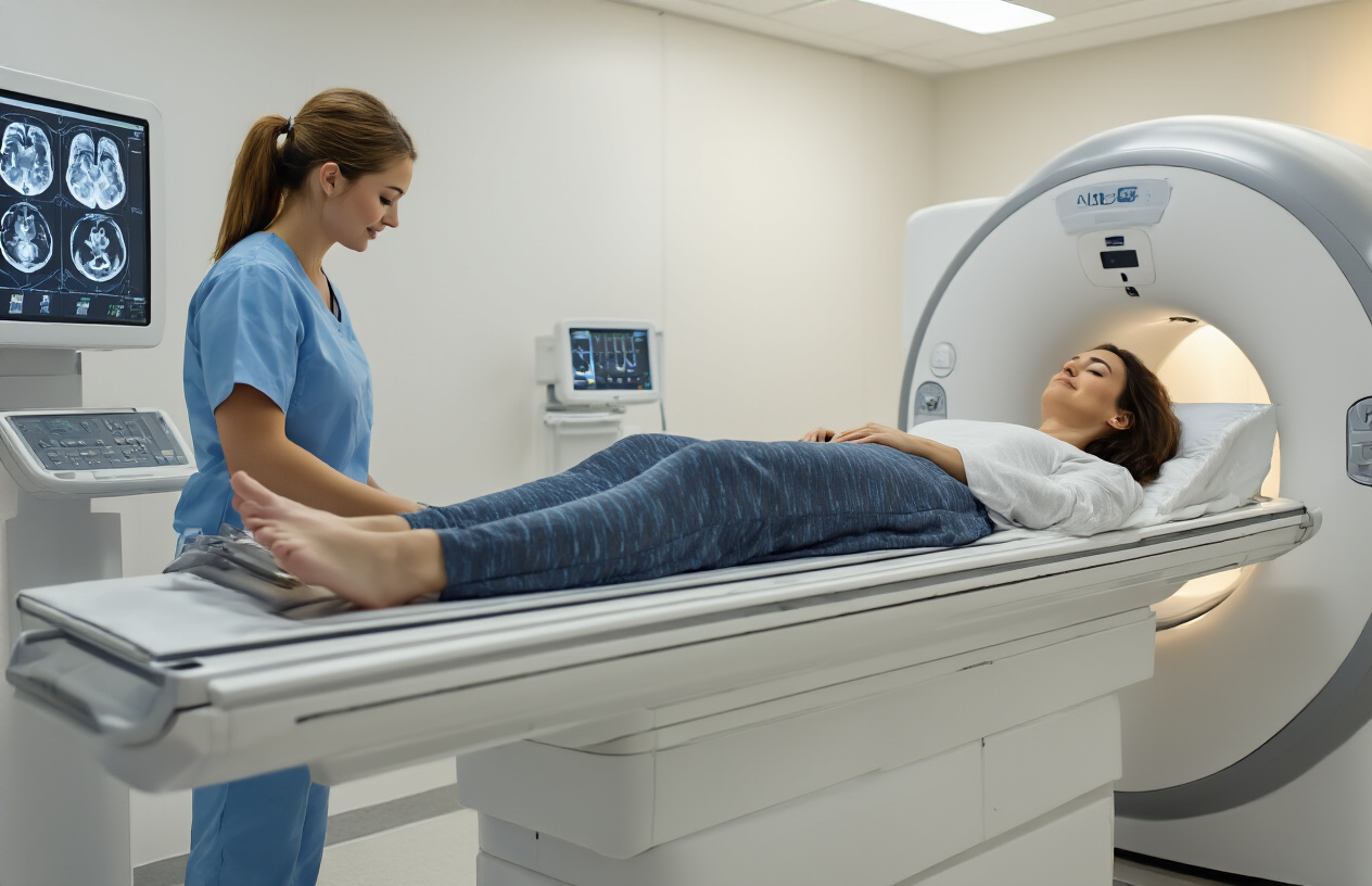
The diffusion MRI brain scan typically takes 30 to 45 minutes, though this can vary depending on the specific sequences needed. You’ll lie on a padded table that slides into the MRI machine, which looks like a large tube or tunnel. A special coil designed for brain imaging will be positioned around your head to capture the detailed images.
During the DWI brain scan, you’ll hear loud knocking, tapping, and buzzing sounds as the machine operates. These noises are completely normal and indicate that the scanner is working properly. Most facilities provide earplugs or headphones to reduce noise levels and may offer music to help you stay relaxed.
The most important thing during your brain MRI preparation is staying as still as possible. Even small movements can blur the images and require repeating certain sequences. The technologist will communicate with you through an intercom system and can stop the scan if you need a break. Some patients find it helpful to focus on breathing slowly and steadily.
The diffusion portion specifically measures how water molecules move through brain tissue, which helps detect conditions like stroke, tumors, and other abnormalities. This technique is particularly valuable for stroke detection MRI because it can identify changes in brain tissue within minutes of an event occurring, often before traditional imaging methods show any problems.
Interpreting Diffusion MRI Results for Better Health Outcomes

Understanding diffusion maps and color-coded images
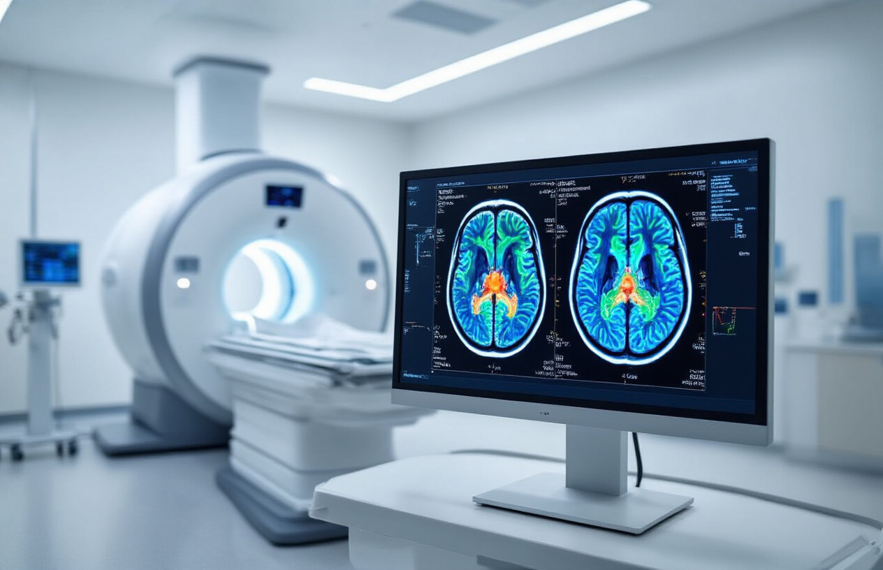
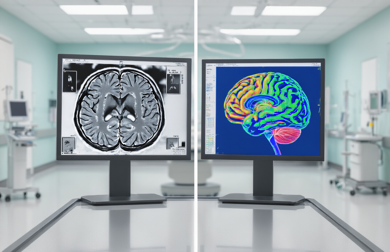
When your radiologist reviews your diffusion MRI results, they’re looking at specialized maps that reveal how water moves through your brain tissue. These aren’t your typical black and white medical images – diffusion weighted imaging produces colorful, detailed pictures that tell a story about your brain’s health at the cellular level.
The most common type you’ll encounter is the ADC (Apparent Diffusion Coefficient) map, which appears in grayscale but provides crucial information about tissue damage. Dark areas on ADC maps often signal restricted water movement, which can indicate acute stroke, infection, or other serious conditions. Bright areas typically show increased water movement, possibly pointing to chronic damage or swelling.
Color-coded directional maps take things a step further, especially in diffusion tensor imaging. These vibrant images use a rainbow of colors to show which direction nerve fibers travel through your brain. Red typically represents left-to-right movement, green shows front-to-back pathways, and blue indicates up-and-down connections. Think of these maps as a GPS system for your brain’s internal highways.
Fractional anisotropy maps display tissue organization using a scale from black to white. White areas show highly organized tissue with strong directional preference – like the major nerve bundles that connect different brain regions. Gray or black areas suggest less organized tissue, which might be normal in certain brain regions or could indicate damage depending on the location.
How radiologists analyze tissue integrity and connectivity
Radiologists approach brain MRI with diffusion analysis like detectives examining evidence. They compare your scans against normal patterns they’ve memorized through years of training, looking for subtle changes that might escape the untrained eye.
The process starts with evaluating tissue microstructure. Your radiologist examines how tightly packed your brain cells are and whether the natural barriers between different tissue types remain intact. When brain tissue gets damaged – whether from stroke, trauma, or disease – these microscopic changes show up clearly on diffusion imaging before they become visible on standard MRI scans.
Connectivity analysis focuses on the white matter tracts that serve as communication cables between different brain areas. Radiologists trace these pathways digitally, checking for interruptions, thinning, or abnormal routing. They pay special attention to critical areas like the corpus callosum, which connects your brain’s left and right hemispheres, and the internal capsule, where motor control fibers travel.
Timing plays a huge role in interpretation. The same brain injury can look completely different on diffusion MRI depending on when the scan happens. Acute strokes show up as bright spots on diffusion weighted imaging within minutes, while chronic damage appears different. Your radiologist considers your symptoms, medical history, and the timing of your scan to piece together the most accurate diagnosis.
They also look for patterns that might suggest specific conditions. Multiple sclerosis creates characteristic lesions, brain tumors alter surrounding tissue in predictable ways, and traumatic brain injuries leave distinct signatures that experienced radiologists can recognize immediately.
Advantages of Diffusion MRI Over Traditional Imaging Methods
Superior sensitivity for detecting acute brain changes
Diffusion weighted imaging stands out as a game-changer when it comes to spotting early signs of brain injury. Traditional MRI sequences often miss the subtle changes that happen in the first few hours after a stroke, but DWI brain scan technology can detect these alterations within minutes of onset. This incredible sensitivity comes from its ability to measure water molecule movement at the cellular level.
When brain cells suffer damage from conditions like stroke, the normal flow of water molecules gets disrupted. Standard MRI scans might show a perfectly normal brain for hours or even days after the initial injury. Diffusion MRI results, however, reveal these microscopic changes almost immediately by tracking how water diffuses through damaged tissue.
Brain imaging technology has come a long way, but diffusion weighted imaging represents a major leap forward in early detection. The technique picks up on reduced water movement in affected areas, appearing as bright spots on the scan. This early warning system gives doctors precious time to intervene with treatments that can prevent permanent damage.
Compared to CT scans, which rely on density differences that develop slowly, diffusion MRI can spot problems while there’s still time to act. This speed advantage has transformed emergency medicine, allowing physicians to distinguish between different types of strokes and start appropriate treatments within the critical time window.
Non-invasive assessment of tissue viability
One of the most impressive features of MRI brain with diffusion technology is its ability to determine whether damaged brain tissue can still be saved. Unlike older imaging methods that simply show where damage has occurred, diffusion weighted imaging reveals which areas might recover with proper treatment.
The technique works by measuring something called the apparent diffusion coefficient, which tells doctors how freely water molecules can move through different brain regions. Healthy tissue allows normal water movement, while severely damaged areas restrict this flow. Most importantly, diffusion MRI can identify the “penumbra” – that crucial zone of threatened but potentially salvageable brain tissue surrounding the core damage.
Traditional imaging methods like standard MRI or CT scans often can’t make this critical distinction between dead tissue and tissue that’s merely stunned. This limitation means doctors have to make treatment decisions based on incomplete information. Diffusion tensor imaging changes this equation by providing a real-time assessment of tissue health.
The non-invasive nature of this assessment means patients don’t need contrast agents or invasive procedures to get this vital information. Doctors can quickly determine whether aggressive treatments like clot-busting medications or mechanical interventions are likely to help. This precision helps avoid unnecessary treatments while ensuring patients who can benefit receive timely intervention.
For stroke detection MRI protocols, this capability has revolutionized patient care by enabling personalized treatment decisions based on each individual’s unique brain response to injury.

Diffusion MRI has transformed how doctors examine brain health, offering detailed insights into conditions like stroke, brain tumors, and neurological disorders that traditional scans might miss. This advanced imaging technique tracks water movement in brain tissue, revealing problems at the cellular level and helping doctors catch issues early when treatment works best.
Getting ready for your diffusion MRI is straightforward – just remove metal objects and follow your doctor’s simple instructions. The scan itself is painless and usually takes about 30-60 minutes. When your results come back, your healthcare team will walk you through the findings and create a treatment plan tailored specifically for you. If your doctor recommends a diffusion MRI brain study, know that you’re getting one of the most precise tools available for understanding what’s happening inside your brain.