

“USG Scrotal Explained: Purpose, Procedure & Benefits of This Ultrasound Test”
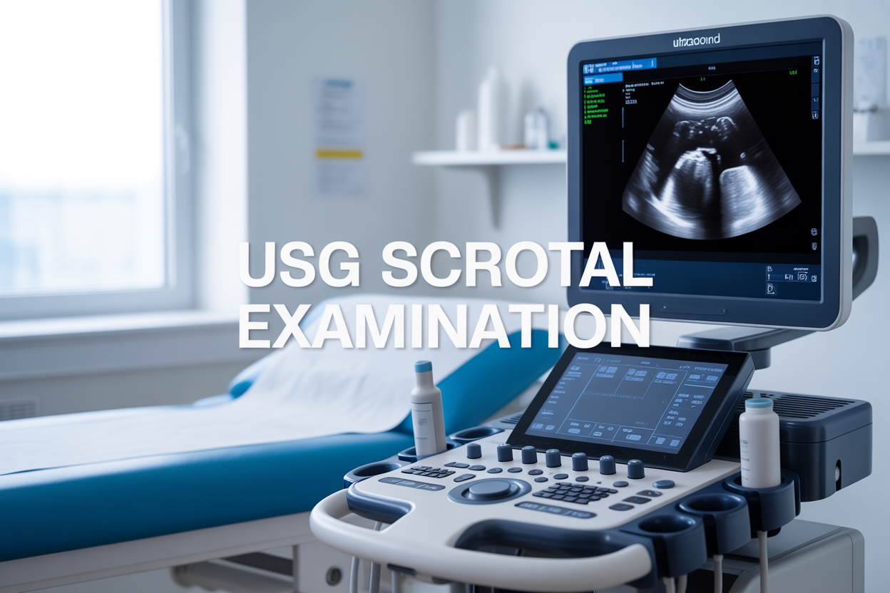
A USG scrotal is a non-invasive ultrasound examination that uses sound waves to create detailed images of the scrotum and testicles. This scrotal ultrasound helps doctors diagnose various testicular conditions and evaluate scrotal pain without discomfort or radiation exposure.
This guide is for men experiencing scrotal symptoms, patients scheduled for testicular ultrasound, and anyone wanting to understand this common diagnostic procedure. Whether you’re dealing with pain, swelling, or lumps, knowing what to expect can ease your concerns.
We’ll walk you through how scrotal imaging technology works and why doctors recommend this examination for different symptoms. You’ll also learn about the simple preparation steps that help ensure clear, accurate results during your ultrasound scrotum procedure. Finally, we’ll explain what happens during the exam and how doctors interpret your results to determine the best treatment plan.
Understanding Ultrasound Technology for Scrotal Examination
How ultrasound waves create detailed images of scrotal structures
USG scrotal imaging relies on high-frequency sound waves that bounce off tissues to create real-time pictures of everything inside the scrotum. When the ultrasound transducer touches your skin, it sends out sound waves at frequencies typically ranging from 7.5 to 15 MHz – much higher than what humans can hear.
These sound waves travel through the scrotal tissues at different speeds depending on what they encounter. When they hit boundaries between different tissue types – like the wall of a blood vessel, the edge of a testis, or fluid in the epididymis – some waves bounce back to the transducer. The ultrasound machine measures how long it takes for these echoes to return and uses this timing to calculate distances and create images.
Different tissues reflect sound waves differently, which is why you can see distinct structures on a scrotal ultrasound. Dense tissues like the testicular tissue appear brighter (hyperechoic), while fluid-filled areas like cysts show up as dark spaces (hypoechoic or anechoic). Blood vessels become visible through Doppler ultrasound, which detects moving red blood cells by measuring slight changes in the frequency of reflected sound waves.
The beauty of scrotal ultrasound lies in its ability to show both structure and function simultaneously. You can see the shape and size of the testes, epididymis, and surrounding tissues while also watching blood flow patterns in real-time. This dual capability makes scrotal imaging incredibly valuable for diagnosing conditions ranging from testicular torsion to varicoceles.
Why high-frequency transducers provide superior scrotal imaging
High-frequency transducers are game-changers for scrotal examination because they deliver exceptional image resolution in superficial tissues. The scrotum sits close to the skin surface, making it perfect territory for these specialized probes that typically operate between 10-15 MHz.
Higher frequencies create shorter wavelengths, which translates directly into better image detail. Think of it like using a fine-tip pen versus a thick marker – the fine tip gives you much more precise lines. With scrotal ultrasound, this precision means you can spot tiny abnormalities that might be missed with lower-frequency equipment.
The trade-off with high-frequency waves is that they don’t penetrate as deeply into the body. But this limitation becomes an advantage for scrotal imaging since the structures you need to examine – the testes, epididymis, and scrotal wall – are all within the first few centimeters from the skin surface.
These transducers excel at showing fine anatomical details like the texture of testicular tissue, small cysts in the epididymis, and even subtle changes in blood vessel patterns. They can detect masses as small as 2-3 millimeters, which is critical for early detection of testicular cancer or other conditions.
Modern high-frequency transducers also offer superior color and power Doppler sensitivity, making it easier to evaluate blood flow to the testicles. This capability proves essential when diagnosing conditions like testicular torsion, where blood flow changes can be the key to saving the testicle.
Medical Conditions Detected Through Scrotal Ultrasound
Identifying testicular masses and potential malignancies
Scrotal ultrasound serves as the primary imaging tool for detecting testicular masses, offering unparalleled accuracy in distinguishing between benign and potentially malignant lesions. When doctors perform a testicular ultrasound, they can identify even small masses that might not be palpable during physical examination, making early detection possible.
The ultrasound scrotum examination reveals critical characteristics of testicular masses including size, location, echogenicity, and blood flow patterns. Malignant tumors typically appear as hypoechoic (darker) lesions with irregular borders and increased vascularity on Doppler imaging. Common testicular cancers like seminomas and non-seminomatous germ cell tumors each have distinct ultrasound appearances that help radiologists make accurate assessments.
USG scrotal technology also excels at differentiating testicular masses from other scrotal conditions. Cysts, hydroceles, and varicoceles all have characteristic ultrasound patterns that prevent unnecessary anxiety and invasive procedures. The examination can identify:
- Solid versus cystic lesions
- Intratesticular versus extratesticular masses
- Bilateral involvement patterns
- Associated complications like hemorrhage or infection
Blood flow assessment through color Doppler ultrasound provides additional diagnostic information. Malignant masses often demonstrate increased blood supply with chaotic flow patterns, while benign conditions typically show normal or decreased vascularity. This vascular mapping helps doctors prioritize cases requiring immediate surgical consultation versus those suitable for monitoring.
Diagnosing epididymitis and inflammatory conditions
Scrotal ultrasound proves invaluable for diagnosing epididymitis and related inflammatory conditions affecting the male reproductive system. The examination provides detailed visualization of the epididymis, allowing doctors to assess inflammation severity and identify potential complications before they become serious.
Acute epididymitis appears on scrotal imaging as an enlarged, hypoechoic epididymis with increased blood flow on Doppler studies. The affected epididymis often measures significantly larger than the contralateral side, with heterogeneous echotexture indicating inflammatory changes. Chronic epididymitis presents differently, showing a thickened epididymis with calcifications and reduced blood flow.
Testicular imaging during inflammatory episodes helps distinguish epididymitis from testicular torsion, a urological emergency requiring immediate surgical intervention. While epididymitis shows increased blood flow, testicular torsion demonstrates absent or decreased flow to the affected testis. This distinction can be life-saving for testicular viability.
The scrotal ultrasound procedure also identifies complications of untreated epididymitis including:
| Complication | Ultrasound Appearance |
|---|---|
| Abscess formation | Hypoechoic collections with debris |
| Chronic scarring | Hyperechoic fibrotic changes |
| Reactive hydrocele | Fluid collection around testis |
| Scrotal wall thickening | Increased skin and soft tissue echogenicity |
Scrotal examination through ultrasound enables doctors to monitor treatment response and adjust antibiotic therapy accordingly. The real-time imaging capability allows for immediate assessment during acute scrotal pain diagnosis, providing rapid answers when patients present with severe symptoms requiring urgent evaluation.
Preparation Steps for Optimal Examination Results
Patient positioning techniques for clear visualization
Proper patient positioning plays a crucial role in obtaining high-quality images during a scrotal ultrasound procedure. The most commonly used position involves having the patient lie supine (on their back) on the examination table with their legs slightly separated. This position allows the sonographer optimal access to the scrotal area while keeping the patient comfortable throughout the scrotal ultrasound procedure.
For enhanced visualization, patients are typically asked to elevate the penis and place it on the lower abdomen, securing it with a towel or medical tape. This technique prevents the penis from obstructing the transducer’s contact with the scrotum and ensures comprehensive imaging of both testicles. The scrotum should be supported using a towel placed between the patient’s thighs, which helps maintain proper positioning and reduces movement artifacts that could compromise image quality.
Some facilities may request alternative positioning depending on the specific clinical indication. Side-lying positions might be used for patients who experience discomfort lying flat or when examining specific areas requires a different angle. The key is maintaining consistent contact between the ultrasound probe and the scrotal skin while minimizing patient discomfort.
Testicular ultrasound examinations benefit greatly from proper draping techniques that maintain patient dignity while allowing full access to the examination area. Medical personnel ensure privacy by exposing only the necessary anatomical structures while keeping the rest of the body covered with appropriate draping materials.
Pre-procedure instructions to enhance image quality
Patients undergoing scrotal imaging receive specific pre-procedure instructions designed to optimize examination results. Unlike many other ultrasound procedures, USG scrotal examinations typically require minimal preparation, making them convenient for both patients and healthcare providers.
The most important instruction involves personal hygiene. Patients should shower or clean the scrotal area thoroughly before their appointment to ensure proper contact between the ultrasound gel and skin. Clean skin allows for better transducer gliding and reduces the risk of infection or contamination during the procedure.
Clothing recommendations include wearing loose-fitting garments that can be easily removed from the waist down. Tight underwear or pants can leave compression marks on the skin that might interfere with initial imaging. Patients should avoid applying lotions, powders, or other topical products to the scrotal area on the day of the examination, as these substances can create barriers between the transducer and skin.
Testicular imaging procedures don’t require fasting or special dietary restrictions, unlike abdominal ultrasounds. Patients can eat and drink normally before their scrotal examination. However, they should inform their healthcare provider about any medications they’re taking, particularly blood thinners or pain medications that might affect their comfort level during the procedure.
Mental preparation is equally important. Patients should understand that the examination involves direct contact with sensitive areas, but trained professionals conduct these procedures with utmost professionalism and care. Discussing any concerns or anxiety with the medical team beforehand helps create a more comfortable experience and often leads to better cooperation during the actual ultrasound scrotum examination.
Examination Process and What Patients Can Expect
Step-by-step walkthrough of the scanning procedure
The USG scrotal procedure begins with the patient lying comfortably on an examination table in a supine position. The sonographer will apply a generous amount of warm ultrasound gel to the scrotum, which helps transmit sound waves clearly between the transducer and skin. This gel might feel cool initially but quickly warms to body temperature.
Using a high-frequency linear transducer, typically 7-15 MHz, the technologist starts the scrotal ultrasound by scanning both testicles systematically. The examination begins with grayscale imaging to assess the overall anatomy and structure of each testicle. The sonographer will gently move the probe across different areas of the scrotum, capturing images from multiple angles to ensure comprehensive coverage.
During the testicular ultrasound, both longitudinal and transverse views are obtained for each testicle. The epididymis, which sits behind each testicle, receives careful attention as this structure commonly shows abnormalities. The scanning process includes evaluation of blood flow using color Doppler and spectral Doppler techniques, which help identify vascular issues or inflammation.
The technologist will ask you to remain still during image acquisition but may request position changes or ask you to hold the scrotum in certain positions for optimal visualization. Communication remains open throughout the scrotal examination, with the sonographer explaining each step and addressing any concerns you might have about the ultrasound scrotum procedure.
Duration and comfort considerations during imaging
A typical scrotal imaging session lasts between 20 to 45 minutes, depending on the complexity of the case and whether additional views are needed. Most patients find the scrotal ultrasound procedure quite comfortable, as the examination involves only external scanning with no invasive techniques or radiation exposure.
The ultrasound gel used during testicular imaging might create a slightly messy sensation, but towels are readily available for cleanup afterward. Some patients experience mild sensitivity when the transducer applies pressure to tender areas, particularly if scrotal pain diagnosis is the reason for the examination. The sonographer will adjust pressure accordingly and work with you to minimize any discomfort.
Temperature control in the examination room helps maintain comfort during the procedure. The room is typically kept at a comfortable temperature, and warming gel is used to prevent any shock from cold substances touching sensitive skin. Privacy is carefully maintained throughout the scrotal ultrasound procedure, with only necessary medical personnel present.
Patients can resume normal activities immediately after the examination, as there are no side effects or recovery time needed. The non-invasive nature of ultrasound technology makes this diagnostic tool both safe and patient-friendly. Most people report that the actual experience is much less uncomfortable than they initially anticipated, especially when they understand the importance of the testicular conditions ultrasound in providing accurate diagnostic information.
Interpreting Results and Next Steps
Understanding normal versus abnormal findings
Reading a scrotal ultrasound report can feel overwhelming, but understanding the basics helps patients make sense of their results. Normal scrotal ultrasound findings show testicles with uniform texture and blood flow patterns, clear visualization of the epididymis, and no fluid collections or masses. The testicles should appear symmetrical in size, with typical measurements ranging from 3-5 cm in length and 2-3 cm in width.
Abnormal findings vary widely depending on the underlying condition. Testicular tumors appear as masses with altered tissue patterns, while infections like epididymitis show increased blood flow and thickening of affected structures. Varicoceles present as dilated veins, often described as having a “bag of worms” appearance on the ultrasound images. Testicular torsion reveals decreased or absent blood flow, making this a medical emergency requiring immediate intervention.
Fluid collections around the testicles, called hydroceles, appear as dark areas on the scan and may indicate infection, trauma, or other underlying issues. Cysts typically show up as well-defined, fluid-filled structures that are usually benign but may need monitoring.
The radiologist’s report will describe these findings using specific medical terminology, but your doctor will translate these results into understandable language during your follow-up appointment. They’ll explain whether the findings are concerning and what steps come next.
When additional testing or specialist referral is recommended
Several ultrasound findings prompt doctors to recommend additional testing or specialist consultation. Suspicious masses or nodules almost always require further evaluation, often including blood tests for tumor markers like AFP and beta-hCG, and potentially a urologist referral for surgical evaluation.
Testicular torsion demands immediate surgical intervention, making emergency urology consultation critical. Even suspected torsion based on ultrasound findings requires urgent specialist evaluation, as delays can result in testicular loss.
Chronic pain with normal ultrasound results might warrant MRI imaging for better soft tissue visualization or referral to a pain management specialist. Recurrent infections detected through scrotal ultrasound often need urology evaluation to identify underlying anatomical issues.
Blood flow abnormalities discovered during the testicular ultrasound procedure may require follow-up imaging studies or specialist assessment. Varicoceles causing symptoms or fertility concerns typically need urology consultation to discuss treatment options.
Your primary care doctor will coordinate these referrals based on your specific ultrasound scrotum results and symptoms. They’ll consider factors like your age, symptom severity, and overall health when determining the urgency of specialist consultation. Don’t hesitate to ask questions about timeline expectations and what each recommended next step involves in your particular situation.
Getting a scrotal ultrasound doesn’t have to be scary or mysterious. This simple, painless procedure gives doctors a clear picture of what’s going on inside the scrotum, helping them spot everything from infections to more serious conditions early on. The best part? You don’t need to do much to prepare, and the whole thing is over pretty quickly.
If your doctor recommends a scrotal ultrasound, trust that it’s a valuable tool for keeping your health on track. The results will guide your next steps, whether that’s treatment, monitoring, or simply peace of mind. Don’t hesitate to ask questions during the process – understanding what’s happening makes the experience much easier for everyone involved.